Introduction
Tetralogy of Fallot is the most common cyanotic CHD. Severe left ventricular dysfunction is uncommon in patients with unrepaired tetralogy of Fallot, and the occurrence of extensive left ventricular intracavitary thrombosis in this setting is exceptionally rare. Advances in early diagnosis and surgical repair have markedly improved survival, allowing most patients to reach adulthood, although residual haemodynamic abnormalities and long-term complications remain common. Reference Baumgartner, de Backer and Babu-Narayan1,Reference Gurvitz, Krieger and Fuller2
Case presentation
A 9-year-old boy from Cameroon was referred to our centre through a humanitarian medical programme for evaluation and treatment of tetralogy of Fallot. The patient had experienced exertional dyspnoea and cyanotic episodes since early childhood and was frequently observed assuming the squatting position. In the following years, the patient developed progressive exercise intolerance and recurrent febrile episodes associated with worsening cyanosis.
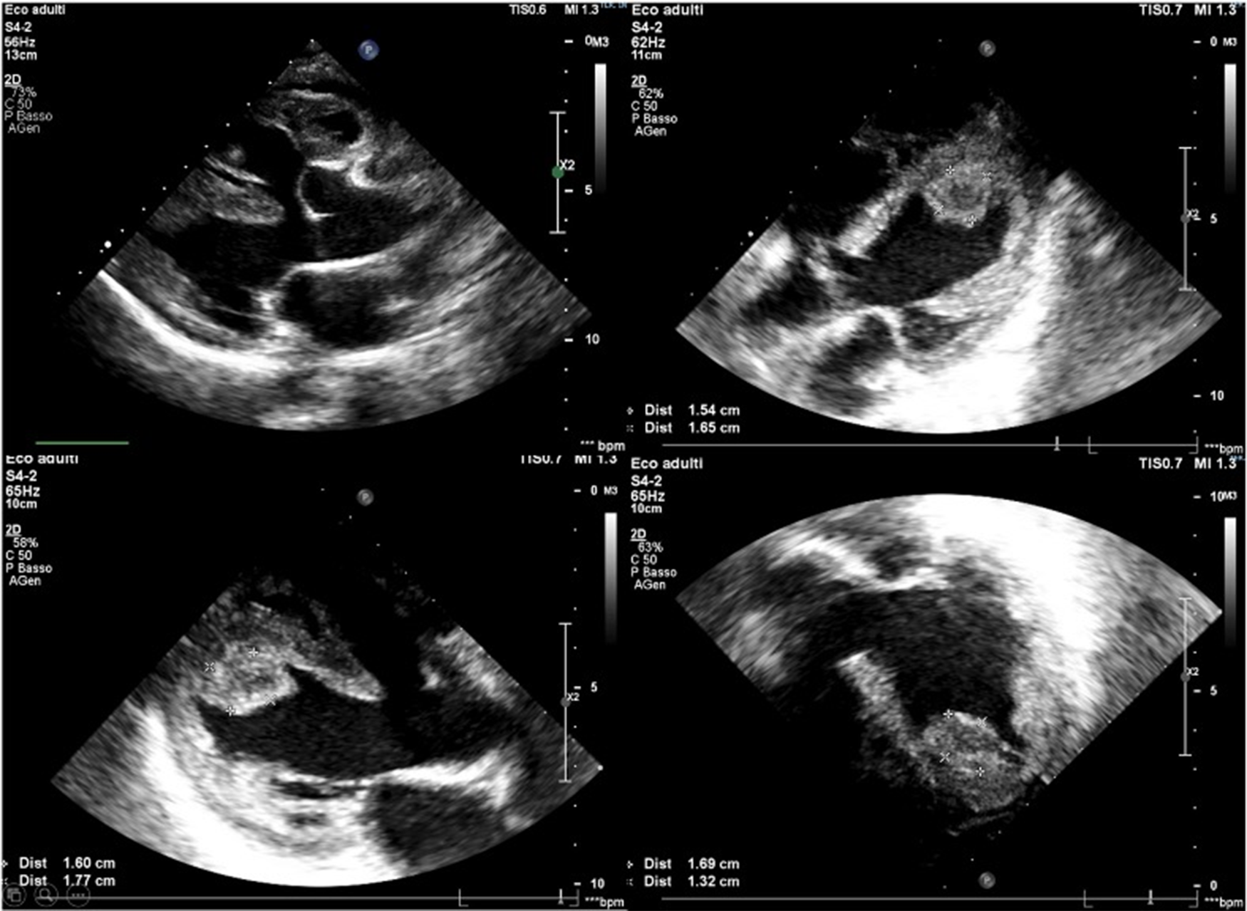
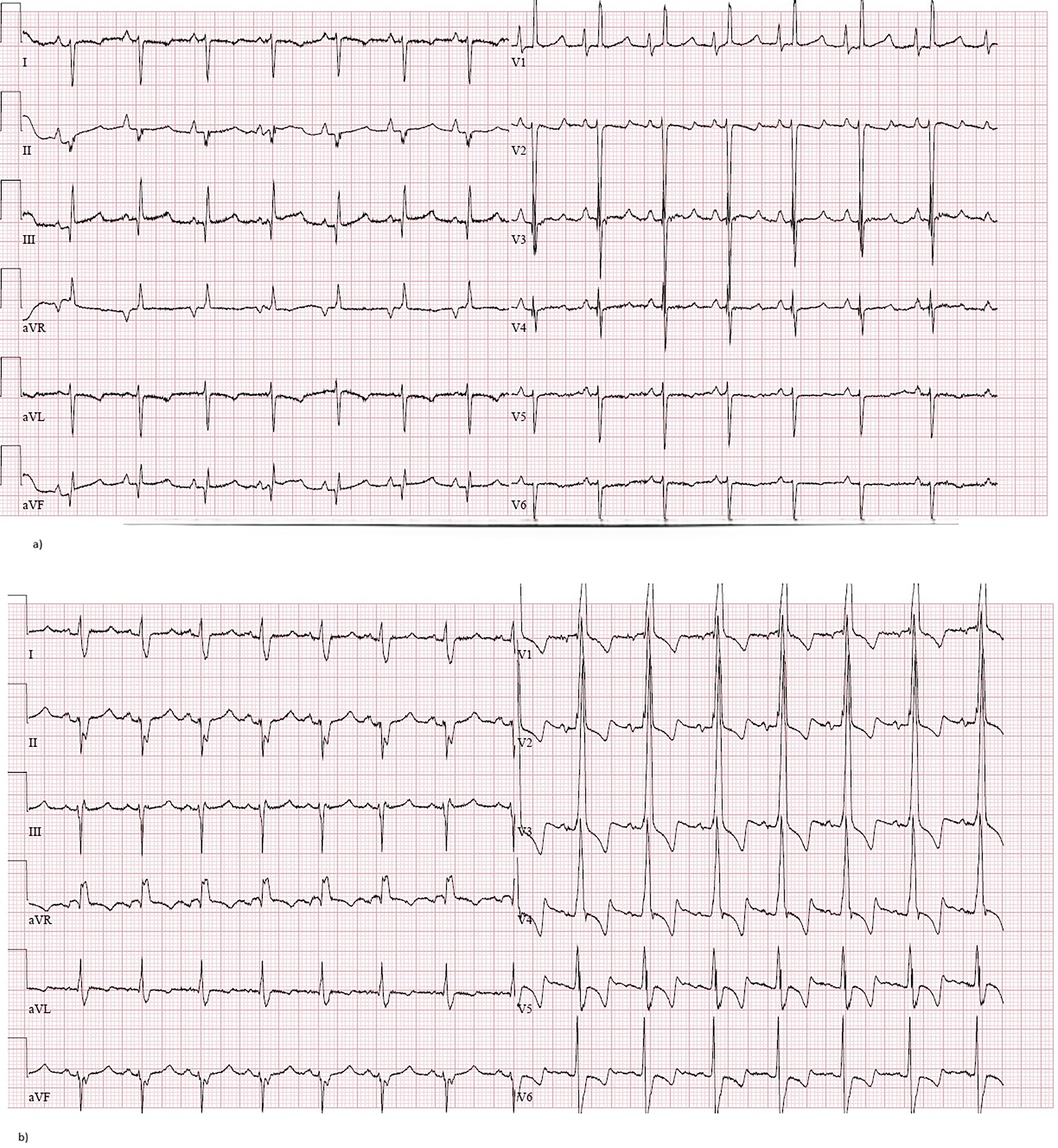
On admission, oxygen saturation was markedly reduced (approximately 60% on room air), blood exams showed severe erythrocytosis (haemoglobin 20.5 g/dL and haematocrit 65.7%), and transthoracic echocardiography demonstrated the typical anatomical features of tetralogy of Fallot, including severe right ventricular outflow tract obstruction due to infundibular stenosis, aorta overriding the interventricular septum, and a subaortic ventricular septal defect with bidirectional shunt (Figure 1). Severe biventricular systolic dysfunction was observed, with a left ventricular ejection fraction of approximately 15% and a suspected apical left ventricular thrombus. Normal coronary anatomy was detected. The ECG demonstrated a sinus rhythm with right chambers hypertrophy (Figure 2a).
Transthoracic echocardiography: the parasternal long-axis view (upper right panel) illustrates a large subaortic ventricular septal defect with overriding aorta. The left ventricle appears dilated with severely reduced systolic function. A rounded echogenic mass is visible in the apical region of the left ventricle, consistent with an intracavitary mass suggestive of thrombus.

Figure 1. Long description
Transthoracic echocardiography images display a large subaortic ventricular septal defect with the aorta positioned above the defect. The left ventricle appears dilated and exhibits severely reduced systolic function. In the apical region of the left ventricle, a rounded echogenic mass is visible, indicative of an intracavitary thrombus. The images are taken from multiple angles to provide a comprehensive view of the heart’s structure and abnormalities.
( a ) Pre-operative ECG shows sinus rhythm, enlarged right atrium, right ventricular hypertrophy and right axis deviation of the QRS. ( b ) Post-operative ECG shows sinus rhythm, enlarged right atrium, right bundle branch block with repolarization abnormalities.

Figure 2. Long description
A contrast-enhanced ECG-gated cardiac CT scan and MRI of the heart. The CT scan shows a large endoluminal filling defect adherent to the anteroseptal and inferolateral walls of the left ventricle, extending to the apical region. The MRI reveals diffuse subendocardial late gadolinium enhancement involving the anterior, lateral, and inferior walls of the mid-apical segments of the left ventricle, consistent with endomyocardial fibrosis/endocardial fibroelastosis pattern. A hypointense intracavitary filling defect in the mid-apical portion of the left ventricle, without contrast uptake, is also visible and is consistent with a left ventricular thrombus.
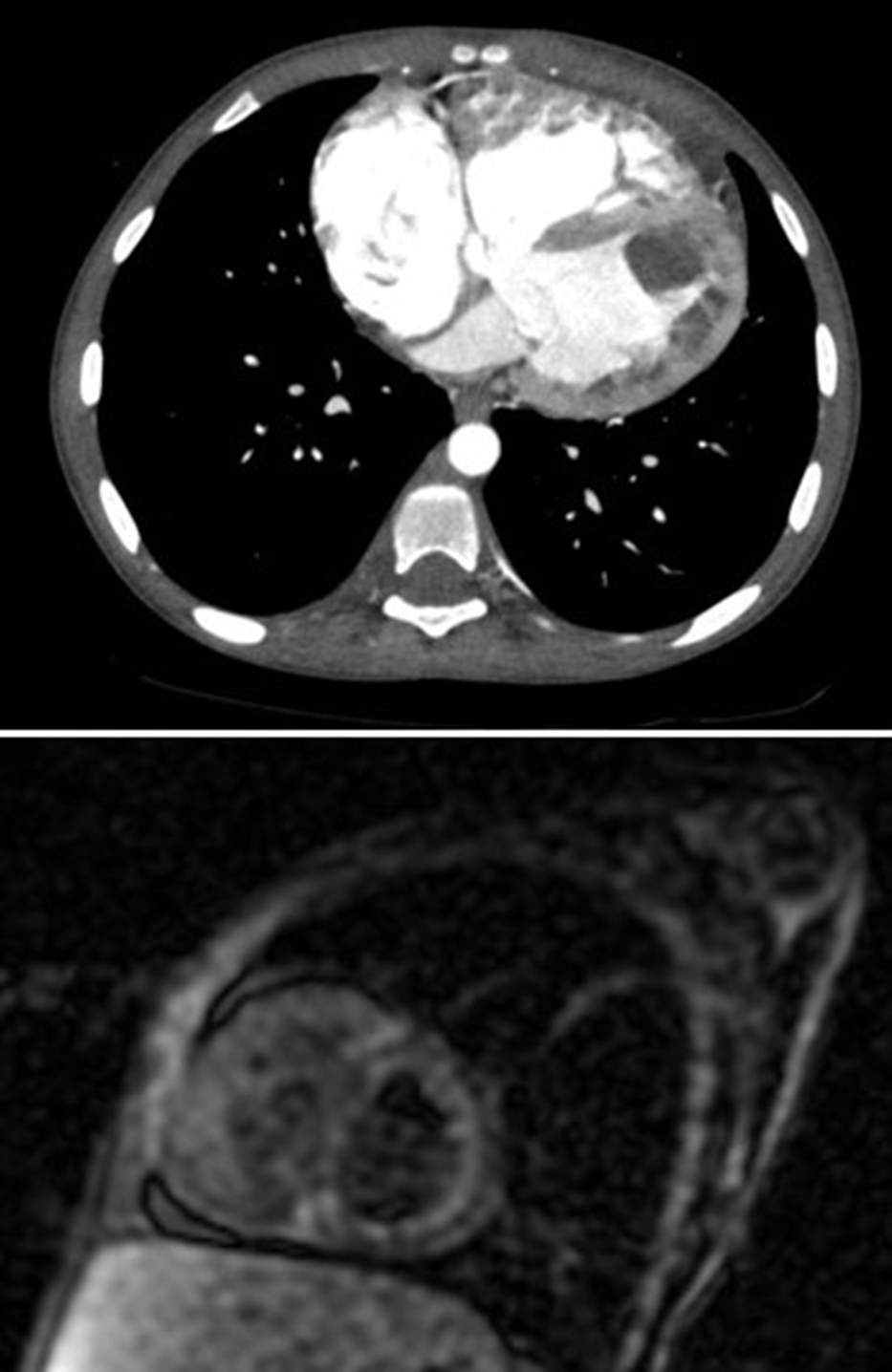
Cardiac CT confirmed the presence of a large endoluminal filling defect consistent with thrombotic material within the left ventricle, adherent to the anteroseptal and inferolateral walls of the mid-ventricular and apical segments, with near-complete obliteration of the mid-apical cavity (Figure 3).
Contrast-enhanced ECG-gated cardiac CT (upper panel) performed during the arterial phase demonstrated a large endoluminal filling defect adherent to the anteroseptal and inferolateral walls of the left ventricle, extending to the apical region. MRI (lower panel) demonstrated diffuse subendocardial late gadolinium enhancement involving the anterior, lateral, and inferior walls of the mid-apical segments of the left ventricle, consistent with endomyocardial fibrosis/endocardial fibroelastosis pattern. A hypointense intracavitary filling defect in the mid-apical portion of the left ventricle, without contrast uptake, is also visible and is consistent with a left ventricular thrombus.

Figure 2. Long description
A contrast-enhanced ECG-gated cardiac CT scan and MRI of the heart. The CT scan shows a large endoluminal filling defect adherent to the anteroseptal and inferolateral walls of the left ventricle, extending to the apical region. The MRI reveals diffuse subendocardial late gadolinium enhancement involving the anterior, lateral, and inferior walls of the mid-apical segments of the left ventricle, consistent with endomyocardial fibrosis/endocardial fibroelastosis pattern. A hypointense intracavitary filling defect in the mid-apical portion of the left ventricle, without contrast uptake, is also visible and is consistent with a left ventricular thrombus.
Cardiac MRI excluded the presence of systemic-to-pulmonary artery collaterals and provided further tissue characterisation. Late gadolinium enhancement imaging showed diffuse subendocardial enhancement of the mid-apical segments, consistent with endomyocardial fibroelastosis (Figure 3).
Anticoagulant therapy with heparin was initiated. Meanwhile, an extensive infectious work-up excluded previous or actual infections, in particular parasitic ones. CT brain scan excluded cerebral embolism.
After multidisciplinary discussion, the patient underwent surgical correction consisting of tetralogy of Fallot repair with placement of a trans-infundibular patch, pulmonary valve sparing, creation of a calibrated atrial septal defect, and surgical removal of the extensive left ventricular thrombus (diagnosis confirmed by the biopsy result).
The post-operative course was complicated by cardiogenic shock with severe biventricular dysfunction and severe mitral and tricuspid regurgitation, requiring temporary mechanical circulatory support with veno-arterial extracorporeal membrane oxygenation. After haemodynamic stabilisation, support was gradually weaned, in combination with inotropic support with levosimendan and milrinone. Subsequently, oral medical therapy for heart failure, including an angiotensin-converting enzyme inhibitor and a beta-blocker, was titrated.
The patient subsequently remained clinically stable with good haemodynamic compensation and was discharged in good general condition. Pre-discharge echocardiography demonstrated improvement in left ventricular systolic function (left ventricular ejection fraction 40–45%), with persistent hypokinesia predominantly involving the apical segments and interventricular septum. A small residual hyperechogenic image at the apical and anterolateral level was still visible, consistent with a laminated mural thrombus. The post-operative ECG showed sinus rhythm with the appearance of right bundle branch block (Figure 2b).
At three-month follow-up, further improvement in left ventricular systolic function was observed with no evidence of residual intracavitary thrombus. Anticoagulant therapy was therefore discontinued, and a six-month follow-up programme was scheduled.
Discussion
Left ventricular dysfunction in patients with tetralogy of Fallot has been most frequently described as a late consequence of surgical repair. Reduced left ventricular systolic function has been identified as an independent predictor of adverse cardiovascular events in this population. Reference Egbe, Adigun and Anand3 However, severe left ventricular dysfunction in unrepaired tetralogy of Fallot remains uncommon and should prompt investigation for additional myocardial abnormalities. Reference Di Salvo, Bulbul and Siblini4
The mechanisms underlying left ventricular dysfunction in this setting are likely multifactorial. Chronic hypoxaemia may lead to myocardial ischaemia and progressive impairment of myocardial contractility. In addition, long-standing right ventricular pressure overload can contribute to ventricular–ventricular interaction and altered left ventricular geometry, further impairing left ventricular filling and systolic performance. Reference Cordina and Celermajer5,Reference Stout, Broberg and Book6
Myocardial remodelling may also play a significant role: chronic ventricular pressure and volume overload have been associated with activation of neurohormonal pathways, including the renin–angiotensin–aldosterone system, promoting myocardial fibrosis and progressive ventricular stiffening. Therefore, diastolic dysfunction may occur and subsequently evolve into systolic dysfunction. Reference Stout, Broberg and Book6
In our case, left ventricular dysfunction resulted in intracardiac blood stasis. The secondary erythrocytosis due to the chronic cyanosis determined a hypercoagulable state. The latter and the intracardiac blood stasis represent two key components of the well-known Virchow’s triad. Reference Bagot and Arya7 To these two factors, the endothelial injury must be added. In our patient, this was suggested by the finding on MRI of diffuse subendocardial late gadolinium enhancement, consistent with endomyocardial fibroelastosis. This condition is characterised by abnormal deposition of collagen and elastin within the endocardium and may develop in response to chronic alterations in intracardiac flow dynamics. In patients with CHD, chronic hypoxia, abnormal flow turbulence, and pressure overload may promote endothelial–mesenchymal transition of endocardial cells, ultimately leading to increased ventricular stiffness and impaired ventricular function. Reference Aldawsari, Alhuzaimi and Alotaibi8,Reference Weixler, Marx and Hammer9
The remarkable recovery of left ventricular systolic function observed in our patient suggests that myocardial impairment associated with chronic cyanosis and endomyocardial fibroelastosis may be reversible following haemodynamic correction.
Multimodal imaging plays a central role in interpreting the complex interplay between congenital anomalies and secondary pathophysiological alterations, as repeatedly emphasised in the literature on CHD, Reference Di Salvo, Sabatino and Babu-Narayan10 and in planning the optimal medical and surgical treatment. Regarding our case, transthoracic echocardiography allowed the identification of the anatomical features of tetralogy of Fallot, documented the presence of severe left ventricular dysfunction, and raised the suspicion of an intracavitary thrombus. This finding was subsequently confirmed by CT and MRI, which represents the diagnostic gold standard for intracardiac thrombus detection.
Tissue characterisation using T1 and T2 mapping sequences together with late gadolinium enhancement revealed an additional abnormality in our patient: endomyocardial fibroelastosis. The identification of this pathological substrate provided further pathophysiological insight into the likely mechanisms underlying thrombus formation in this case.
Overall, this case highlights the complex interplay between chronic cyanosis, myocardial remodelling, and hypercoagulability in unrepaired tetralogy of Fallot and emphasises the pivotal role of multimodal imaging in identifying rare but clinically relevant complications.
This research received no specific grant from any funding agency, commercial or not-for-profit sectors.
Supplementary material
The supplementary material for this article can be found at https://doi.org/10.1017/S1047951126113432
Competing interests
The authors declare none.