Introduction
The beef tapeworm, Taenia saginata, is the most common taeniid of humans, with an estimated 60–70 million carriers worldwide (Andreassen, Reference Andreassen, Cox, Wakelin, Gillespie and Despommier2005). Infection of humans with this large worm has been reported since antiquity in Greek, Arabian and Chinese writings (Hoeppli, Reference Hoeppli1953). It was formerly described by Goeze in 1782; however, its connection with eating beef was probably first noted by Knox in South Africa in 1819, and its two-host–parasite life-cycle was subsequently demonstrated in 1861 by Leuckart, who fed gravid tapeworm segments (proglottids) to calves and recovered cysticerci at post-mortem (Viljoen, Reference Viljoen1937). Bovine cysticercosis can be of significant economic importance, due to downgrading or condemnation of infected beef (e.g. Kebede, Reference Kebede2008). In Europe, T. saginata remains endemic but with sporadic autochthonous human cases and low prevalence in cattle (usually < 0.1%), although higher bovine rates have been reported, e.g. in Slovakia and Turkey (Cabaret et al., Reference Cabaret, Geerts, Madeline, de Borchgrave, Ducheyne and Dorny2002; Dorny & Praet, Reference Dorny and Praet2007). In many world regions, however, prevalence of bovine cysticercosis is significantly greater than 5% (Oryan et al., Reference Oryan, Moghaddar and Gaur1995; Kebede, Reference Kebede2008). Human taeniosis due to T. saginata is usually at low prevalence but is >>1% in parts of East Africa, China and Indonesia (Tesfa-Yohannes, Reference Tesfa-Yohannes1990; Li et al., Reference Li, Craig, Ito, Chen, Qiu, Qiu, Sato, Wandra, Bradshaw, Yang and Wang2006; Wandra et al., Reference Wandra, Depary, Sutisna, Margono, Suroso, Okamoto, Craig and Ito2006).
Human taeniosis due to T. saginata is generally of low pathogenicity, mainly because it lacks the ability to cause human cysticercosis (in contrast to T. solium). However, tapeworm carriers may report symptoms that are most commonly associated with abdominal discomfort and independent migration of segments from the anus (Tesfa-Yohannes, Reference Tesfa-Yohannes1990; Craig & Ito, Reference Craig and Ito2007). Contrary to popular belief, weight loss does not occur as frequently as assumed, and was reported in less than 20% of>2000 taeniosis cases (Pawlowski & Schultz, Reference Pawlowski and Schultz1972). Classical screening for T. saginata or T. solium taeniosis may include a questionnaire about symptoms, stool (or perianal swab) microscopy for taeniid eggs, followed by subsequent anthelmintic treatment/purgation and the examination of mature proglottids for characteristic uterine branch morphology (Pawlowski & Schultz, Reference Pawlowski and Schultz1972; Hall et al., Reference Hall, Latham, Crompton and Stephenson1981; Fan et al., Reference Fan, Chung, Lin and Chan1992; Li et al., Reference Li, Craig, Ito, Chen, Qiu, Qiu, Sato, Wandra, Bradshaw, Yang and Wang2006). Laboratory-based diagnostic tests include copro-antigen detection by enzyme-linked immunosorbent assay (ELISA) (Allan et al., Reference Allan, Avila, Garcia Noval, Flisser and Craig1990; Deplazes et al., Reference Deplazes, Eckert, Pawlowski, Machowska and Gottstein1991) as well as polymerase chain reaction (PCR) to amplify species-specific DNA directly from parasite segments (Rishi & McManus, Reference Rishi and McManus1987; Harrison et al., Reference Harrison, Delgado and Parkhouse1990; Chapman et al., Reference Chapman, Vallejo, Mossie, Ortiz, Agabian and Flisser1995) or from stool samples (Yamasaki et al., Reference Yamasaki, Allan, Sato, Nakao, Sako, Nakaya, Qiu, Mamuti, Craig and Ito2004; Nunes et al., Reference Nunes, Lima, Manoel, Pereira, Nakano and Garcia2006). Copro-antigen ELISAs are usually of good sensitivity (>80%) and exhibit high Taenia genus specificity, furthermore their ease of use with formalin-treated stools and high throughput is advantageous in community-based studies compared to conventional copro-microscopy (Allan & Craig, Reference Allan and Craig2006). A further advantage is that Taenia copro-antigen levels usually become negative in ELISA within 7 days of successful treatment of human carriers (Deplazes et al., Reference Deplazes, Eckert, Pawlowski, Machowska and Gottstein1991; Allan et al., Reference Allan, Craig, Garcia Noval, Mencos, Liu, Wang, Wen, Zhou, Stringer, Rogan and Zeyle1992) and can also indicate treatment failure (Bustos et al., Reference Bustos, Rodriguez, Jiminez, Moyano, Castillo, Ayvar, Allan, Craig, Gonzalez, Gilman, Tsang and Garcia2012).
One problem in assessment of copro-antigen diagnostic tests for human taeniosis is the lack of any data on pre-patent detection, i.e. prior to the appearance of eggs and/or gravid proglottids in stool samples. In contrast, experimental taeniosis infections of dogs with canid Taenia species (Deplazes et al., Reference Deplazes, Gottstein, Stingelin and Eckert1990; Allan et al., Reference Allan, Craig, Garcia Noval, Mencos, Liu, Wang, Wen, Zhou, Stringer, Rogan and Zeyle1992) or of immunosuppressed hamsters with T. solium (Allan et al., Reference Allan, Avila, Garcia Noval, Flisser and Craig1990; Maravilla et al., Reference Maravilla, Avila, Cabrera, Aguilar and Flisser1998) showed that Taenia copro-antigens could be detected in animals during development of immature tapeworms (i.e. pre-patent infection), and as early as 5–18 days post-infection.
Experimental human self-infection with Taenia species has, unsurprisingly, not been widely reported, even from the early scientific literature. The biology and behaviour of T. saginata and T. solium taeniosis was reported during self-infections by Perroncito in 1877 and Yoshino in 1933, respectively (see Grove, Reference Grove1990). In order to assess metacestode infectivity in humans by ‘Asian Taenia’ and to record any associated taeniosis symptomatology, two separate voluntary self-infections with T. asiatica were reported by Chao et al. (Reference Chao, Wong and Fan1988) and Eom & Rim (Reference Eom and Rim1992). Taenia asiatica is morphologically very similar, and taxonomically closely related, to T. saginata (Hoberg, Reference Hoberg2002). Human voluntary self-infection with the beef tapeworm (T. saginata) has not been formerly reported, as far as we know, in the medical/parasitological literature, and copro-antigen tests have not been assessed in human taeniosis time-course studies.
The current investigation of a voluntary T. saginata self-infection was primarily undertaken in order to determine the time-course post-infection of copro-antigen production in host faeces. Additionally, data were recorded about general symptomatology during an approximate 6-month infection period with T. saginata taeniosis.
Materials and methods
Infection and faecal samples
Viable cysticerci of T. saginata were grown intraperitoneally in non-obese, diabetic, severe combined immunodeficient (NOD/Shi-scid) mice in the laboratory of Professor A. Ito, Asahikawa Medical College, Japan, according to Ito et al. (Reference Ito, Chung, Chen, Ito, Endo, Okamoto and Fan1997) and Nakaya et al. (Reference Nakaya, Mamuti, Xiao, Sato, Wandra, Nakao, Sako, Yamasaki, Ishikawa, Craig, Schantz and Ito2006). Briefly, mice were inoculated with in vitro hatched oncospheres from eggs of a confirmed T. saginata tapeworm recovered from an Indonesian carrier. Mice were sacrificed 8 weeks post-infection and cysts were removed from the peritoneal cavity, checked for viability (movement) and two cysticerci were used to infect a volunteer (P.S.C.) by oral ingestion with a saline drink.
Stool samples were collected prior to infection, every 1–2 weeks during the course of infection, and for 2 weeks post-treatment. Faecal samples were frozen raw in labelled screw-capped plastic specimen tubes at − 20°C until processed for copro-ELISA testing or PCR/DNA analysis. PCR was not carried out on every stool sample.
For copro-antigen ELISA assessment, panels of human faecal samples were available from natural infections with: T. saginata (n= 33, from taeniosis patients in Peru), T. solium (n= 33 taeniosis cases from Peru) or T. asiatica (n= 3, from China); also from two control panels: endemic controls without taeniosis (n= 21, from Guatemala) and UK negative controls (n= 19 from UK).
Taeniosis time-course and treatment
During the voluntary self-infection period (total 180 days) a daily record of any symptoms was kept and weight was recorded weekly. Any proglottids produced by spontaneous release from the anus (i.e. in underwear or from the groin, inguinal, thigh or other body regions) and/or visually present in faeces, were collected using plastic forceps, counted and stored in 95% ethanol. Complete daily total faecal output, however, was not routinely collected nor sieved/processed rigorously for segments or egg counts. The taeniosis infection was terminated by taking praziquantel, 2 g orally, followed after 2 h with 50 ml of 2% magnesium sulphate solution as a purgative, and 2 litres of drinking water. Total faeces were collected in a bucket for 5 h after treatment and any tapeworm material was removed and fixed in 90% ethanol.
Taenia saginata antigen extracts and rabbit hyperimmune antisera
Whole-worm crude somatic extracts (WWE) were prepared from non-gravid segments (proglottids) of T. saginata essentially as described by Allan & Craig (Reference Allan and Craig1989). Briefly, segments were washed in phosphate-buffered saline (PBS), cut into small pieces, freeze–thawed three times ( − 80°C to 4°C) and homogenized, then sonicated on ice using a 150 W sonicator for 20 s. The suspension was incubated on ice for 1 h and centrifuged at 5000 g for 15 min at 4°C, then the supernatant was stored in 0.5-ml aliquots at − 80°C, and designated T. saginata WWE.
Excretory/secretory (ES) antigens were produced from live T. saginata tapeworm proglottids that had been washed in PBS and incubated in a 25-ml tissue-culture flask at 37°C containing 15 ml of DMEM (Dulbecco's Modified Eagle's Medium) supplemented with 1% sterile glucose, 100 units/ml penicillin and 100 μg/ml streptomycin (after Wilkins et al., Reference Wilkins, Allan, Verastegui, Acosta, Eason, Garcia, Gonzalez, Gilman and Tsang1999). Conditioned medium was collected daily for 1 week (replenished with fresh culture medium), pooled and stored at 4°C. Pooled media were concentrated tenfold in a stirred Amicon unit, using a 10,000 molecular weight cut-off PM10 membrane, then stored at − 80°C until used. This preparation was designated T. saginata ES.
Four New Zealand (NZ) white rabbits were hyperimmunized with 0.5 mg of WWE or ES preparations using Freund's adjuvant over a period of 6–7 weeks, and rabbit serum was harvested at post-mortem as previously described (Allan et al., Reference Allan, Avila, Garcia Noval, Flisser and Craig1990). Immunoglobulin G (IgG) from the rabbits with the highest titres was purified from the sera using Protein A Sepharose CL4B (Sigma, Poole, Dorset, UK) and either stored concentrated in 0.5 ml aliquots at − 80°C, or conjugated to horseradish peroxidase as described by Allan & Craig (Reference Allan and Craig1989).
T. saginata copro-antigen ELISA
Human faecal samples were thawed and diluted 1:2 (w/v) with 0.05 m PBS, pH 7.2, containing 0.3% Tween 20 detergent (PBS Tween) and mixed vigorously by hand to form a slurry which was centrifuged at 3000 g for 5 min at room temperature. The supernatant was removed and used immediately in ELISA or stored at − 20°C prior to testing.
The copro-antigen ELISA was developed for T. saginata following the general approach of Allan et al. (Reference Allan, Craig, Garcia Noval, Mencos, Liu, Wang, Wen, Zhou, Stringer, Rogan and Zeyle1992), but using the modification described by Guezala et al. (Reference Guezala, Rodriguez, Zamora, Garcia, Gonzalez, Tembo, Allan and Craig2009) to improve specificity and sensitivity, which incorporated both anti-somatic (capture) and anti-ES (detection antibody) polyclonal antibodies. Thus we developed rabbit anti-T. saginata adult-derived antibodies (IgG) raised against either: (1) immature T. saginata proglottid somatic extracts (WWE) (capture antibodies) or (2) T. saginata mature proglottid ES products (secondary antibody conjugated to horseradish peroxidise).
The T. saginata copro-antigen ELISA was carried out as follows. Polystyrene microtitre plates (Immulon 4HBX, ThermoLabsystems, Altrincham, Cheshire, UK) were incubated overnight at 4°C (100 μl/well) with rabbit IgG anti-T. saginata WWE (6.5 μg/ml) in bicarbonate–carbonate buffer (pH 9.6). Plates were washed three times in PBS containing 0.1% Tween 20, then incubated for 1 h with PBS 0.3% Tween (on an orbital shaker). Wells were emptied and 50 μl/well of heat-inactivated fetal calf serum (HIFCS) was added, followed by 50 μl/well of faecal supernatant (in duplicate) and incubated at 20°C for 1 h on an orbital shaker. Plates were washed as above, and incubated with 100 μl/well of horseradish peroxidase-conjugated rabbit IgG anti-T. saginata ES (10 μg/ml) diluted in PBS 0.3%Tween 20 containing 15% (v/v) HIFCS, for 1 h at 20°C on an orbital shaker. After plate washing, substrate TMB solution (3,3′,5,5′-tetramethylbenzidine; SureBlue, BD Biosciences Pharmingen, San Diego, California, USA) was added at 100 μl/well and plates were incubated in the dark for 5 min, then the optical density (OD) of each well was read at 630 nm using an automatic ELISA reader (Multiskan Ascent, Labsystems, Hemel Hempstead, Hertfordshire, UK).
ELISA positive–negative cut-off determination was calculated using two methods: (1) mean value of control negatives plus three standard deviations (after Allan et al., Reference Allan, Craig, Garcia Noval, Mencos, Liu, Wang, Wen, Zhou, Stringer, Rogan and Zeyle1992); or (2) by calculating an ELISA index based on the test OD as a percentage of maximum absorbance value of a positive reference, taking into account the negative mean (after Gottstein et al., Reference Gottstein, Lengeler, Bachmann, Hagemann, Kocher, Brossard, Witassek and Eckert1987): ELISA index = (OD value of test sample/(positive reference OD value − mean negative +3SD)) × 100
The effective copro-antigen detection limit in ELISA was measured using twofold serial dilutions of either WWE extract or ES preparation in normal human faeces.
Copro-PCR
Taeniosis time-course selected faecal samples were tested for presence of T. saginata DNA using a uniplex PCR based on oligonucleotide primers targeting the cox1 gene (Yamasaki et al., Reference Yamasaki, Allan, Sato, Nakao, Sako, Nakaya, Qiu, Mamuti, Craig and Ito2004). The positive control was a human negative faecal sample spiked with tissue-derived DNA from a T. saginata proglottid. DNA was extracted from faecal samples using the QIAmp DNA Stool Mini Kit (Qiagen, Hilden, Germany) as described previously (Yamasaki et al., Reference Yamasaki, Allan, Sato, Nakao, Sako, Nakaya, Qiu, Mamuti, Craig and Ito2004; Li et al., Reference Li, Craig, Ito, Chen, Qiu, Qiu, Sato, Wandra, Bradshaw, Yang and Wang2006)
Results
T. saginata taeniosis infection, symptoms and time-course
Following oral infection with two viable cysts of T. saginata the infection was voluntarily maintained until termination (with praziquantel) at 180 days post-infection (dpi). No symptoms attributable to the infection occurred until 42 dpi when 3 days of mild diarrhoea occurred. On day 75 post-infection daily stool frequency changed from 1 to 2 stools/day, and that frequency continued (unless constipation occurred) until the end of the experiment on 180 dpi.
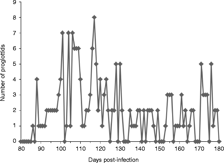
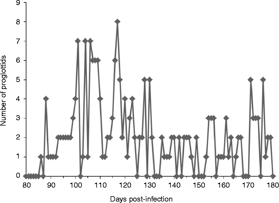
From 82 to 85 dpi the volunteer reported the first (of many) painless involuntary rectal sensations, or rectal ‘flutters’ and pruritis ani, followed on 86 dpi by the spontaneous emergence from the anus of a single motile proglottid. Day 86 can therefore be considered the patency period for this particular individual infection. From day 86 to 98 dpi, on average 1–2 individual, motile proglottids per day were passed/voided spontaneously or collected from the stool; with mild abdominal pain or discomfort reported over a total of 7 days during that period. From day 99 to 130 dpi an average of 3.5 segments (proglottids) were spontaneously voided (individually) or passed per day (range 1–8 proglottids) (fig. 1). Rarely 2 or 3 joined proglottids were found in the faeces. Abdominal pain was reported nine times during that period and was usually preceded the day before by constipation. From day 132 to 180 dpi daily proglottid output decreased to 1–2 per day (range 1–5 proglottids) with normal stool most of the time and only 2 days of mild abdominal discomfort reported. Spontaneous release of motile proglottids occurred only during the daytime (not at night).
The number of T. saginata proglottids collected per day after spontaneous release and/or from stool samples. The first proglottid was voided on day 86 post-infection and the infection terminated on day 180 post-infection.
On day 180 pi the infection was terminated by 2 g of oral praziquantel followed by a magnesium sulphate purge. Stool samples collected over the following 24 h produced worm fragments only and no intact worm or scolex was recovered. Diarrhoea was reported on days 2 and 3 post-treatment, then stools became normal from day 4 post-treatment. The last worm fragments were observed in stool samples on day 3 post-treatment.
During the course of infection the body weight of the volunteer did not change significantly from normal (75 kg) with fluctuation between 74 and 76 kg. However, appetite was reported to increase, in particular with increased intake of foods rich in carbohydrates.
T. saginata copro-antigen ELISA
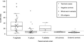
A copro-antigen ELISA was developed using rabbit polyclonal antibodies against T. saginata proglottid somatic (WWE) or ES extracts, as capture and conjugated antibodies respectively. The lowest calculated limit of copro-antigen detection in ELISA using serial twofold dilutions of T. saginata WWE or ES extracts, was 0.6 μg/ml and 1.8 μg/ml, respectively. The test sensitivity of the copro-antigen ELISA for faecal antigen detection from a panel of natural T. saginata stool samples was 81.8% (27/33, using ELSA index cut-off). Copro-antigen ELISA species specificity for proven T. saginata stool samples was 85.5% (i.e. 65/76, non-T. saginata were negative) versus panels of stool samples from other infections (fig. 2). Seven of 33 (21%) T. solium and 2 of 3 T. asiatica stool samples cross-reacted in the T. saginata copro-antigen ELISA.
ELISA values expressed as an index for T. saginata coproantigen panels of stool samples from taeniasis cases, controls or for antigen extracts. Taenia saginata cases (n= 33); T. solium cases (n= 33); T. asiatica cases (n= 3); UK controls (n= 19); endemic controls without taeniosis (n= 21); antigen extracts (T. saginata ES, T. solium ES, T. saginata WWE, T. asiatica WWE). The ‘boxes’ of the box and whisker plots show the median and upper and lower quartiles for each group (excluding antigen extracts), and the ‘whiskers’ cover all points within 1.5 times the interquartile range from the upper or lower quartile. The positive–negative cut-off (estimated as the point three standard deviations above a panel of known negative samples) is shown as a horizontal dotted line.

Copro-antigen ELISA time-course in a voluntary T. saginata infection
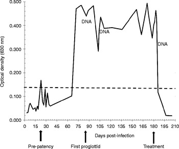
Copro-antigen was first detected (using the novel T. saginata copro-antigen ELISA described above) on day 23 pi; however, this appeared to be a temporary minor spike in copro-antigen because levels did not become consistently positive until after 72 dpi, just prior to patency (proglottid release) at 86 dpi. This spike in antigen in faeces at 23 dpi may be associated with release of worm fragments or even loss of a juvenile tapeworm (the volunteer took two cysticerci). After 72 dpi copro-antigen levels remained significantly elevated for approximately 15 weeks until termination of infection (fig. 3). Copro-antigens were detectable by ELISA up to 3 days post-treatment but not thereafter. Application of a standard Taenia-genus specific copro-antigen ELISA based on the method of Allan et al. (Reference Allan, Craig, Garcia Noval, Mencos, Liu, Wang, Wen, Zhou, Stringer, Rogan and Zeyle1992) gave a very similar time-course profile of copro-antigen levels, with first significant detection of copro-antigen only after 79 dpi (data not shown).
Time-course of optical density (OD) values at 630 nm for T. saginata coproantigen ELISA tests of weekly stool samples over 80–180 days post-infection (dpi) for a voluntary self-infection with T. saginata. The first proglottid was released (patency) on day 86, treatment with praziquantel was on day 180. OD cut-off (––) and DNA denote when T. saginata coproantigens or coproDNA were detected unequivocally in stool samples.

CoproDNA could be amplified in the T. saginata time-course stool samples only at 86 and 104 dpi, and in a stool at 5 days post-treatment. Surprisingly, coproDNA of T. saginata could not be detected in faeces tested (n= 5 samples) from the pre-patent period, i.e. before 86 dpi.
Discussion
The development of copro-antigen ELISAs for the detection of human taeniosis (Allan et al., Reference Allan, Avila, Garcia Noval, Flisser and Craig1990, Reference Allan, Craig, Garcia Noval, Mencos, Liu, Wang, Wen, Zhou, Stringer, Rogan and Zeyle1992; Deplazes et al., Reference Deplazes, Eckert, Pawlowski, Machowska and Gottstein1991) was an important advance in diagnosis that enabled more accurate community screening and, in particular, a better understanding of the epidemiology of the pathogenic species T. solium (Garcia-Noval et al., Reference Garcia-Noval, Allan, Fletes, Moreno, de Mata, Torres-Alvarez, Soto de Alfaro, Yurrita, Higueros-Morales, Mencos and Craig1996; Allan et al., Reference Allan, Velasquez-Tohom, Fletes, Torres-Alvarez, Lopez Virula, Yurrita, de Alfaro, Rivera and Garcia-Noval1997; O'Neal et al., Reference O'Neal, Moyano, Ayvar, Gonzalvez, Diaz, Rodriguez, Wilkins, Tsang, Gilman, Garcia and Gonzalez2012). Copro-antigen testing also enabled more sensitive and practical surveillance during and after T. solium cysticercosis control interventions that were applied in endemic communities (e.g. Allan et al., Reference Allan, Velasquez-Tohom, Fletes, Torres-Alvarez, Lopez Virula, Yurrita, de Alfaro, Rivera and Garcia-Noval1997; Garcia et al., Reference Garcia, Gonzalez, Gilman, Moulton, Verastegui, Rodriguez, Gavidia and Tsang2006). Mass treatment of human taeniosis is recommended as an important component of T. solium control, and should contribute to reduction of taeniosis prevalence and incidence (Murrell, Reference Murrell2005; Lightowlers, Reference Lightowlers2013). At low incidence following successful intervention, human taeniosis cases will be much harder to detect, and new tapeworm carriers might include a higher relative proportion of early or pre-patent infections.
In the current study, a voluntary self-infection with human T. saginata taeniosis was used as a ‘model’ for copro-antigen detection potential in relation to pre-patent detection in human taeniosis. Surprisingly, copro-antigens could not be reliably detected (despite a small spike in activity at 23 dpi) up to 71 dpi, which was during most of the pre-patent period. The first active proglottid was passed (spontaneously) at 86 dpi, which may thus be considered the pre-patent period. Taenia saginata copro-antigen levels only became readily detectable 2 weeks before first proglottid release or patency (on 72 dpi), but thereafter copro-antigen levels were maintained at high positivity until termination of the infection (at 180 dpi). The lack of detectable pre-patent copro-antigens is surprising because copro-antigen ELISA studies in dogs experimentally infected with the related canid tapeworms, Taenia hydatigena or T. pisiformis, were able to detect copro-antigens by 11–18 dpi, which is well before patency at 40–57 dpi in those taeniid species (Deplazes et al., Reference Deplazes, Gottstein, Stingelin and Eckert1990; Allan et al., Reference Allan, Craig, Garcia Noval, Mencos, Liu, Wang, Wen, Zhou, Stringer, Rogan and Zeyle1992).
Lack of pre-patent copro-antigen in the human volunteer self-infection is hard to explain. Taenia saginata tapeworm growth is rapid – for example a 3.5 m tapeworm was recovered at 86 dpi in a previous self-infection study, which indicated a growth rate of approximately 4 cm/day (Craig & Ito, Reference Craig and Ito2007). Release and/or turnover of surface carbohydrate-rich antigens in taeniid tapeworms were considered the main constituents of copro-antigens (Elayoubi & Craig, Reference Elayoubi and Craig2004) and would be expected to occur during the growing pre-patent phase, as detected in experimental canid Taenia infections (e.g. Deplazes et al., Reference Deplazes, Gottstein, Stingelin and Eckert1990). The copro-antigen ELISA test was developed in the current study using capture and conjugated antibodies against T. saginata adult tapeworm somatic and excretory/secretory (ES) antigens, respectively, and gave reasonable sensitivity (81.8%) against a panel of stools from naturally infected T. saginata cases, as well as an antigen detection equivalent limit of 1.8 μg T. saginata ES antigen/g of stool. This level is within the detectable range reported for T. hydatigena adult ES antigen in a coproELISA that detected pre-patent infections in dogs (Deplazes et al., Reference Deplazes, Gottstein, Stingelin and Eckert1990).
Assuming that the T. saginata coproELISA we developed was of appropriate sensitivity, and the fact that we obtained similar data using the standard Taenia copro-antigen ELISA of Allan et al. (Reference Allan, Craig, Garcia Noval, Mencos, Liu, Wang, Wen, Zhou, Stringer, Rogan and Zeyle1992) (data not shown), then the self-infection time-course results we obtained suggest that no, or only low levels of, T. saginata strobila or proglottid antigens (copro-antigens) were produced/released during the first 10 weeks of the infection. The dramatic rise in detectable copro-antigens 2 weeks before patency of the T. saginata self-infection, could indicate that antigen release or turnover was associated or linked with apolysis (i.e. the process of proglottid separation from the strobila). Ambrosio et al. (Reference Ambrosio, Cruz-Rivera, Allan, Moran, Ersfeld and Flisser1997) detected myosin-like protein on the surface of detached proglottids of T. solium and in human faeces in natural infections, which could also be associated with apolysis. This is also supported in the current study by the occurrence of high levels of copro-antigen throughout the patent period (when almost daily release of proglottids occurred) and until termination of the infection. Furthermore, parasite DNA was not detected in stool samples until after patency. From a practical viewpoint, these observations indicate that copro-antigens and/or tegumental surface turnover in T. saginata infection (and possibly by inference also T. solium) may not be detected reliably until close to, or after, patency. Detection of copro-antigens in pre-patent canid infections (Deplazes et al., Reference Deplazes, Gottstein, Stingelin and Eckert1990; Allan et al., Reference Allan, Craig, Garcia Noval, Mencos, Liu, Wang, Wen, Zhou, Stringer, Rogan and Zeyle1992) may be associated with the much shorter pre-patent periods for T. hydatigena and T. pisiformis.
Regarding symptomatology of T. saginata taeniosis over the individual's infection time-course, i.e. 180 days (approximately 26 weeks), there appeared to be four broad phases of symptoms over that period: (1) an initial asymptomatic period for the first 10 weeks post-infection (wpi); (2) onset of patency around 12 wpi (i.e. 86 dpi) with spontaneous proglottid release of 1–2 individual segments/day and intermittent mild abdominal discomfort up to 14 wpi; (3) an increased output of proglottids (1–8 per day, average 3.5 per day) from 15–19 wpi associated with more frequent bouts of diarrhoea and moderate to severe abdominal discomfort; (4) from 20 wpi a dramatic decline in abdominal symptoms/discomfort with normal stool output and release of 1–5 proglottids/day until termination of the infection. Grove (Reference Grove1990) recounts the T. saginata human infection studies of Perroncito (in 1877), who recorded first proglottids passed at 54 dpi, with a calculation of 13–14 proglottids produced/day, which is much earlier and a greater proglottid output than described in the current self-infection. Our previous early data in three volunteers with T. saginata self-infection gave pre-patent periods of 94, 98 or 106 dpi (P.S. Craig and A. Ito, unpublished observations). In the current study, the latter period from 20 wpi (described in (4) above), when greatly reduced abdominal discomfort and lower proglottid output occurred, might be interpreted as the parasite–host relationship settling into a status quo. Weight loss did not occur in the current individual experimental infection, but segment voiding and abdominal discomfort were the main symptoms after patency, as reported in large case studies on natural infections with T. saginata and T. asiatica taenioses (Pawlowski & Schultz, Reference Pawlowski and Schultz1972; Fan et al., Reference Fan, Chung, Lin and Chan1992).
Spontaneous voiding of T. saginata tapeworm proglottids appears to be a distinctive sign and a main symptom in humans (Craig & Ito, Reference Craig and Ito2007). This was commented on by Hippocrates, and much later by Leuckart in the mid-19th century, who remarked that this behaviour was more usual for T. saginata compared to T. solium (see Viljoen, Reference Viljoen1937). In 26 cases of T. saginata studied in Ethiopia, 69% reported spontaneous segment discharge and 92% had abdominal pain (Tesfa-Yohannes, Reference Tesfa-Yohannes1990). The largest data set collected on T. saginata taeniosis symptoms (>2000 cases) indicated that most cases reported discharge of 1–3 proglottids daily, 35.6% of cases reported abdominal discomfort and 15%, weight loss (Pawlowski & Schultz, Reference Pawlowski and Schultz1972). Fan et al. (Reference Fan, Chung, Lin and Chan1992) studied 1661 cases of Asian Taenia (T. asiatica) in Taiwan and reported main symptoms as segments in faeces/discharge (95%), pruritis ani (77%), abdominal pain (45%) and weight loss (6%). During a self-infection with Asian Taenia, Chao et al. (Reference Chao, Wong and Fan1988) recorded that patency occurred on 122 dpi and then regular discharge of 1–4 proglottids/day, but also an associated 7-week period of abdominal pain/discomfort. That self-infection case has similarities with the current T. saginata self-infection study.
In summary, a single voluntary self-infection with T. saginata was followed over a 180-day infection time-course using copro-antigen ELISA. Copro-antigen levels were not reliably detected until 72 dpi (2 weeks before segment release) but remained elevated thereafter until termination, and became negative by 4 days post-treatment. Although these observations are based on a single voluntary infection, they suggest that copro-antigen ELISA for human taeniosis detection may not be reliable during the early pre-patent growth period (approximately 3 months). The main symptoms reported were spontaneous voiding of proglottids (1–8 per day) over the whole post-patent period, and variably associated abdominal discomfort, especially over the 15- to 19-week post-infection period, but much less thereafter.
Acknowledgements
Great thanks are due to Professor Akira Ito and Dr Kazuhiro Nakaya, Parasitology Department, Asahikawa Medical College, Asahikawa, Japan, for provision of viable cysticerci from scid mice. Thanks also to Professor Hugo Garcia, Department of Microbiology, Universidad Peruana Cayetano Heredia, Lima, Peru; and Professor Li Tiaoying, Parasitic Diseases Unit, Sichuan CDC, Chengdu, China, for providing stool samples from taeniosis cases; also to Dr Jim Allan for provision of a panel of endemic normal stool samples from Guatemala. Thanks also to Dr Belgees Boufana for assisting with the PCR test, and to Alex Mastin for checking the manuscript and producing the figures.
Financial support
A.T. received a Commonwealth Commission post-graduate studentship.
Conflict of interest
None.
Ethical standards
The authors assert that all procedures contributing to this work comply with the ethical standards of the relevant national guidelines on human experimentation and with the Helsinki Declaration of 1975, as revised in 2008, and have been approved by the Ethics Committee of the University of Salford. The authors assert that all procedures contributing to this work comply with the ethical standards of the Japanese Regulations on Animal Experiments and have been approved by Asahikawa Medical College Ethics Committee.