Introduction
The in vitro maturation (IVM) technique is widely used in assisted animal reproduction. It is one of the most efficient methods to produce mature oocytes (Smitz et al., Reference Smitz, Thompson and Gilchrist2011) in procedures such as in vitro fertilization (IVF), intracytoplasmic injection (ICSI) and cloning. However, IVM is clinically limited by its poor efficiency in terms of fertilization and embryo viability compared with conventional in vitro fertilization that uses mature oocytes produced in vivo (Gremeau et al., Reference Gremeau, Andreadis, Fatum, Craig, Turner, McVeigh and Child2012; Lonergan and Fair, Reference Lonergan and Fair2016).
It is known that oocytes require the maturation of both cytoplasm and nucleus to support successful fertilization and subsequent embryo development (Trounson et al., Reference Trounson, Anderiesz and Jones2001; Kuzmina et al., Reference Kuzmina, Alm, Denisenko, Tuchscherer, Kanitz and Torner2007). Nuclear maturation refers to the initiation of meiosis from the germinal vesicle (GV)-stage oocytes and progression to MII. The techniques used to stimulate oocyte development, and the culture medium employed, significantly influenced oocyte characteristics and growth (Lenie et al., Reference Lenie, Cortvrindt, Adriaenssens and Smitz2004; Miki et al., Reference Miki, Ogonuki, Inoue, Baba and Ogura2006). There have been, in recent times, considerable research focused on the establishment of optimal conditions for IVM in terms of altering the type of medium used, addition of hormones etc. for better maturation of oocytes (De La Fuente et al., Reference De La Fuente, O’Brien and Eppig1999; Nakano and Kubo, Reference Nakano and Kubo2000; Miki et al., Reference Miki, Ogonuki, Inoue, Baba and Ogura2006).
In in vivo follicular development, growth factors and cytokines act as intra-ovarian regulators and gonadotropin modulation is affected by paracrine and autocrine growth factors produced in the ovary (Sakaguchi et al., Reference Sakaguchi, Dominko, Yamauchi, Leibfried-Rutledge, Nagai and First2002; Gilchrist and Thompson, Reference Gilchrist and Thompson2007). GCCM, obtained from the third passage of surrounding granulosa cells of cultured preantral follicles of mouse, is known to contain various cytokines and growth factors such as epidermal growth factor (EGF), IGF and TGFβ (Dirnfeld et al., Reference Dirnfeld, Goldman, Gonen, Koifman, Calderon and Abramovici1997; Malekshah et al., Reference Malekshah, Moghaddam and Daraka2006). Given that cytokines and growth factors stimulate resumption of the meiotic process, especially in the activation of the maturation promoting factor (MPF) subunits (CyclinB1 and CdK1) that play a dominant role in inducing the GVBD stage, it seems logical that conditioned medium derived from cultured granulosa cells could improve maturation outcomes of in vitro oocytes.
Apart from the factors contained in the granulosa cells, it is known that an increase in the levels of luteinizing hormone (LH) in females leads to resumption of oocyte meiosis. One of the leading molecular cascades that propagate LH induction is a key membrane component named EGF and its subfamily member proteins known as EGF-like peptides (Park et al., Reference Park, Su, Ariga, Law, Jin and Conti2004; Richani et al., Reference Richani, Sutton-McDowall, Frank, Gilchrist and Thompson2014). An increase in LH levels also results in rapid expression of EGF-like peptides in follicle granulosa cells. These peptides act as transmembrane precursors that diffuse and become attached to oocyte membrane-bound EGF receptors (EGFR). This process, in turn, triggers subsequent signalling pathways (Conti et al., Reference Conti, Hsieh, Park and Su2006). In addition, meiosis resumes following FSH and EGF effects, as too many important inducers, on EGF and FSH receptors.
This research aimed to investigate the possibility of improving IVM outcomes through the use of conditioned medium for culture rather than conventional supplementary materials, based on the hypothesis that GCCM enhanced activation and maturation of GV-stage oocytes from mouse germinal vesicles under in vitro conditions.
Materials and methods
Animals and samples preparation
NMRI mice, originally derived from the Royan Institute, were housed in a conditioned environment (20–25°C), humidity (40–60%) and a 12 h light:12 h dark cycle. The GV-stage oocytes were obtained from 4–6-week-old females ovaries. The procedure was performed on Royan ethical committee guideline.
Granulosa cells culture and providing the conditioned medium
Preantral follicles (100–120 µm) were isolated mechanically using 29-gauge insulin needles, placed into α-MEM supplemented with 10% fetal bovine serum (FBS) and 1% FSH, and incubated at 37°C and 5% CO2 in air. After 3 days, non-adherent cells were removed after washing twice with phosphate-buffered saline (PBS) and the remaining adherent cells were cultured in complete medium until passage 3. When cultured cells reached 70% confluency, GCCM was collected for using in further IVM.
In vitro maturation of germinal vesicles
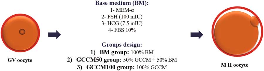
In total, 324 granulosa denuded GV oocytes were obtained from the ovaries of 6–8-week-old NMRI female mice after mechanical dissection of the antral follicles. GV-stage oocytes were collected and categorized into three groups based on different culture medium components: 108 GV-stage oocytes in base medium (BM) (Control), 108 in GCCM50 and 108 in GCCM100. α-MEM supplemented with HCG (7.5 IU), FSH (100 mIU) and FBS (10%) was used as BM. For the GCCM50 group, 50% GCCM was added to BM. In the GCCM100 group, BM was completely replaced with GCCM (Fig. 1).
Design of the groups based on different concentrations of granulosa cell conditioned medium (GCCM) in in vitro maturation (IVM) medium.

The GV-stage oocytes were evaluated using the trypan blue staining for survival assay. Non-trypan blue-positive GV-stage oocytes were randomly divided and incubated in drops of the three media under mineral oil for 24 h. The in vitro cultured oocytes were observed using an inverted microscopy and first polar body extrusion was considered to be the maturation criterion (MII rate).
Trypan blue staining (oocyte survival assay)
Trypan blue (TB) is a diazo dye that is widely used to stain dead tissues or cells. The principle of trypan blue staining (TBS) is that TB is negatively charged and binds only to damaged membranes. Intact cells allow the passage of very few select compounds through the membrane, and therefore do not absorb TB. Before IVM, GV-stage oocytes treated with TB (0.4%) and found non-positive or viable GV-stage oocytes were selected for IVM. Eighteen hours after in vitro maturation (IVM), the oocytes were stained again with TB and non-TB positive MII oocytes were considered to be viable cells. In contrast, cells with damaged membranes were stained a distinctive blue colour, as readily observed under a microscope.
Mitochondrial activity potential (JC-1 staining)
JC-1, a sensitive cationic carbocyanine fluorescence dye that accumulates in the mitochondria, was applied to investigate the mitochondria potential activity in in vitro matured oocytes. At low concentrations, JC-1 is a monomer and emits green fluorescence (~530 nm), similar to that of fluorescein, when excited at 490 nm. At higher concentrations, driven by membrane polarization, the dye reversibly forms JC-1 aggregates, which exhibit an emission maximum at ~590 nm (orange colour) when the membrane potential is 80–100 mV. Thus, the colour of the dye changes from green to red (Wilding et al., Reference Wilding, Dale, Marino, di Matteo, Alviggi, Pisaturo, Lombardi and De Placido2001; Smiley et al., Reference Smiley, Reers, Mottola-Hartshorn, Lin, Chen, Smith, Steele and Chen1991). Accordingly, an increase in red/green ratio, indicates an enhancement in mitochondria potential activity. After 18 h, the oocytes were collected and treated in 0.25 µg/ml JC-1 at 37°C for 30 min. They were then washed three times with PBS and finally analyzed using a laser-scanning confocal microscope (TCS SP5 II Lecia). For quantification, the red/green ratio was analyzed using Image J software.
Gene expression following in vitro maturation
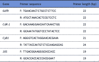
For evaluation of genes involved in resumption of meiosis (CyclinB1 and Cdk1) and an oocyte-specific gene (Gdf9), total RNA was extracted from all samples. The PicoPure Kit (TaKaRa Bio Inc., Shiga, Japan) was used for RNA extraction, and then qualified and quantified in each group containing 45 MII oocytes, with a NanoDrop 2000 spectrophotometer (Thermo Scientific) at 260/280 nm (a 1.8–2 ratio indicating a suitable sample). Finally, gene expression level was assessed with the qPCR technique following cDNA synthesis, which was done using the Takamed kit (TaKaRa Bio Inc.; Shiga, Japan) and stored at −20°C until subsequent analysis. Reactions were run with Step One Plus Real-Time PCR detection system for qPCR analysis of each 20 µl reaction. The primer sequences are provided in Table 1. Following this, a melt curve analysis was conducted for each reaction to verify that one product was synthetized with no primer dimer and relative gene expression was calculated using 2−ΔΔCt methods. Also, the 18S gene was considered as a ‘housekeeping’ gene and for each reaction a non-template control (NTC) with primer and without cDNA was set for approving the results derived from the other reactions with cDNA.
Sequences of primers for real-time PCR test

Statistical analysis
Continuous data were initially tested for normal distribution using the Kolmogorov–Smirnov test (UNIVARIATE procedure). Data associated with JC-1 and expression of CdK-1 and CyclinB-1, had a normal distribution and were analyzed using the GLM procedure; however, the data for GDF-9 expression did not have a normal distribution and were analyzed using the Kruskal–Wallis test. Binary data including the proportion of GV and MII were analyzed using the GENMOD procedure including function link logit in the model. The LSMEANS statement was used for implementation of multiple comparisons. All analyses were conducted in SAS (User’s guide version 9.4: statistics. Cary, NC: SAS Institute; 2013 SAS). Data are presented as proportions or mean ± standard error of the mean (SEM). Differences with P < 0.05 were considered significant.
Results
Mouse GV in vitro maturation
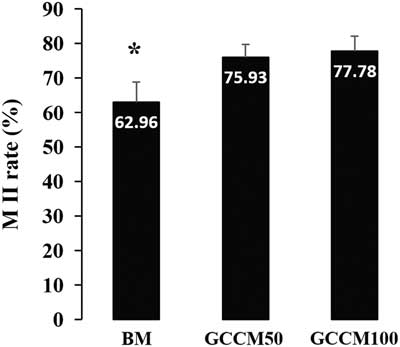
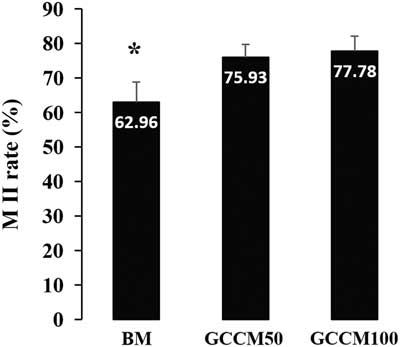
Figure 2 shows the proportion of oocytes that have released their polar bodies and reached the MII stage after 18 h of culture. In this case, the maturation rates of both GCCM100 (77.78%; odds ratio [OR] = 2.06, 95% confidence interval [CI] = 1.13–3.75; P = 0.02) and 50 75.93%; OR = 1.86, 95% CI = 1.03–3.34; P = 0.04) groups were significantly higher than that of the control (62.96%) (P < 0.05). In this regard, the difference in MII rate between GCCM50 and GCCM100 groups (P > 0.05) was insignificant. At various intervals from the onset of incubation, oocytes were observed by confocal microscopy and evaluated for morphological changes in the nucleus and polar body as criteria for nuclear maturation of GV-stage oocytes.
Percentage of polar body released oocytes (MII rate) in all control (base medium, BM) and experimental groups (GCCM50 and GCCM100). *Significant with other groups: P < 0.05.
Trypan blue staining (oocyte viability)
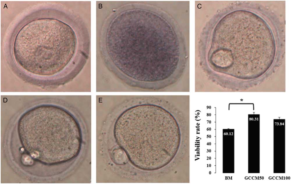
Trypan blue staining was applied to assess the viability of GV-stage oocytes and then of in vitro matured oocytes. Following the first staining, viable GVs were selected for further IVM. The next staining by TB was carried out to evaluate oocytes matured in vitro (Fig. 3).
Trypan blue staining to select the viable germinal vesicles (GV) for in vitro maturation (IVM) and viable IVM oocytes (MII oocytes). Viable GV (A) and dead GV (B) before IVM. (C) Viable MII oocyte after maturation in base medium (control group), viable MII oocytes after maturation in GCCM100 (D) and GCCM50 (E). Graph indicates different viability rates after IVM; P = 0.012.
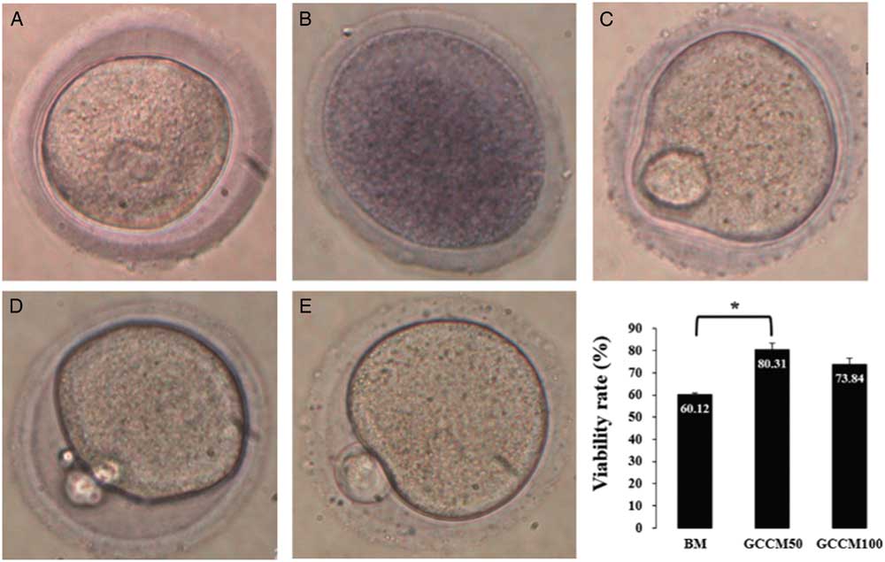
The TBS technique showed that the highest viability rate of in vitro matured oocytes occurred in the samples that used 50% GCCM (GCCM50) and this result was comparable with the control BM group (OR = 2.67, 95% CI = 1.09–6.52; P = 0.03) and non-comparable with the GCCM100 group.
Mitochondrial potential activity (JC-1 staining)
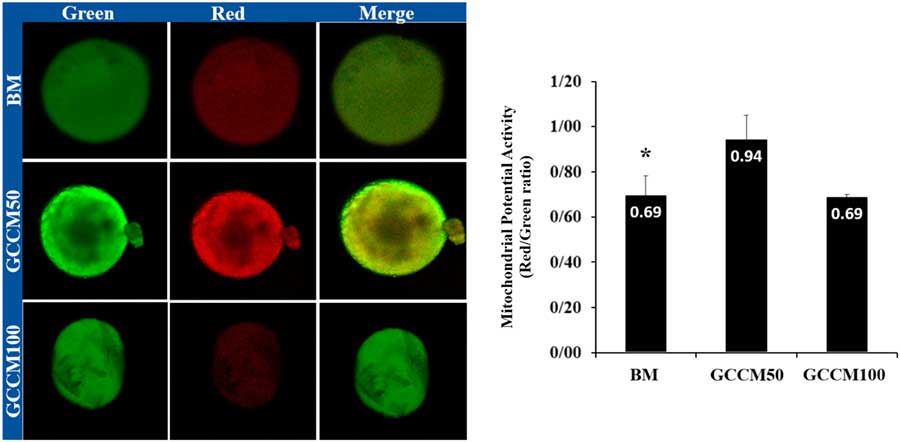
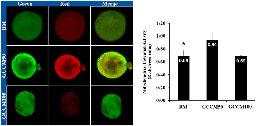
The mitochondria potential activity (MPA) was detected by confocal microscopy (Olympus Invert microscope) measured by Image J software (Image J 1.46r, Java 1.6.0_20) in in vitro matured MII oocytes. MPA was analytically higher in GCCM50 than both the control and GCCM100 groups (P < 0.05), but 100% concentration GCCM (GCCM100) did not have comparable effects on IVM rate against the BM group (P < 0.05) (Fig. 4).
JC-1 staining for mitochondrial membrane potential activity; red to green: increasing of mitochondrial potential activity (MPA). Green: JC-1 monomeric form (low intensity), Red: JC-1-aggregated form (high intensity). Graph: *Significant with other groups: P < 0.05.
Expression level of genes following in vitro maturation
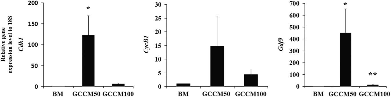
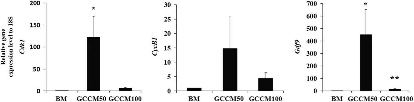
The relative mRNA expression of MPF complex showed that the level of Cdk1 expression in GCCM50 was significantly higher than that of the control (BM) group (P < 0.05), but there was no significant difference between GCCM100 and with GCCM50 and control groups (P > 0.05) (Fig. 5). However the expression of the CyclinB1 gene was not different among all groups (P > 0.05). Gdf9 gene expression increased for both 50 and 100% GCCM groups; expression levels in the GCCM50 group were higher than those of both control and GCCM100 groups (P < 0.05). In addition, Gdf9 expression was higher in the GCCM100 group than in control group (P < 0.05) (Fig. 5).
Gene expression level of CdK1, CycB1 and Gdf9 in in vitro matured (IVM) oocytes (MII) of all control (BM) and experimental (GCCM50 and GCCM100) groups. *Significant with BM. **Significant with BM and GCCM50, P < 0.05.
Discussion
The present study is the first report to our knowledge on the ability of GCCM to support IVM of GV-stage oocytes to the MII stage. The results showed that GV-stage oocytes isolated from 6–8-week-old NMRI female mice, 18 h after culture in 50% GCCM (GCCM50 group), showed better maturation rates than both BM (without GCCM) and GCCM100 (100% GCCM) groups. The benefits of granulosa cells on oocyte maturation and subsequent embryo development have been demonstrated earlier (Kobayashi et al., Reference Kobayashi, Takagi, Satoh, Hoshi and Oikawa1992; Malekshah et al., Reference Malekshah, Moghaddam and Daraka2006). Malekshah and his co-workers investigated the in vitro culture of mouse embryos in medium conditioned with human granulosa cells and showed a small improvement in outcome, albeit not comparable with co-culture. The effectiveness of granulosa cells to support embryo development until the blastocyst stage has been reported by Myers and colleagues (Myers et al., Reference Myers, Broussard, Menezo, Prough, Blackwell, Godke and Thibodeaux1994). Among the growth factors that have been used to induce better maturation and fertilization outcomes in oocytes, EGF has been known to induce meiotic resumption in various mammalian species such as rat (Lanuza et al., Reference Lanuza, Fischman and Baranao1998), mouse (Das et al., Reference Das, Phipps, Hensleigh and Tagatz1992), cattle (Nandi et al., Reference Nandi, Raghu, Ravindranatha and Chauhan2002), pig (Sirotkin et al., Reference Sirotkin, Dukesova, Makarevich, Kubek and Bulla2000), dog (Bolamba et al., Reference Bolamba, Russ, Harper, Sandler and Durrant2006) and buffalo (Singhal et al., Reference Singhal, Prasad, Singh, Prasad and Gupta2009).
This study aimed to investigate the effects of GCCM on upregulation of maturation genes and enhancement of mitochondrial activity in mouse oocytes matured in vitro. Here, we showed that GCCM induces meiosis resumption and improves the GVBD rate. Interestingly, use of BM combined with GCCM significantly improved IVM rates of mouse GVs.
To gain a better understanding of the GCCM improvement mechanism at the nuclear level, expression levels of both subunits (CdK1, CyclinB1) of MPF were assessed. CdK-1 mRNA was upregulated in the GCCM50 group more than in the control group (Fig. 5). But the level of CyclinB1 gene expression did not change among all groups.
The results of the present study showed that IVM rate increased in the presence of GCCM compared with the control group, which did not contain granulosa cells. It seems that the presence of growth factors, such as EGF and IGF1, is responsible for better in vitro oocyte maturation. The synergistic effects of FSH along with granulosa cells secretions in GCCM50, led to the increased effect of medium on meiosis resumption. Although there are a few reports that mentioned the inhibitory effect of IGF1 on oocyte maturation (Guler et al., Reference Guler, Poulin, Mermillod, Terqui and Cognie2000), some studies have proposed positive effects of IGF on oocyte stimulation. Liang et al. (Reference Liang, Su, Fan, Schatten and Sun2007) showed that IGF activates MAPK followed by PI3K/Akt induction after attaching to its receptors on the surface of the oocyte. These events activate MPF after removing the stopping forces from Cdc2 tracked by P90rsk activation and Myt1 inhibition. The IGF1 role in IVM of Xenopus oocytes has also been mentioned in the literature (Schmitt and Nebreda, Reference Schmitt and Nebreda2002).
The results of the present study confirmed earlier reports that GCCM contains factors such as IGF1, which could induce and sustain oocyte meiosis. The other effective factor is EGF, which provides a large amount of GCCM. EGF induces another ERK1/2 and then steroidogenesis and MOS signalling pathways (Shimada et al., Reference Shimada, Hernandez-Gonzalez, Gonzalez-Robayna and Richards2006). The last factors activate MPF and meiosis resumption through contact with the oolemma. Richani et al. (Reference Richani, Sutton-McDowall, Frank, Gilchrist and Thompson2014) first reported mitochondrial activity improvement using an EGF-like peptide. Our study showed similar results in the increase in mitochondrial membrane potential activity by the granulosa cell secretions. It seems that this effect may enhance the BM containing FSH in the GCCM50 group. We also analyzed gene expression of oocyte-specific marker, Gdf9, in all groups. Gdf9 was found to be upregulated in the GCCM50 group more than in the control and GCCM100 groups. GDF9 is a growth factor with a robust effect on proliferation and viability of granulosa cells and follicles. Gdf9 upregulation mentions to higher responsivity of oocyte to GCCM components. Increasing oocyte activity leads to an increase in the quality and viability of oocytes. This statement approved by progressive oocyte viability in both GCCM50 and GCCM100 groups compared with BM group. This situation indicates the influential role of GCCM on oocytes during IVM.
As mitochondria are the major source of ATP, the energy molecule, and play an important role in cellular metabolism, their membrane potential activity is closely related to both the in vivo and in vitro developmental potential of oocyte and embryo (May-Panloup et al., Reference May-Panloup, Chretien, Malthiery and Reynier2007). The quantity, localization and activity of mitochondria in oocyte have been shown (Wilding et al., Reference Wilding, Dale, Marino, di Matteo, Alviggi, Pisaturo, Lombardi and De Placido2001). In the present study, we used JC-1 staining methods to show a marked difference in mitochondrial membrane potential activity between the BM and GCCM50 groups.
To conclude, this work showed that maturation and viability rates and mitochondrial membrane potential activity of in vitro cultured oocytes are significantly enhanced on using a combination of GCCM as seen in the GCCM50 group. It was also shown that the presence of FSH in the BM in the GCCM50 sample had a synergetic effect on signalling pathways and oocyte maturation.
Acknowledgements
The authors greatly acknowledge their colleagues in the Embryology Department of the Royan Institute (Tehran, Iran), especially Mrs Abed Heidari for her help with real-time PCR techniques.
Conflict of interest
There are no conflicts of interest.