Introduction
Modular coral-like fossils in Lower Ordovician (Tremadocian) strata of the St. George Group in western Newfoundland, Canada, were initially identified as the tabulate coral Lichenaria Winchell and Schuchert, Reference Winchell and Schuchert1895 (Pratt and James, Reference Pratt and James1982, Reference Pratt and James1989a, Reference Pratt and Jamesb). The lowermost occurrence of such fossils, just above the Cambrian-Ordovician boundary as currently placed (Boyce et al., Reference Boyce, McCobb and Knight2013, fig. 2), was considered to record the earliest tabulates (Pratt and James, Reference Pratt and James1989a; Webby et al., Reference Webby, Elias, Young, Neuman, Kaljo, Webby, Paris, Droser and Percival2004). In a recent analysis, however, it was determined that Tremadocian material from Newfoundland and elsewhere in North America, which had long been attributed to Lichenaria, actually belongs to the Ordovician genus Amsassia Sokolov and Mironova, Reference Sokolov and Mironova1959 (Elias et al., Reference Elias, Lee and Pratt2021). The only other Tremadocian record of Amsassia is A. argentina Carrera, Astini, and Gomez, Reference Carrera, Astini and Gomez2017, from the La Silla Formation in the Argentine Precordillera (Carrera et al., Reference Carrera, Astini and Gomez2017), which is approximately coeval with the lowermost occurrence of the genus in the St. George Group (Elias et al., Reference Elias, Lee and Pratt2021). Amsassia is more widely distributed in the Middle and especially Upper Ordovician, occurring in North America and numerous places in Asia (e.g., M. Lee et al., Reference Lee, Sun, Choh and Lee2014, Reference Lee, Park, Tien, Choh, Elias and Lee2016, Reference Lee, Elias, Choh and Lee2018; Sun et al., Reference Sun, Elias and Lee2014).
In the present paper, occurrences of Amsassia in the St. George Group of western Newfoundland are documented with respect to their geologic and paleoecologic settings. Two species are recognized: A. terranovensis n. sp. from the Watts Bight Formation and A. diversa n. sp. from the Boat Harbour Formation. In addition, two rare forms providing limited morphologic information are described: Amsassia? spp. A and B from the Watts Bight and Boat Harbour formations, respectively. Based on an analysis of morphologic features that characterize Amsassia, the genus is interpreted as a calcareous alga that may represent an extinct group of green algae. The new data on Amsassia from Newfoundland, along with compiled information about other definite and probable Tremadocian occurrences, allow the origin and early dispersal of the genus to be appreciated in a framework of paleogeography and paleoecology. Considered together with updated information on younger Ordovician occurrences, a better understanding of the overall geologic history of Amsassia is presented, with regard to its temporal and paleogeographic ranges and paleoecology, and relation to major events involving global biodiversity and reef development.
Geologic setting
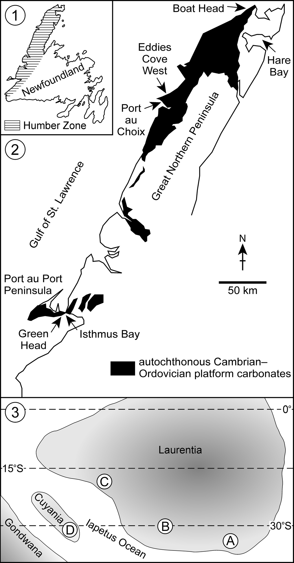
The uppermost Cambrian to lowermost Middle Ordovician St. George Group of western Newfoundland crops out discontinuously from the Port au Port Peninsula in the southwestern part of the island to Cape Norman on the northern tip of the Great Northern Peninsula (Knight and James, Reference Knight and James1987). This unit, up to ~500 m thick, belongs to the Humber tectonic zone of the northern Appalachians (Fig. 1.1). It is part of the autochthonous lower Paleozoic platform carbonate succession (Fig. 1.2), which was deposited on the passive margin of Laurentia on its south-facing side (Allen et al., Reference Allen, Thomas, Lavoie, Tollo, Bartholomew, Hibbard and Karabinos2010; Lavoie et al., Reference Lavoie, Desrochers, Dix, Knight and Salad Hersi2012), bordering the Iapetus Ocean (Torsvik and Cocks, Reference Torsvik and Cocks2013; Fig. 1.3). The St. George Group consists of peritidal limestones and dolostones represented by a variety of shallow-water facies, including bioturbated lime mudstone and wackestone, wave-rippled cross-laminated lime mudstone, laterally linked hemispheroidal stromatolites, and microbial laminites (Pratt and James, Reference Pratt and James1986). Numerous beds contain microbially dominated patch reefs (i.e., thrombolite mounds) up to ~2 m in thickness (Pratt and James, Reference Pratt and James1982, Reference Pratt and James1989a, Reference Pratt and Jamesb). Larger reef complexes are present at Green Head and in Hare Bay (Pratt and James, Reference Pratt and James1982).
Geologic and paleogeographic maps. (1, 2) Maps of Newfoundland and western Newfoundland showing geologic features and locations (based on Knight et al., Reference Knight, Azmy, Greene and Lavoie2007, fig. 1, Reference Knight, Azmy, Boyce and Lavoie2008, fig. 1; Boyce et al., Reference Boyce, McCobb and Knight2011, fig. 1). The Humber Zone of the northern Appalachians includes autochthonous lower Paleozoic platform carbonates, which occur from Port au Port Peninsula to the northern tip of the Great Northern Peninsula. Samples of Tremadocian-age Amsassia were collected from the Watts Bight Formation at Green Head and the Boat Harbour Formation at Boat Head and Eddies Cove West. (3) Paleogeography at the beginning of Tremadocian time (adapted from Scotese, Reference Scotese2014, map 83; Albanesi et al., Reference Albanesi, Barnes, Trotter and Williams2020, fig. 4). Definite and probable occurrences of Tremadocian-age Amsassia are located in the paleocontinent Laurentia (A, western Newfoundland; B, Maryland; C, Texas) and in the Cuyania Terrane (D, Argentine Precordillera).

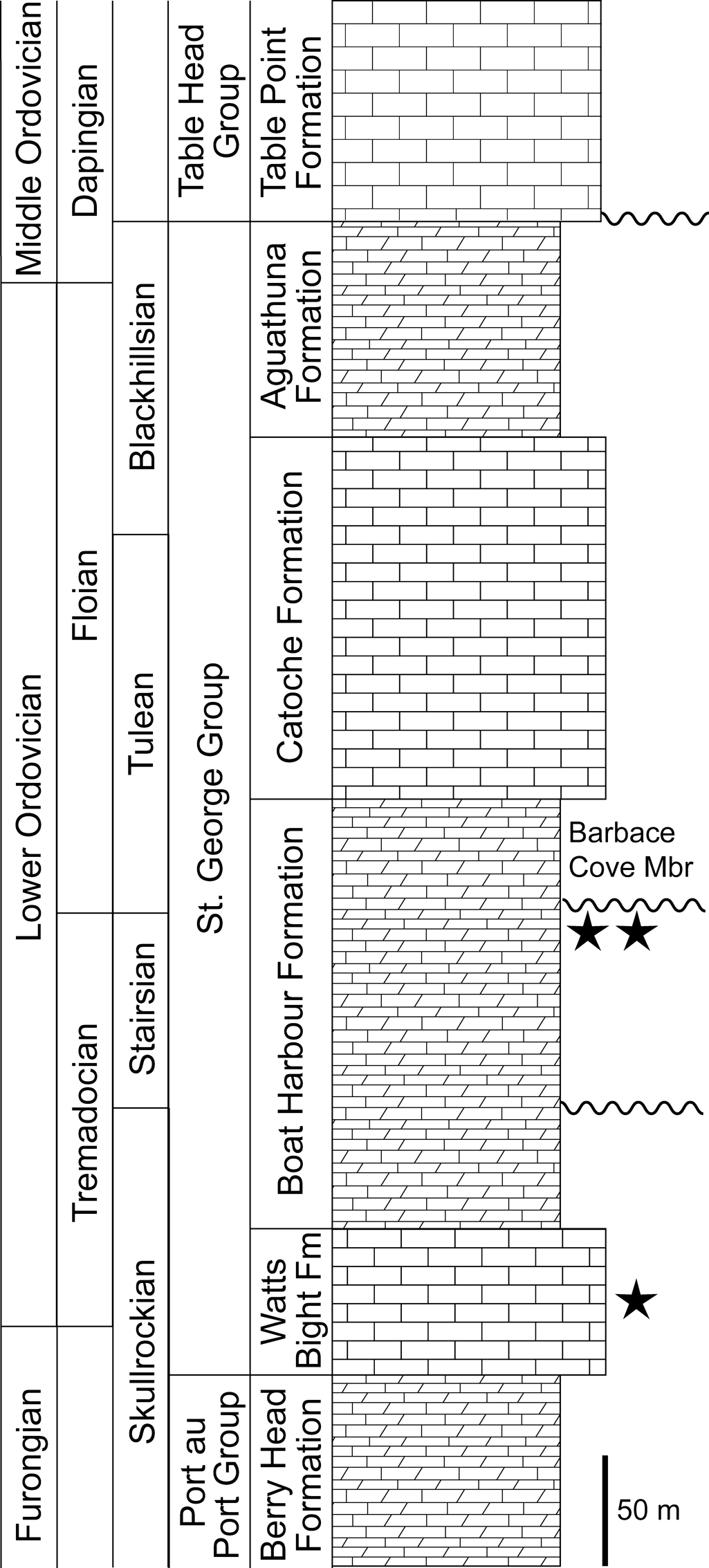
The St. George Group is subdivided into four formations: the Watts Bight, Boat Harbour, Catoche, and Aguathuna formations, in ascending stratigraphic order (Fig. 2). Two regional, temporally significant disconformities are present in the Boat Harbour Formation (Knight et al., Reference Knight, Azmy, Boyce and Lavoie2008). These units have been subjected to considerable paleontologic study and are well dated biostratigraphically using conodonts and trilobites (Fortey, Reference Fortey1979; Boyce, Reference Boyce1989; Ji and Barnes, Reference Ji and Barnes1994; Boyce and Stouge, Reference Boyce and Stouge1997).
Generalized stratigraphic section of the St. George Group in western Newfoundland (based on Knight et al., Reference Knight, Azmy, Boyce and Lavoie2008; with updated chronostratigraphy in Boyce et al., Reference Boyce, McCobb and Knight2011, Reference Boyce, McCobb and Knight2013). The North American Skullrockian through Blackhillsian stages comprise the Ibexian Series. The Barbace Cove Member is shown, but other members are not. In addition to the contact between the St. George Group and the overlying Table Head Group, there are two significant regional disconformities in the Boat Harbour Formation (shown with wavy lines). Stratigraphic positions of the three occurrences of Tremadocian-age Amsassia are marked with stars (~26–33 m above the base of the Watts Bight Formation, and ~18 m and ~13.5 m below the Barbace Cove Member in the Boat Harbour Formation).

Coral-like fossils, initially identified as Lichenaria by Pratt and James (Reference Pratt and James1982, Reference Pratt and James1989a, Reference Pratt and Jamesb), occur in lower Lower Ordovician (Tremadocian) strata of the St. George Group. They were observed in microbial patch reefs within the Watts Bight Formation at Green Head, and in the Boat Harbour Formation at Eddies Cove West and Boat Head (Figs. 1.2, 2). Those fossils were recently reassigned to Amsassia (Elias et al., Reference Elias, Lee and Pratt2021). Sampling and petrographic study as part of Pratt's (Reference Pratt1979) research was intensive and, with the exception of a different genus in the Boat Harbour Formation of Isthmus Bay (unpublished data), it is reasonably certain that these coral-like fossils are not present in other patch reef-bearing beds in the measured sections of the St. George Group from Isthmus Bay and both sides of The Gravels connecting Port au Port Peninsula with the mainland, Port au Choix, Eddies Cove West, and Boat Head (Fig. 1.2). Knight and James (Reference Knight and James1987) reported corals as occurring rarely in the Catoche Formation, but none was observed by Pratt (Reference Pratt1979). Samples of large reefs in the allochthonous Catoche Formation in Hare Bay (Knight and James, Reference Knight and James1987), while containing siliceous sponges, also lack coral-like fossils (Pratt, Reference Pratt1979). However, there are other exposures in western Port au Port Peninsula that were not examined for the present study (Boyce et al., Reference Boyce, McCobb and Knight2011, Reference Boyce, McCobb and Knight2013); fossils identified as Lichenaria were reported by Kröger and Penny (Reference Kröger and Penny2020) from a reef complex in the Watts Bight Formation at Ship Cove, ~20 km west of Green Head.
Occurrences of Amsassia in Newfoundland
The three occurrences of Amsassia in western Newfoundland are described in ascending stratigraphic order.
Green Head, Port au Port Peninsula
Amsassia terranovensis n. sp. and Amsassia? sp. A occur in a reef complex ~26–33 m above the base of the Watts Bight Formation at Green Head in Isthmus Bay, on the southeastern coast of Port au Port Peninsula (Figs. 1.2, 2; for detailed map and stratigraphic column see Knight and James, Reference Knight and James1987, figs. 3d, 4; Knight et al., Reference Knight, Azmy, Boyce and Lavoie2008, fig. 4, column b; Boyce et al., Reference Boyce, McCobb and Knight2011, fig. 4). The age is early Tremadocian (Ibexian, Skullrockian; Boyce et al., Reference Boyce, McCobb and Knight2013), just above the Cambrian-Ordovician boundary.
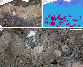
Amsassia-bearing biolithite forms the lower part of the Green Head reef complex in the Watts Bight Formation (Pratt and James, Reference Pratt and James1982, fig. 19, Reference Pratt and James1989a, fig. 3; Kröger and Penny, Reference Kröger and Penny2020, fig. 5). This low-relief reef complex is dominated by thrombolite frameworks composed of fenestral, clotted, and peloidal micrite plus matrix of sparse biomicrite. In some places, there are scallop-shaped erosion surfaces onlapped by wackestone (Fig. 3.1, 3.2). Individual and grouped Amsassia skeletons are incorporated in parts of the framework, but not evenly throughout; the skeletons are mostly silicified (Fig. 4.1, 4.2). Amsassia also occurs variably intergrown with bulbous masses and fringes of Renalcis Vologdin, Reference Vologdin1932 (Pratt and James, Reference Pratt and James1982, figs. 20–22, 25a, Reference Pratt and James1989a, figs. 4a–c, 5, 8; Kröger and Penny, Reference Kröger and Penny2020, fig. 6b; Fig. 3.3). The biomicritic matrix surrounding the framework elements contains incompletely filled tubular burrows assignable to Palaeophycus Hall, Reference Hall1847; variably silicified, whole, disarticulated, and broken orthid brachiopod valves, both smooth, thin-shelled as well as thick, ribbed valves (including forms that have been identified as Finkelnburgia Walcott, Reference Walcott1905; Flower, Reference Flower1978); straight- and curved-shelled nautiloids (see also Flower, Reference Flower1978); complete and fragmented turriform, turbiform, and planispiral gastropods (see also Knight et al., Reference Knight, Azmy, Boyce and Lavoie2008); trilobite sclerites, some of which are broken; rare stylophoran echinoderm ossicles; and rare blunt-tipped tetraradiate and hexaradiate calcareous sponge spicules, large calcitized monaxons, local accumulations of small monaxons, and patches of vermiform networks up to a few millimeters across, which are referable to non-spicular keratose demosponges (Luo and Reitner, Reference Luo and Reitner2016; Lee and Riding, Reference Lee and Riding2021). Many modules in skeletons of Amsassia contain uniformly sized peloids and micritic clots, likely of microbial origin.
Biolithite in the lower part of the reef complex in the Watts Bight Formation at Green Head, western Newfoundland. Scale bars = 20 mm. (1) Surface cut parallel to bedding, showing inner part of thrombolite framework with mostly silicified Amsassia (appearing bluish in color), passing outward into a zone lacking Amsassia, which is cut by a scalloped erosion surface onlapped by burrowed, fossiliferous wackestone containing nautiloids. Part of the thrombolite (upper left in photograph) exhibits an invagination representing a crevice in the reef filled with burrowed lime mudstone. (2) Tracing of (1): light blue = thrombolite consisting of microbial framework and surrounding lime mudstone matrix; magenta = Amsassia; dark blue = fossiliferous wackestone (nautiloids outlined). (3) Surface cut parallel to bedding, showing silicified Amsassia (appearing bluish in color; in upper center and lower right), thrombolite framework (appearing darker colored and speckled; incorporating two gastropods in lower right), bulbous masses of Renalcis (appearing light-colored and speckled; left of center, lower right of center, and upper right), and erosion surfaces onlapped by lighter-colored wackestone with burrows, gastropods, and nautiloids (center).

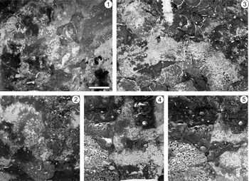
Representative Tremadocian-age biolithite from western Newfoundland, featuring modular skeletons of Amsassia: back-lit photographs of thin sections oriented horizontally (1) and vertically (2–5) with respect to bedding. Scale bar for all (in 1) = 10 mm. (1, 2) Watts Bight Formation at Green Head: (1) mostly silicified Amsassia with clotted microbial micrite surrounded by burrowed biopelmicrite containing gastropod and brachiopod valve (upper center); (2) Amsassia and clotted microbial micrite surrounded by burrowed, sparsely bioclastic micrite and pelmicrite. (3) Boat Harbour Formation at Boat Head: large mass of upward radiating Amsassia surrounded by burrowed biomicrite, with brachiopod valves, gastropod, and nautiloid (upper center). (4, 5) Boat Harbour Formation at Eddies Cove West: serial sections spaced 3 mm apart, showing Amsassia and clotted microbial micrite cut by an undulating, vertical erosion surface, and flanked by grainstone consisting of intrapelsparite passing upward into burrowed biopelmicrite with broken brachiopod valves, trilobite sclerites, and gastropod shells.

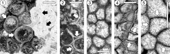
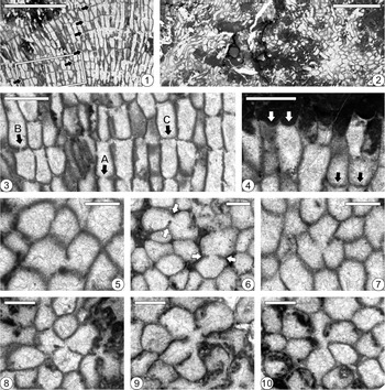
The module walls of Amsassia terranovensis n. sp. from the Watts Bight Formation at Green Head have been altered to various degrees by multiple diagenetic processes. Silicification affected most specimens, usually resulting in complete obliteration of microstructure (black arrows in Fig. 5.1). In other places, the walls were dissolved and replaced by a coarse mosaic of neomorphic calcite spar (white arrows in Fig. 5.1, 5.2). The latter type of preservation is considered to result from replacement of the original aragonitic skeleton with calcite during early diagenesis, following interpretations based on Ordovician tetradiids (Steele-Petrovich, Reference Steele-Petrovich2009a). Less commonly, walls of A. terranovensis n. sp. (Fig. 5.3) and Amsassia? sp. A are preserved in the form of micritic fabric having variable thickness, retaining no trace of original microstructure.
Thin-section photomicrographs showing different types of preservation in transverse views of Amsassia terranovensis n. sp. from the Watts Bight Formation at Green Head (1–3) and A. diversa n. sp. from the Boat Harbour Formation at Boat Head (4, 5), western Newfoundland. Scale bars = 0.5 mm. (1) Partially silicified skeleton showing complete obliteration of module walls by silicification (black arrows), and walls dissolved and replaced with a coarse mosaic of neomorphic calcite spar (white arrows). (2) Module walls dissolved and replaced with a coarse mosaic of neomorphic calcite spar (arrows). (3) Preservation of walls in the form of micritic fabric having variable thickness, retaining no trace of original microstructure. (4) Walls between adjoining modules dissolved and replaced with a coarse mosaic of neomorphic calcite spar (arrows). (5) Walls between adjoining modules represented by a thin, dark micritic line with a thick zone of isopachous fibrous calcite cement on both sides, having fibers oriented perpendicular to the dark line (arrows).

Boat Head, Great Northern Peninsula
Amsassia diversa n. sp. and Amsassia? sp. B occur in a bed of patch reefs, ~18 m below the Barbace Cove Member in the Boat Harbour Formation at Boat Head, southwest of Cape Norman, ~130 km to the northeast of Eddies Cove West (Figs. 1.2, 2; for detailed map and stratigraphic column see Knight and James, Reference Knight and James1987, figs. 3a, 6). The age is Tremadocian (Ibexian, Stairsian; Boyce et al., Reference Boyce, McCobb and Knight2013).
Amsassia-bearing patch reefs at Boat Head occur in a 1.6-m-thick unit consisting of superimposed, individual thrombolite mounds up to 1 m thick, flanked by grainstone, and overlain by laterally adjacent, domical stromatolites up to 30 cm in diameter. Centimeter-scale erosion surfaces locally cross-cut Amsassia skeletons. Microbial components of the biolithite are difficult to discern on outcrop and sawn surfaces, but in thin section consist of dense micrite and patches of clotted micrite with fenestral porosity, in a less dense micrite matrix that is variably peloidal. Bioclasts include turriform and planispiral gastropods, nautiloids, brachiopods, trilobite sclerites, monaxon and calcareous sponge spicules, rare ostracodes, and scarce stylophoran ossicles, along with patches of keratose sponge networks 2–5 mm across, and scattered tubular burrows conforming to Palaeophycus (Fig. 4.3). Trilobite sclerites and gastropods are commonly broken. In skeletons of Amsassia, many modules contain uniformly sized peloids and micritic clots of probable microbial origin.
The module walls of Amsassia diversa n. sp. from the Boat Harbour Formation at Boat Head are commonly preserved in the form of micritic fabric having variable thickness, obscuring the original microstructure. In places, the wall between adjoining modules was dissolved and replaced with a coarse mosaic of neomorphic calcite spar (arrows in Fig. 5.4). Rarely, the wall between adjoining modules is represented by a thin, dark micritic line with a thick zone of isopachous fibrous calcite cement on both sides having fibers oriented perpendicular to the dark line (Fig. 5.5). Carrera et al. (Reference Carrera, Astini and Gomez2017, fig. 6.5) described similar diagenetic features in the best-preserved material of A. argentina from the La Silla Formation in the Argentine Precordillera. The walls of Amsassia? sp. B are micritic with variable thickness.
Eddies Cove West, Great Northern Peninsula
Amsassia diversa n. sp. occurs in microbial patch reefs, ~13.5 m below the Barbace Cove Member in the Boat Harbour Formation at Eddies Cove West, just northeast of Port au Choix, ~275 km north-northeast of Isthmus Bay (Figs. 1.2, 2; for detailed map see Knight and James, Reference Knight and James1987, fig. 3b; for detailed stratigraphic column see Knight, Reference Knight1991, fig. 7). The age is Tremadocian (Ibexian, Stairsian; Boyce et al., Reference Boyce, McCobb and Knight2013).
Amsassia-bearing patch reefs cropping out at Eddies Cove West consist of individual and laterally coalesced thrombolite mounds 1.0–1.2 m in diameter and 0.4 m thick, with two episodes of reef growth superimposed to form a unit 0.8 m thick. The mounds are flanked by moderately sorted peloidal and intraclastic grainstone containing gastropods; subvertical scoured surfaces occur locally in the mounds and are onlapped by grainstone. Individual microbial framework elements of the biolithite are not well defined on outcrop and sawn surfaces, but in thin section are seen to consist of clotted micrite with fenestral pores, whereas the matrix is less dense biopelmicrite with localized accumulations of sand-size peloids (Fig. 4.4, 4.5). Associated bioclasts include sporadically articulated, large, thick-shelled brachiopods; small, thin-shelled brachiopods, some with distinct ribbing; trilobite sclerites belonging to several species; turriform, turbinate, and planispiral gastropods; rare ostracodes; sexiradiate, blunt-tipped spicules with distal and proximal rays belonging to the Calcarea Bowerbank, Reference Bowerbank1864; and possible tetraxons. Patches of keratose sponge networks up to 10 mm across are present. Many of the brachiopod valves and trilobite sclerites are fragmented. Uniformly sized peloids and micritic clots, likely of microbial origin, are abundant in the modules of Amsassia skeletons. Spar-filled tubular burrows, assignable to Palaeophycus, are sporadically present. Internal erosion surfaces are locally present and flanked by intraclastic peloidal grainstone.
The walls of Amsassia diversa n. sp. from the Boat Harbour Formation at Eddies Cove West are commonly preserved in the form of micritic fabric having variable thickness, obscuring the original microstructure.
Definite and probable Tremadocian occurrences of Amsassia
In addition to three confirmed occurrences of Amsassia in western Newfoundland, Tremadocian material that definitely or probably belongs to Amsassia is known from four occurrences described in ascending stratigraphic order.
Cerro La Silla, Argentine Precordillera
Amsassia argentina occurs ~95 m above the base of the La Silla Formation, within the Cordylodus angulatus Conodont Zone, in a 2-m-thick unit, apparently representing a low-relief carbonate mound (Carrera et al., Reference Carrera, Astini and Gomez2017). The unit consists of matrix-rich boundstone with peloidal mudstone and grainstone. Amsassia was a main framework builder, along with a complex microbial-algal consortium including thrombolites, small stromatolites, renalciforms, and calcareous Nuia Maslov, Reference Maslov and Shatskiy1954. The age is Tremadocian (Ibexian, Skullrockian; Keller, Reference Keller2012), slightly above the Cambrian-Ordovician boundary.
San Saba, Texas, USA
Lichenaria cloudi Bassler, Reference Bassler1950, probably belongs to Amsassia (Elias et al., Reference Elias, Lee and Pratt2021). Based on the original collection label (included with USNM 113998), these silicified specimens are from the calcitic facies near the top of the Staendebach Member, which is the upper unit of the Tanyard Formation. Cloud and Barnes (Reference Cloud and Barnes1948) reported that the upper 27–30 m of the formation is characterized by fossils including this species, the calcareous alga Rhabdoporella Stolley, Reference Stolley1893, cephalopods, rostroconchs, and trilobites. The age is Tremadocian (Ibexian, late Skullrockian to early Stairsian; Miller et al., Reference Miller, Loch and Taylor2012).
Hagerstown, Maryland, USA
Tetradium? simplex Bassler, Reference Bassler1919, was recognized in the original description as the earliest recorded coral (Bassler, Reference Bassler1919). This species was subsequently assigned to Lichenaria (Bassler, Reference Bassler1950), but probably belongs to Amsassia (Elias et al., Reference Elias, Lee and Pratt2021). It occurs just above the Stonehenge Formation (Bassler, Reference Bassler1950), in what is now known as the Rockdale Run Formation (Taylor, Reference Taylor2015). According to Bassler (Reference Bassler1919), this species is abundant in limestone at the base of the Cryptozoon steeli Zone, occurring in lamellose masses in association with gastropods. The age is Tremadocian (Ibexian, Stairsian; Taylor, Reference Taylor, Loch, Repetski and Brezinski2015).
Williamsport, Maryland, USA
Material described by Sando (Reference Sando1957) as Lichenaria cloudi was reassigned to Amsassia by Elias et al. (Reference Elias, Lee and Pratt2021). The species identification, however, is unconfirmed because the type specimens of L. cloudi from San Saba, Texas, are poorly preserved and cannot be assigned with certainty to Amsassia (Elias et al., Reference Elias, Lee and Pratt2021). Sando's (Reference Sando1957) material is from 159 m above the base of the Rockdale Run Formation, within the Lecanospira Zone, where Amsassia occurs in a 2-m-thick unit of limestone with gymnosolenid stromatolites, gastropods including Lecanospira Butts, Reference Butts1926, and cephalopods. The age is Tremadocian (Ibexian, Stairsian; Taylor et al., Reference Taylor, Loch, Repetski and Brezinski2015).
Biologic affinity of Amsassia
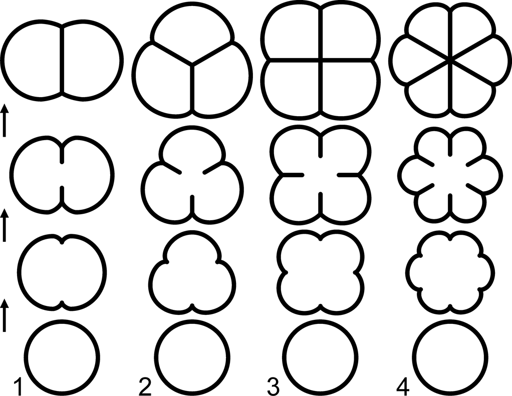
Amsassia, traditionally treated as a tabulate coral, has been assigned by various authors to lichenariids and tetradiids, as well as chaetetids (see Sun et al., Reference Sun, Elias and Lee2014). Although lichenariids are tabulate corals (Elias et al., Reference Elias, Lee and Woo2008, Reference Elias, Lee and Pratt2021), tetradiids have been excluded from tabulates (Scrutton, Reference Scrutton1997) and interpreted as calcareous red algae (Steele-Petrovich, Reference Steele-Petrovich2009a, Reference Steele-Petrovichb, Reference Steele-Petrovich2011), while chaetetids have been accepted as calcareous sponges (West, Reference West2011a). Numerous morphologic characters have been considered in attempts to determine the biologic affinity of Amsassia (Sun et al., Reference Sun, Elias and Lee2014; M. Lee et al., Reference Lee, Park, Tien, Choh, Elias and Lee2016, Reference Lee, Elias, Choh and Lee2018; Elias et al., Reference Elias, Lee and Pratt2021). Of particular significance are the way modules increase in number during growth, and the resulting arrangement and size of modules (Figs. 6–9). Module increase by fission occurs in Amsassia, tetradiids, some chaetetids, and a few corals. In the latter three groups, module increase involves the development of one or more longitudinal skeletal partitions inside the module. The process is fundamentally different in Amsassia, where infolding of the module wall (Figs. 6, 7.9–7.12, 9.5–9.10) is consistent with simple di-, tri-, quadri-, and hexachotomous branching of the module occupant (black arrow in Fig. 7.8, white arrows in Fig. 8.5). Di- to polychotomous branching is common in algae. Module increase in Amsassia resulted in a closely packed to loose, phacelocerioid arrangement of modules having separate walls, indicating individuality of occupants (Fig. 7.4), which would not be expected in sponges. The modules in chaetetid skeletons are contiguous, with shared walls. West (Reference West2011b) rejected Amsassia from chaetetid-type sponges. Some calcimicrobes have a modular appearance and branching tubules. Garwoodia Wood, Reference Wood1941, for example, has been regarded by some authors as green algal rather than cyanobacterial, because of its relatively large tubules with diameters up to 0.07 mm (Liu et al., Reference Liu, Wu, Bao, Jiang, Zheng and Chen2021). Modules attain even larger sizes in Amsassia, with the smallest and largest species having maximum module diameters of 0.28 mm and 1.66 mm, respectively (M. Lee et al., Reference Lee, Park, Tien, Choh, Elias and Lee2016, fig. 4a). In summary, fundamental biologic characters suggest that Amsassia can be excluded from cyanobacteria, calcareous sponges, corals, and tetradiids, but point to an algal affinity.
Diagrams showing successive transverse serial sections of modules in Amsassia undergoing bi-, tri-, quadri-, and hexapartite fission (1–4, respectively). The actual expansion rate and shape of outer walls are determined by the degree of crowding of modules within the phacelocerioid skeleton. The appearance of features in thin sections varies if sections are not perpendicular to the modules (Sun et al., Reference Sun, Elias and Lee2014, fig. 5).

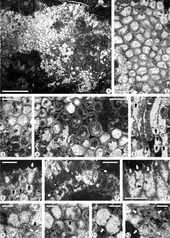
Thin-section photomicrographs showing transverse (1–4, 9–12) and longitudinal (5–8) views of Amsassia terranovensis n. sp. from the Watts Bight Formation at Green Head, western Newfoundland. (1) Partially silicified skeleton; note module undergoing bipartite fission (outlined; enlarged in 9); holotype GSC 141439a; scale bar = 5 mm. (2) Cerioid area showing densely packed, predominantly polygonal to subpolygonal modules; note module undergoing bipartite fission (outlined); holotype GSC 141439b; scale bar = 1 mm. (3) Extreme size and shape variability of modules; paratype GSC 141438; scale bar = 1 mm. (4) Phaceloid area showing loosely packed, predominantly subcircular to circular modules partially separated by micrite; GSC 141448; scale bar = 1 mm. (5) Tabula-like structures (black arrows at A–D); note tabula-like structures at the same level in several modules (three black arrows at level A); note constriction of a module followed by regeneration (arrow at B); note constriction of two modules (white arrows) and incomplete tabula-like structure in another module at the same level (black arrow at D); GSC 141447; scale bar = 1 mm. (6) Tabula-like structures developed at the same level in adjacent modules (black arrows); note calice with concave-upward bottom (white arrow); GSC 142628; scale bar = 1 mm. (7) Curved growth surface showing partial mortality (white arrows) and rejuvenation/regeneration with tabula-like structures (between black arrows); GSC 142629; scale bar = 2 mm. (8) Calices (white arrows); note a module undergoing longitudinal fission (black arrow); GSC 142630; scale bar = 1 mm. (9) Enlargement of outlined area in 1, showing module undergoing bipartite fission (center of photograph); note subtriangular space between modules (black arrow), and convergence of walls resulting in back-to-back walls between adjoining modules (white arrows); holotype GSC 141439a; scale bar = 0.5 mm. (10) Module undergoing bipartite fission (arrow); note micrite-filled spaces between some modules, and back-to-back walls between adjoining modules; GSC 142632; scale bar = 0.5 mm. (11) Module undergoing bipartite fission; note infolding of wall (arrow); GSC 142631; scale bar = 0.5 mm. (12) Probable result of recently completed quadripartite fission involving four walls (arrows); GSC 142631; scale bar = 0.5 mm.

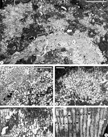
Thin-section photomicrographs showing transverse (2), longitudinal (4, 5), and both transverse and longitudinal (1, 3) views of Amsassia diversa n. sp. from the Boat Harbour Formation at Boat Head (1, 2, 4, 5) and Eddies Cove West (3), western Newfoundland. (1) Skeleton showing phacelocerioid structure; note surface of partial mortality (black arrow) and area of rejuvenation/regeneration (white arrow); modules have shallow calices with concave-upward bottoms (outlined areas A and B; B is enlarged in Fig. 9.4); holotype GSC 141441c; scale bar = 10 mm. (2) Extreme size and shape variability of modules; note outlined areas of small polygonal to subpolygonal modules (A), large polygonal to subcircular modules (B), variably sized subpolygonal to subcircular modules (C), and large irregularly shaped modules (D); GSC 141437; scale bar = 5 mm. (3) Modules densely packed, predominantly polygonal to subpolygonal, in cerioid area of skeleton, showing relatively small variability in size and shape of modules; calices shallow to moderately deep with concave-upward bottoms (outlined); many modules contain peloids and micritic clots of probable microbial origin (e.g., arrows); GSC 141445; scale bar = 2 mm. (4) Skeleton showing growth interruptions and partial mortality associated with peloids and micritic clots likely of microbial origin (dark areas); GSC 142638; scale bar = 2 mm. (5) Modules undergoing longitudinal fission (white arrows); note constriction of modules at certain levels, followed by rejuvenation and, in some cases, regeneration with development of tabula-like structures (black arrows and dotted lines); holotype GSC 141441b; scale bar = 1 mm.

Thin-section photomicrographs showing longitudinal (1, 3, 4), transverse (5–10), and both longitudinal and transverse (2) views of Amsassia diversa n. sp. from the Boat Harbour Formation at Boat Head, western Newfoundland. (1) Tabula-like structures conspicuously developed at same levels in adjacent modules, possibly showing cyclomorphism (arrows); outlined area enlarged in 3; GSC 141446; scale bar = 5 mm. (2) Rare example showing wavy to irregular modules in oblique to longitudinal views; GSC 142636; scale bar = 5 mm. (3) Enlargement of outlined area in 1, showing features associated with rejuvenation/regeneration and formation of many tabula-like structures; note constriction of module with incomplete tabula-like structure (arrow A), nearly complete tabula-like structure (arrow B), and complete tabula-like structure (arrow C); GSC 141446; scale bar = 1 mm. (4) Enlargement of outlined area B in Figure 8.1, showing shallow calices with concave-upward bottoms (white arrows) having same shape as complete tabula-like structures within modules (black arrows); holotype GSC 141441c; scale bar = 1 mm. (5, 6) Modules undergoing bipartite fission; note two examples showing infoldings of wall in 6 (arrows); holotype GSC 141441a and GSC 141444, respectively; scale bars = 0.5 mm. (7) Module undergoing tripartite fission; holotype GSC 141441a; scale bar = 0.5 mm. (8) Module undergoing quadripartite fission; GSC 141442; scale bar = 0.5 mm. (9, 10) Modules undergoing hexapartite fission; GSC 141443 and GSC 142637, respectively; scale bars = 0.5 mm.

The unmineralized extant green alga Cladophora Kützing, Reference Kützing1843, possesses characters comparable to Amsassia. Its thallus is composed of branching filaments made up of large, elongate cells separated by transverse walls located at constrictions of the longitudinal walls (see photographs in Titlyanov et al., Reference Titlyanov, Titlyanova, Li and Huang2017, p. 93, 94; Michalak and Messyasz, Reference Michalak and Messyasz2021). Transverse walls grow in from the longitudinal walls towards the interior, closing to divide cells (van den Hoek, Reference van den Hoek1963). Maximum cell diameters range from 0.07 mm in a small species such as C. albida (Nees, Reference Nees1820), to 1.00 mm in a large species such as C. ohkuboana Holmes, Reference Holmes1896 (Sakai, Reference Sakai1964). Branching is commonly dichotomous, but also polychotomous in species such as C. flagelliformis (Suhr, Reference Suhr1840), which has whorls of up to six branches (Lluch, Reference Lluch2002, fig. 217).
If the branching filaments of an alga resembling Cladophora were tightly to loosely arranged and the cell walls became calcified during growth, it seems conceivable that phacelocerioid skeletons consisting of modules with tabula-like structures at constrictions could result, as seen in Amsassia (Figs. 7.5, 9.1, 9.3). The scale is consistent, with overlap between maximum cell diameters in species of Cladophora (0.07–1.00 mm) and maximum module diameters in species of Amsassia (0.28–1.66 mm). Di- to hexachotomous branching occurs in Cladophora; in Amsassia, the types of module fission indicate di-, tri-, quadri-, and hexachotomous branching. The tops of modules in Amsassia would have remained uncalcified to permit elongation of filaments during growth, and for liberation of gametes and zoospores, as seen in C. surera Parodi and Cáceres, Reference Parodi and Cáceres1995 (Parodi and Cáceres, Reference Parodi and Cáceres1995, figs. 21, 28). Thus, upon death and decomposition, concave-upward calices with tabula-like structures at their base would have been present at the tops of skeletons and become filled with sediment, as seen in Amsassia (Figs. 7.6, 7.8, 9.4). Amsassia is therefore interpreted as a calcareous alga, possibly representing an extinct group of green algae.
Geologic history of Amsassia
The new and compiled data on Tremadocian occurrences of Amsassia, together with updated information on the age of younger occurrences, contribute to an understanding of the overall geologic history of the genus. The focus is on its temporal and paleogeographic ranges and paleoecology.
Temporal and paleogeographic ranges
The earliest known representatives of Amsassia are A. terranovensis n. sp. from western Newfoundland and A. argentina from the Argentine Precordillera (Carrera et al., Reference Carrera, Astini and Gomez2017; Elias et al., Reference Elias, Lee and Pratt2021). Both appeared in the early Tremadocian, just after the beginning of the Ordovician Period. The genus evidently originated in the vicinity of the southern margin of Laurentia and the Cuyania Terrane (Fig. 1.3). The occurrence of these two, similar species (see Systematic paleontology) is consistent with the hypothesis of a close paleogeographic relation between Laurentia and Cuyania in the earliest Ordovician (Martin et al., Reference Martin, Collins and Spencer2020). The probable appearance of Amsassia in Texas in the mid-Tremadocian (Elias et al., Reference Elias, Lee and Pratt2021), followed by definite occurrences in Maryland and western Newfoundland in the late Tremadocian (Elias et al., Reference Elias, Lee and Pratt2021), indicate that the genus spread geographically near the shelf margin of southern Laurentia during Tremadocian time (Fig. 1.3; for lithofacies map see Vachard et al., Reference Vachard, Clausen, Palafox, Buitrón, Devaere, Hayart and Régnier2017, fig. 19).
Amsassia is unknown from the late Early Ordovician (Floian) to early Middle Ordovician (Dapingian). In the late Middle Ordovician, there is a probable occurrence in the early Darriwilian Lehman Formation in Utah, USA (Elias et al., Reference Elias, Lee and Pratt2021, table 1). Coral-like fossils in the Sandaogou Formation in western North China, initially assigned to Lichenaria (Li and Lin, Reference Li and Lin1982; Lin, Reference Lin1983), are now recognized as Amsassia (Sun et al., Reference Sun, Elias and Lee2014); the age is Darriwilian (Zhang et al., Reference Zhang, Zhan, Zhen, Wang, Yuan, Fang, Ma and Zhang2019, fig. 5). In the Taebaeksan Basin of Korea, A. koreanensis M. Lee et al., Reference Lee, Hong, Woo, Oh, Lee and Choh2016, occurs in the Duwibong Formation (M. Lee et al., Reference Lee, Hong, Woo, Oh, Lee and Choh2016), which is mid–late middle Darriwilian (Cho et al., Reference Cho, Lee, Lee and Choh2021). Thus, the paleogeographic range of Amsassia expanded in the Darriwilian to include the Sino-Korean Block (for paleogeographic map, see M. Lee et al., Reference Lee, Hong, Woo, Oh, Lee and Choh2016, fig. 4b).
All other definite and probable occurrences of Amsassia (e.g., see M. Lee et al., Reference Lee, Sun, Choh and Lee2014, table 3, Reference Lee, Hong, Woo, Oh, Lee and Choh2016, fig. 4a, Reference Lee and Riding2018; Sun et al., Reference Sun, Elias and Lee2014), including some previously considered to be Middle Ordovician, are regarded as Late Ordovician based on current age assignments of the stratigraphic units in which they were found. The greatest diversity of Amsassia was reached in the early–middle Late Ordovician (Sandbian, Katian), when its paleogeographic range expanded to include the South China Block, Tarim Block, Kazakhstan, and Siberia, as well as the Sino-Korean Block and Laurentia (for paleogeographic map, see M. Lee et al., Reference Lee, Hong, Woo, Oh, Lee and Choh2016, fig. 4b). In the late Late Ordovician (Hirnantian), Amsassia is known only from the South China Block. Amsassia minima Lin and Chow in Yang et al., Reference Yang, Jin and Zhou1978, and A. tudiaoensis Chow in Yang et al., Reference Yang, Jin and Zhou1978, are from the Kuanyinchiao Formation (Yang et al., Reference Yang, Jin and Zhou1978), which is mid-Hirnantian (Wang et al., Reference Wang, Wei, Luan, Wu, Percival and Zhan2020, fig. 8). The youngest known occurrence of Amsassia is in the Wulipo Formation (J.-W. Jeon, personal communication, 2021), considered to be latest Hirnantian in age (Wang et al., Reference Wang, Wei, Luan, Wu, Percival and Zhan2020, fig. 8).
In summary, the origin and initial dispersal of Amsassia occurred during the Tremadocian, at the beginning of the major rise in global diversity during the Great Ordovician Biodiversification Event (see Fan et al., Reference Fan, Shen, Erwin, Sadler and MacLeod2020, fig. 1). Amsassia attained its greatest diversity and paleogeographic extent in the Sandbian–Katian, during the culmination of the Great Ordovician Biodiversification Event. The diversity and distribution of Amsassia were reduced in the Hirnantian, leading to termination during the end-Ordovician mass extinction. Thus, Amsassia occurs over a time span of ca. 40 Myr.
Paleoecology
In the Tremadocian Watts Bight and Boat Harbour formations of western Newfoundland, Amsassia occurs in small, low-relief patch reefs that developed in shallow-marine environments. Amsassia does not form an interlocking reef framework by itself (Fig. 3.1–3.3); rather, its skeletons appear to encrust the sides and tops of thrombolitic microbial masses of the accreting reef surface (Pratt and James, Reference Pratt and James1982, fig. 18c, d, Reference Pratt and James1989b, fig. 6f, g). Growth directions range from sideways to obliquely upward to vertical, providing evidence for local synoptic relief of ~5 to 10 cm. The accompanying reef-dwelling biota is moderately diverse, in all cases including gastropods, nautiloids, trilobites, sponges, and other groups with hard parts, as well as burrowers. This diversity, however, is not captured in the petrographic data from the Boat Harbour Formation presented in Pruss et al. (Reference Pruss, Finnegan, Fischer and Knoll2010) and Pruss and Knoll (Reference Pruss and Knoll2017). Modules in skeletons of Amsassia commonly contain peloids and micritic clots, probably of in situ microbial origin.
Amsassia evidently originated in a shallow-marine reefal environment in association with thrombolites, as indicated by its early Tremadocian appearance in Newfoundland (Figs. 3, 4.1, 4.2) and the Argentine Precordillera (Carrera et al., Reference Carrera, Astini and Gomez2017, fig. 4). Mid-Tremadocian material from Texas that probably represents Amsassia (Elias et al., Reference Elias, Lee and Pratt2021) was derived from a unit that includes algae, mollusks, and trilobites (Cloud and Barnes, Reference Cloud and Barnes1948), but it is unknown whether the environment was reefal. In Maryland, late Tremadocian occurrences of probable and definite Amsassia (Elias et al., Reference Elias, Lee and Pratt2021) are in stromatolitic limestone with gastropods (Bassler, Reference Bassler1919; Sando, Reference Sando1957). One of these occurrences is at the base of the Rockdale Run Formation, where thin reefs have been described as thrombolitic rather than stromatolitic (Brezinski et al., Reference Brezinski, Taylor and Repetski2012). In the late Tremadocian of Newfoundland, Amsassia is present in small thrombolitic patch reefs (Fig. 4.3–4.5). During the Tremadocian, it is apparent that Amsassia was almost exclusively associated with small-scale thrombolitic (or perhaps in some cases stromatolitic) reefal structures.
The environmental range of Amsassia expanded in the Darriwilian to include non-reefal, restricted- and open-marine settings. In the Lehman Formation in Utah, the probable occurrence of Amsassia is in an interval of silty limestone (Rigby and Hintze, Reference Rigby and Hintze1977), considered to represent a restricted lagoonal environment (Boyer and Droser, Reference Boyer and Droser2003; Hofmann and Kehl, Reference Hofmann and Kehl2020). In the Duwibong Formation in Korea, transported skeletons of Amsassia koreanensis occur in packstones and grainstones (M. Lee et al., Reference Lee, Hong, Woo, Oh, Lee and Choh2016, fig. 2a) deposited on an open-marine platform (Lee et al., Reference Lee, Hyeong and Yoo2001). A small stromatoporoid-bryozoan reef is present in the Duwibong Formation at one locality (J.-H. Lee et al., Reference Lee, Hong, Woo, Oh, Lee and Choh2016), but it does not contain Amsassia.
Several examples demonstrate that Amsassia inhabited a variety of environments in the Late Ordovician. Among them are microbial and stromatolitic reefs, at least some of which had a higher diversity of associated organisms than those in the Early Ordovician. In the southwestern Sino-Korean Block, Lee et al. (Reference Lee, Sun, Choh and Lee2014) reported Amsassia from the Jinghe Formation, which is currently considered Sandbian in age (Fan et al., Reference Fan, Deng, Lu, Tan, Ma, Lu and Song2019). Amsassia, with some skeletons attached to one another, was a subordinate framework builder (Lee et al., Reference Lee, Sun, Choh and Lee2014, figs. 2, 3). It occurs in small patch reefs consisting of vertically stacked, 0.3–0.9-m-thick tabular reefs of microbial boundstone. In addition, there are calcimicrobes, including Girvanella-like forms and Ortonella Garwood, Reference Garwood1914, calcareous dasycladalean green algae, tetradiids, gastropods, and cephalopods. Also in the Jinghe Formation, there is an interval of “laterally coalescent, knobby and tabular patch reefs up to 28 m thick” (Sun et al., Reference Sun, Elias and Lee2014, p. 1068), which includes clusters of Amsassia shaanxiensis Sun, Elias, and Lee, Reference Sun, Elias and Lee2014, in association with stromatolites (Sun et al., Reference Sun, Elias and Lee2014, fig. 3a–c) and microbial organisms identified as Trichophyton Bian and Zhou, Reference Bian and Zhou1990, and Epiphyton Bornemann, Reference Bornemann1886. Amsassia shaanxiensis is also present in the Beiguoshan Formation (Sun et al., Reference Sun, Elias and Lee2014), currently assigned a Katian age (Zhang et al., Reference Zhang, Zhan, Zhen, Wang, Yuan, Fang, Ma and Zhang2019). It occurs in reefal limestone (Sun et al., Reference Sun, Elias and Lee2014, fig. 3d), intergrown with algae and stromatolites, in a stratigraphic interval containing sponges and algae bound by stromatolites.
In the South China Block, Amsassia occurs in the middle mixed-lithology member of the Xiazhen Formation (Lee et al., Reference Lee, Park, Woo, Kwon and Lee2012, fig. 12A.a), which is Katian in age. It is present in numerous stratigraphic intervals and various lithologies, including wackestones, packstones, and grainstones (Lee et al., Reference Lee, Elias, Choh and Lee2018, fig. 1c; Park et al., Reference Park, Lee, Liang and Choh2021, table 1). In one example, Amsassia yushanensis Lee et al., Reference Lee, Elias, Choh and Lee2018, formed a small patch reef in a lime-mudstone interval, in association with Amsassia koreanensis, Tetradium Dana, Reference Dana1846 (now Prismostylus Okulitch, Reference Okulitch1935, considered to be a calcareous red alga; Steele-Petrovich, Reference Steele-Petrovich2011), and Ortonella (Lee et al., Reference Lee, Elias, Choh and Lee2018, fig. 2a–d). Amsassia occurs in the mid-Hirnantian Kuanyinchiao Formation (Yang et al., Reference Yang, Jin and Zhou1978), but reefs are absent (Yue and Kershaw, Reference Yue and Kershaw2003). The youngest known occurrence of Amsassia is in bioclastic floatstone of the late Hirnantian Wulipo Formation (J.-W. Jeon, personal communication, 2021), where it is associated with gastropods, trilobites, brachiopods, bryozoans, and corals (Wang et al., Reference Wang, Wei, Luan, Wu, Percival and Zhan2020, fig. 2b).
In summary, Amsassia originated in the early Tremadocian in a shallow-marine reefal environment, in small mounds constructed in association with microbes. At that time, reefs in general were predominantly microbial and seldom included skeletal constructors (see Webby, Reference Webby2002; Adachi et al., Reference Adachi, Ezaki and Liu2011, fig. 7; J.-H. Lee et al., Reference Lee, Park, Tien, Choh, Elias and Lee2016, fig. 9; Lee and Riding, Reference Lee and Riding2018, fig. 2). The environmental range of Amsassia expanded to include non-reefal, open- and restricted-marine settings in the Darriwilian, but the affinity of Amsassia for small microbial mounds continued in the Sandbian and Katian. During that time, reefs in general were primarily metazoan-dominated. Although reefs were present in the mid to late Hirnantian (Webby, Reference Webby2002; Wang et al., Reference Wang, Zhan and Percival2019, fig. 2), Amsassia was restricted to non-reefal settings, and became extinct in the latest Hirnantian.
Materials and methods
Materials referred to in Systematic paleontology (see below) were obtained from three rock samples having dimensions of ~10 × 10 × 20 cm. Each sample is representative of an occurrence of Amsassia in western Newfoundland: the Watts Bight Formation at Green Head, and the Boat Harbour Formation at Boat Head and Eddies Cove West. The samples were cut into slabs and many large-format thin sections (51 × 76 mm), including some serial sections spaced ~1.5 mm apart, were prepared: 35 from Green Head, 42 from Boat Head, and eight from Eddies Cove West. The thin sections revealed numerous small skeletons of Amsassia, which were studied petrographically.
Repositories and institutional abbreviations
Types, figured specimens, and other materials from Newfoundland examined in this study are deposited in the Geological Survey of Canada (GSC), Ottawa, Ontario, Canada. Specimens from Maryland, identified as Lichenaria cloudi by Sando (Reference Sando1957; reassigned to Amsassia by Elias et al., Reference Elias, Lee and Pratt2021), are deposited in the National Museum of Natural History (USNM), Washington, D.C., USA.
Systematic paleontology
Amsassia is considered to be a calcareous alga, possibly belonging to the green algae (Phylum Chlorophyta Reichenbach, Reference Reichenbach1828).
Genus Amsassia Sokolov and Mironova, Reference Sokolov and Mironova1959
Type species
Amsassia raduguini Mironova in Sokolov and Mironova, Reference Sokolov and Mironova1959. Amsass Suite (Upper Ordovician); Shoria Mountains, western Siberia.
Diagnosis
Growth form massive, structure phacelocerioid. Transverse shape of modules circular in loosely packed areas to polygonal in densely packed areas. Adjoining modules have back-to-back walls. Species have maximum module diameters ranging from 0.28–1.66 mm. Module increase by longitudinal fission involving infolding(s) of wall: bipartite, tripartite, quadripartite, and hexapartite. Tabulae absent. Tabula-like structures may be present, forming incomplete to complete, concave-upward bases of rejuvenated or regenerated modules. Calice of module shallow to moderately deep, with concave-upward bottom.
Remarks
The diagnosis, which is emended from Sun et al. (Reference Sun, Elias and Lee2014) and M. Lee et al. (Reference Lee, Hong, Woo, Oh, Lee and Choh2016), takes into account the observation that adjoining modules have back-to-back walls. Hexapartite fission is recognized, in addition to bipartite, tripartite, and quadripartite fissions previously known from species of Amsassia. True tabulae, which are platforms with edges attached to the inner surface of the module wall, as in corals, chaetetids, and tetradiids (Hill, Reference Hill and Teichert1981), are absent in this genus. However, the concave-upward, incomplete to complete bases of rejuvenated or regenerated modules, observed in some species of Amsassia, superficially resemble tabulae, but are structurally continuous with the vertical module wall. If present, such tabula-like structures are usually sparse and distantly spaced, may be associated with prominent constrictions of the module, and may occur at the same levels in adjacent modules. Skeletons of Amsassia are generally poorly preserved, but the nature of tabula-like structures has been confirmed in specimens of A. shaanxiensis (Sun et al., Reference Sun, Elias and Lee2014, fig. 4c), A. argentina (Carrera et al., Reference Carrera, Astini and Gomez2017, fig. 6.8, 6.9), Lichenaria cloudi of Sando (Reference Sando1957; assigned to Amsassia by Elias et al., Reference Elias, Lee and Pratt2021; observed in USNM 123850b, c), and A. terranovensis n. sp. and A. diversa n. sp. (see descriptions below). The same sort of tabula-like structures may be present in the holotype of the type species, A. raduguini, but the quality of the relevant published figure is inadequate for confirmation with certainty (Sokolov and Mironova, Reference Sokolov and Mironova1959, fig. 1b).
Amsassia terranovensis new species
Figure 7
- Reference Pratt and James1982
Lichenaria sp. unident. Pratt and James, fig. 21b.
- Reference Pratt and James1989a
Lichenaria sp. unident. Pratt and James, figs. 4b, 5 (top of p. 228).
- Reference Elias, Lee and Pratt2021
Amsassia sp. unident. Elias et al., fig. 1b, c, p.
Types
Holotype GSC 141439: GSC 141439a (thin section; Fig. 7.1, 7.9) and 141439b (thin section; Fig. 7.2). Paratype GSC 141438 (thin section; Fig. 7.3). Reef complex, ~26–33 m above base of Watts Bight Formation (Tremadocian; Ibexian, Skullrockian); Green Head in Isthmus Bay, eastern Port au Port Peninsula, Newfoundland, Canada.
Diagnosis
Amsassia with maximum module diameter 1.06 mm. Module increase bipartite, probable quadripartite increase rare. Tabula-like structures generally sparse.
Description
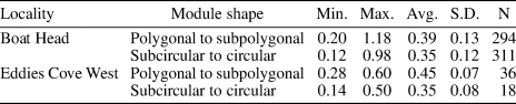
Growth form massive, shape predominantly domical or irregular; structure phacelocerioid; size up to 25 × 25 mm across, 20 mm high (Fig. 7.1). In transverse section, modules generally polygonal to subpolygonal and densely packed (Fig. 7.1–7.3), less commonly subcircular to circular and partially to entirely separated by micrite in loosely packed areas (Fig. 7.3, 7.4); adjoining modules have back-to-back walls (Fig. 7.9, 7.10); maximum module diameter 1.06 mm (Table 1). Module increase by longitudinal fission (Fig. 7.8): typically bipartite (Fig. 7.2, 7.9–7.11), one probable quadripartite example observed (Fig. 7.12). In longitudinal section, module walls relatively straight to slightly undulate (Fig. 7.5). Tabulae absent. Constriction of modules associated with rejuvenation or regeneration, in some cases following partial mortality of growth surface, commonly accompanied by concave-upward tabula-like structures forming incomplete or usually complete bases of subsequent module growth, generally sparse and distantly spaced, usually developed at same levels in adjacent modules (Fig. 7.5–7.7). Calices of modules shallow to moderately deep, with concave-upward bottoms having same shape as complete tabula-like structures (Fig. 7.6, 7.8).
Diameter (mm) of modules of Amsassia terranovensis n. sp. from the Watts Bight Formation, based on four skeletons (including holotype and paratype) from Green Head, western Newfoundland. In transverse sections, diameter was measured internally in the largest 20% of modules; module diameter = (long axis + short axis) ÷ 2 (see Scrutton, Reference Scrutton1981). Abbreviations: Min., minimum; Max., maximum; Avg., average; S.D., standard deviation; N, number of modules.

Etymology
The new species is named for Newfoundland, where it occurs.
Materials
GSC 141448 (thin section; Fig. 7.4), GSC 141447 (thin section; Fig. 7.5), GSC 142628 (thin section; Fig. 7.6), GSC 142629 (thin section; Fig. 7.7), GSC 142630 (thin section; Fig. 7.8), GSC 142632 (thin section; Fig. 7.10), GSC 142631 (thin section; Fig. 7.11, 7.12). Materials came from a rock sample providing nine skeletons (including holotype and paratype), collected from a reef complex ~26–33 m above base of Watts Bight Formation (Tremadocian; Ibexian, Skullrockian); Green Head in Isthmus Bay, eastern Port au Port Peninsula, Newfoundland, Canada.
Remarks
The materials described above, originally identified as Lichenaria and considered to be the earliest tabulate corals (Pratt and James, Reference Pratt and James1982, Reference Pratt and James1989a), were transferred to Amsassia by Elias et al. (Reference Elias, Lee and Pratt2021) and are here assigned to a new species, A. terranovensis. This species is most similar in both geologic age and morphology to A. argentina, from reefal mounds in the early Tremadocian La Silla Formation in the Argentine Precordillera (Carrera et al., Reference Carrera, Astini and Gomez2017, p. 80, 82, figs. 5, 6, table 1). Maximum module diameters in the two species are similar: 1.06 mm in A. terranovensis n. sp. and 1.12 mm in A. argentina. In A. terranovensis n. sp., polygonal modules tend to be slightly smaller than circular modules. Amsassia argentina, however, is unusual for the genus in that the average size of polygonal modules is substantially greater than the average size of circular modules. In addition to bipartite module increase, which is predominant in both A. terranovensis n. sp. and A. argentina, tripartite increase was reported in A. argentina, whereas a probable case of quadripartite increase was observed in A. terranovensis n. sp. The tabula-like structures in A. terranovensis n. sp. are similar to those in A. argentina.
Amsassia diversa new species
Figures 8, 9
- Reference Elias, Lee and Pratt2021
Amsassia sp. unident. Elias et al., fig. 1a, e–i, l, n, o.
Types
Holotype GSC 141441: GSC 141441c (thin section; Figs. 8.1, 9.4), 141441b (thin section; Fig. 8.5), and 141441a (thin section; Fig. 9.5, 9.7). Paratype GSC 141437 (thin section; Fig. 8.2). Mound, ~18 m below Barbace Cove Member in Boat Harbour Formation (Tremadocian; Ibexian, Stairsian); Boat Head, Great Northern Peninsula, Newfoundland, Canada.
Diagnosis
Amsassia with maximum module diameter 1.18 mm. Module increase predominantly bipartite and tripartite, less commonly quadripartite, rarely hexapartite. Tabula-like structures generally sparse, development at same levels in adjacent modules may be conspicuous.
Description
Growth form massive, shape hemispherical to irregular; structure phacelocerioid; size up to 50 × 50 mm across, 32 mm high (Fig. 8.1). Modules in some skeletons at both localities (Boat Head, Eddies Cove West) contain various amounts of peloids and micritic clots, likely of in situ microbial origin, commonly associated with growth interruptions and partial mortality (Fig. 8.3, 8.4). In transverse section, module size and shape vary significantly, with areas of small polygonal to subpolygonal modules, large polygonal to subcircular modules, variably sized subpolygonal to subcircular modules, and large, irregularly shaped modules (outlined areas A–D, respectively, in Fig. 8.2). Modules tend to be polygonal to subpolygonal in densely packed areas (Fig. 8.3) and subcircular in loosely packed areas, where separated by micrite (outlined areas B and C in Fig. 8.2; Fig. 9.6). Maximum module diameter 1.18 mm (Table 2). Module increase by longitudinal fission (white arrows in Fig. 8.5): predominantly bipartite (Fig. 9.5, 9.6) and tripartite (Fig. 9.7), less commonly quadripartite (Fig. 9.8), rarely hexapartite (Fig. 9.9, 9.10). In longitudinal section, module walls mostly straight to slightly undulate (Fig. 8.4, 8.5), rarely wavy to irregular in loosely packed areas (Fig. 9.2). Tabulae absent. Constriction of modules associated with rejuvenation or regeneration, commonly accompanied by concave-upward tabula-like structures forming incomplete or usually complete bases of subsequent module growth (black arrows in Figs. 8.5, 9.3). Tabula-like structures generally sparse and distantly spaced (Fig. 8.5); development at same levels in adjacent modules may be conspicuous and possibly cyclomorphic (Fig. 9.1, 9.3). Calices of modules shallow to moderately deep, with concave-upward bottoms having same shape as complete tabula-like structures (Figs. 8.1, 8.3, 9.4).
Diameter (mm) of modules of Amsassia diversa n. sp. from the Boat Harbour Formation, based on four skeletons (including holotype and paratype) from Boat Head and two skeletons from Eddies Cove West, western Newfoundland. In transverse sections, diameter was measured internally in the largest 20% of modules; module diameter = (long axis + short axis) ÷ 2 (see Scrutton, Reference Scrutton1981). Abbreviations: Min., minimum; Max., maximum; Avg., average; S.D., standard deviation; N, number of modules.

Etymology
The name of the new species refers to the diverse types of module increase, including bipartite, tripartite, quadripartite, and hexapartite.
Materials
GSC 142638 (thin section; Fig. 8.4), GSC 141446 (thin section; Fig. 9.1, 9.3), GSC 142636 (thin section; Fig. 9.2), GSC 141444 (thin section; Fig. 9.6), GSC 141442 (thin section; Fig. 9.8), GSC 141443 (thin section; Fig. 9.9), GSC 142637 (thin section; Fig. 9.10); mound, ~18 m below Barbace Cove Member in Boat Harbour Formation (Tremadocian; Ibexian, Stairsian); Boat Head, Great Northern Peninsula, Newfoundland, Canada. GSC 141445 (thin section; Fig. 8.3); mound, ~13.5 m below Barbace Cove Member in Boat Harbour Formation (Tremadocian; Ibexian, Stairsian); Eddies Cove West, Great Northern Peninsula, Newfoundland, Canada. Materials came from a rock sample collected at Boat Head, providing eight skeletons (including holotype and paratype), and a rock sample collected at Eddies Cove West, providing one skeleton.
Remarks
Hexapartite fission is rare in the skeletons described above from Boat Head and has not been recognized in those from Eddies Cove West, but this may reflect the relatively small amount of material available from the latter locality. Otherwise, the average diameters of modules (Table 2) and ranges of variability in all other morphologic features are closely similar in specimens from Boat Head and Eddies Cove West. All things considered, the specimens from both localities are regarded as conspecific and are identified as a new species, Amsassia diversa. This species is distinguished from others of the genus by its diverse types of module increase. Bipartite, tripartite, and quadripartite fissions previously have been recognized in many species of Amsassia (Sun et al., Reference Sun, Elias and Lee2014; M. Lee et al., Reference Lee, Park, Tien, Choh, Elias and Lee2016, Reference Lee, Elias, Choh and Lee2018), but A. diversa n. sp. is the first to show that hexapartite fission also occurred in the genus.
Specimens from Maryland, identified by Sando (Reference Sando1957) as Lichenaria cloudi, were reassigned to Amsassia by Elias et al. (Reference Elias, Lee and Pratt2021). They are from the Rockdale Run Formation (Tremadocian; Ibexian, Stairsian), and are therefore comparable in age to A. diversa n. sp. Sando (Reference Sando1957) reported an average module diameter of 0.4950 mm for his specimens, which is greater than the average for A. diversa n. sp. (see Avg. column in Table 2), although it falls within the overall range of values for A. diversa n. sp. (0.12–1.18 mm). We observed only bipartite increase in Sando's material (USNM 123850a; Elias et al., Reference Elias, Lee and Pratt2021, fig. 1j, k), whereas increase in A. diversa n. sp. is predominantly bipartite and tripartite, less commonly quadripartite, and rarely hexapartite. The species of Amsassia in Maryland is considered different from A. diversa n. sp.
Amsassia? sp. A
Figure 10.1–10.3
Description
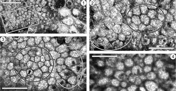
Growth form massive; structure phacelocerioid; size up to 2.1 mm across, height unknown (Fig. 10.1–10.3). One skeleton is attached to a co-occurring species of Amsassia having comparatively large modules (A. terranovensis n. sp.; Fig. 10.1), the other skeleton is unattached. In transverse section, modules polygonal to subpolygonal in densely packed areas, subcircular in less densely packed areas, with modules partially separated by micrite (Fig. 10.1–10.3). Maximum module diameter 0.24 mm (Table 3). One possible example of bipartite fission observed (arrow in Fig. 10.3).
Thin-section photomicrographs showing transverse views of Amsassia? sp. A (1–3) from the Watts Bight Formation at Green Head, and Amsassia? sp. B (4) from the Boat Harbour Formation at Boat Head, western Newfoundland. (1) Amsassia? sp. A (GSC 142633; left and center) attached to Amsassia terranovensis n. sp. having comparatively large modules (right side); outlined area enlarged in 2; scale bar = 1 mm. (2) Enlargement of outlined area in 1, showing phacelocerioid structure of Amsassia? sp. A; note polygonal modules in densely packed area (black outline) and subpolygonal modules partially separated by micrite in less densely packed area (white outline); GSC 142633; scale bar = 0.5 mm. (3) Phaceocerioid structure of Amsassia? sp. A, showing variability in size and shape of modules; note polygonal modules in densely packed area (black outline) and large, subpolygonal modules partially separated by micrite in less densely packed area (white outline); note possible result of recently completed bipartite fission (arrow); GSC 142634; scale bar = 0.5 mm. (4) Phacelocerioid structure of Amsassia? sp. B; note predominantly subcircular to circular modules partially separated by micrite; GSC 142635; scale bar = 0.5 mm.

Diameter (mm) of modules of Amsassia? sp. A from the Watts Bight Formation, based on two skeletons (GSC 142633, GSC 142634) from Green Head, and Amsassia? sp. B from the Boat Harbour Formation, based on one skeleton (GSC 142635) from Boat Head, western Newfoundland. In transverse sections, diameter was measured internally in the largest 80% of modules within the central growth area of these small skeletons (i.e., excluding peripheral modules distorted by divergent growth); module diameter = (long axis + short axis) ÷ 2 (see Scrutton, Reference Scrutton1981). Abbreviations: Min., minimum; Max., maximum; Avg., average; S.D., standard deviation; N, number of modules.

Materials
GSC 142633 (thin section; Fig. 10.1, 10.2), GSC 142634 (thin section; Fig. 10.3). Materials came from a rock sample providing two skeletons of Amsassia? sp. A (as well as skeletons of Amsassia terranovensis n. sp., described above), collected from a reef complex ~26–33 m above base of Watts Bight Formation (Tremadocian; Ibexian, Skullrockian); Green Head in Isthmus Bay, eastern Port au Port Peninsula, Newfoundland, Canada.
Remarks
The massive growth form with phacelocerioid structure resembles that of Amsassia, and the size and shape of modules fall within the range of the genus. However, module increase by fission is unconfirmed and features in longitudinal section are unknown. Inclusion in Amsassia is therefore uncertain, and a formal name is not proposed for this rare species. Although module size is comparable to that of Amsassia? sp. B (see below), Amsassia? sp. A differs in having modules that are predominantly polygonal to subpolygonal, rather than subcircular to circular. The maximum module diameter in Amsassia? sp. A (0.24 mm) is similar to that in the smallest known species of Amsassia, A. koreanensis (0.28 mm), from the Middle Ordovician (Darriwilian) of Korea and Upper Ordovician (Katian) of South China (M. Lee et al., Reference Lee, Park, Tien, Choh, Elias and Lee2016). Bipartite, tripartite, and quadripartite module increase have been confirmed in A. koreanensis.
Amsassia? sp. B
Figure 10.4
Description
Growth form massive; structure phacelocerioid; size up to 2.5 mm across, height unknown (Fig. 10.4). In transverse section, modules predominantly subcircular to circular, partially separated by micrite. Maximum module diameter 0.18 mm (Table 3).
Materials
GSC 142635 (thin section; Fig. 10.4). Materials came from a rock sample providing one skeleton of Amsassia? sp. B (as well as skeletons of Amsassia diversa n. sp., described above), collected from a mound, ~18 m below Barbace Cove Member in Boat Harbour Formation (Tremadocian; Ibexian, Stairsian); Boat Head, Great Northern Peninsula, Newfoundland, Canada.
Remarks
The massive growth form with phacelocerioid structure resembles that of Amsassia, and the size and shape of modules fall within the range of the genus. However, module increase by fission is unconfirmed and features in longitudinal section are unknown. Inclusion in Amsassia is therefore uncertain, and a formal name is not proposed for this rare species. Although module size is comparable to that of Amsassia? sp. A (Table 3), Amsassia? sp. B differs in having modules that are predominantly subcircular to circular, rather than polygonal to subpolygonal.
Acknowledgments
This study was supported by funding from the National Research Foundation of Korea (grant 2018R1A2B2005578) to D.-J. Lee, and the Natural Sciences and Engineering Research Council of Canada to B.R. Pratt. Pratt is grateful to N.P. James for supervising his M.Sc. thesis work, during which specimens from western Newfoundland were discovered. We thank H.M. Park for assistance in preparing thin sections, and J.-W. Jeon for sharing his unpublished data regarding Amsassia in the Wulipo Formation. The reviewers (M.G. Carrera, G.A. Young) and Associate Editor (A. Pisera) provided helpful comments on the manuscript.