Introduction
Originating in southwest and central Europe, Camelina sativa (Linnaeus) Crantz (Brassicaceae) is a cold-tolerant, annual brassicaceous oilseed crop that is well adapted to varied climate conditions and nutritionally poor soils. Currently, Camelina is being promoted as a new cropping opportunity for prairie grain farmers because of its agronomic plasticity as a short season, fast-growing crop with minimal input requirements, and its multiple uses. It has high levels of oils rich in OMEGA-3 fatty acids (Gugel and Falk Reference Gugel and Falk2006), and has potential as a source of biofuel and jet fuel. Few insects are regarded as pests of the plant. Flea beetles (Phyllotreta cruciferae (Goeze)) (Coleoptera: Chrysomelidae) have been observed on Camelina in Minnesota, United States of America, but in multi-year, small-plot trials the insect injury observed was not sufficient to warrant control (Robinson Reference Robinson1987). Likewise, Pachagounder et al. (Reference Pachagounder, Lamb and Bodnaryk1998) reported little feeding by P. cruciferae on C. sativa accessions compared with seven other Brassicaceae in laboratory trials. These researchers observed flea beetles sitting but not feeding on Camelina in the field near Winnipeg, Manitoba, Canada, but did not run comparative field trials with C. sativa and canola. Henriksen et al. (Reference Henriksen, Lundon, Prestløkken, Abrahamsen and Eltun2009) found no detectable damage to C. sativa accessions from striped flea beetle, Phyllotreta striolata (Fabricius) (Coleoptera: Chrysomelidae), in field trials in Norway. A report by the Scottish Rural Colleges (2013) stated but did not offer evidence that treatment for the control of the crucifer-specialist pollen beetle Meligethes aeneus (Fabricius) (Coleoptera: Nitidulidae) in C. sativa may be necessary if the beetles are present in the crop before the flowers open. However, studies in Ireland (Crowley and Frölich Reference Crowley and Frölich1998) and Norway (Henriksen et al. Reference Henriksen, Lundon, Prestløkken, Abrahamsen and Eltun2009) found no detectable damage to C. sativa by the pollen beetle. In trials at Lethbridge, Alberta, Canada, C. sativa was a poor host for cabbage seedpod weevil Ceutorhyncus obstrictus (Marsham) (Coleoptera: Curculionidae) (Cárcamo et al. Reference Cárcamo, Olfert, Dosdall, Herle, Beres and Soroka2007), but asynchrony between crop and insect may have been the reason, rather than host resistance. Lygus hesperus Knight (Hemiptera: Miridae) will feed on C. sativa, but spends more time walking, resting, and grooming on the plant than probing or feeding on it (Naranjo and Stefanek Reference Naranjo and Stefanek2012). Ehrensing and Guy (Reference Ehrensing and Guy2008) found that the brassicaceous aphid complex of Brevicoryne brassicae (Linnaeus) (Hemiptera: Aphididae) and Lipaphis erysimi (Kaltenbach) (Hemiptera: Aphididae) does not cause economic injury to C. sativa. The crop appears to exhibit antibiosis and antixenosis to diamondback moth Plutella xylostella (Linnaeus) (Lepidoptera: Plutellidae); larvae of diamondback moth feed less, have lower survival, lower pupation rates and weights, and shorter longevity of subsequent adult moths when fed on C. sativa compared with cabbage Brassica oleracea Linnaeus (Deng et al. Reference Deng, Yun, Zhang, Xu and Cai2004).
As well as reducing plant vigour by extracting photosynthate, the migratory leafhopper Macrosteles quadrilineatus (Forbes) (Hemiptera: Cicadellidae) is the main vector of aster yellows in canola crops grown on the Canadian prairies (Olivier et al. Reference Olivier, Lowery and Stobbs2009a). Aster yellows, triggered by phytoplasma belonging to the taxon “Candidatus phytoplasma asteris” (Lee et al. Reference Lee, Gundersen-Rindal, Davis, Bottner, Marcone and Seemüller2004), can cause severe production losses in canola (Olivier et al. Reference Olivier, Lowery and Stobbs2009a). Aster yellows outbreaks in canola crops on the prairies were noted in 2000, 2007, and 2012. The highest aster yellows incidence was observed in 2012, with the disease present in 77% of surveyed canola fields, incidence ranging from 0% to 64% (Miller et al. Reference Miller, Anderson, Bassendowski, Britz, Buitenhuis and Campbell2013), and production losses from aster yellows estimated to average 10%. Although resistant to common diseases of Brassica oilseeds, such as blackleg and alternaria black spot, C. sativa is susceptible to aster yellows (Khadhair et al. Reference Khadhair, Tewari, Howard and Paul2001; Séguin-Swartz et al. Reference Séguin-Swartz, Eynck, Gugel, Strelkov, Olivier and Li2009; Olivier et al. Reference Olivier, Séguin-Swartz, Gugel and Gossen2009b). Typical symptoms of aster yellows in C. sativa are chlorosis (yellow/red leaves), phyllody (green petals), and virescence (presence of sterile bladder-like pods containing malformed, green seeds, no seeds or a mixture of normal-looking and malformed seeds). Previous surveys showed that aster yellows incidence in C. sativa varied depending on C. sativa accessions (Olivier et al. Reference Olivier, Galka, Murza, Hegedus, Peng and Séguin-Swartz2007, Reference Olivier, Séguin-Swartz, Galka and Olfert2011). However, biodiversity and infectivity of leafhopper species were not assessed in these studies, nor was correlation between leafhopper infectivity and aster yellows incidence in C. sativa. Whether leafhoppers and other insect pests common in canola on the Canadian prairies could become economically important pests in C. sativa is unknown.
The objective of this investigation was to determine the susceptibility of C. sativa to feeding by common insect pests of cruciferous crops on the Canadian prairies. This was achieved by assessing the damage to C. sativa by natural infestations of flea beetles (Phyllotreta Chevrolat species), root maggots (Delia Robineau-Desvoidy (Diptera: Anthomyiidae) species), and leafhoppers (Hemiptera: Cicadellidae) in field trials and the suitability of C. sativa as a host of bertha armyworm Mamestra configurata and diamondback moth P. xylostella in laboratory bioassays.
Materials and methods
Camelina sativa accessions
Seed for the C. sativa accessions tested in the study was obtained from Dr. Robert Bodnaryk, Agriculture and Agri-Food Canada Winnipeg Research Station, and from Plant Gene Resources Canada, Saskatoon, Saskatchewan, Canada.
Flea beetles
Laboratory bioassays
Adult P. cruciferae were collected from field-placed modified mustard oil traps (Soroka et al. Reference Soroka, Bartelt, Zilkowski and Cossé2005) or by sweeping commercial canola fields, hedgerows, or winter canola Brassica napus cultivar Casino plots grown at Agriculture and Agri-Food Canada’s Saskatoon Research Centre (SRC) Farm (52°09'N, 106°34'W) (orthic dark brown chernozemic soil). Beetles were fed a diet of cabbage leaves and water in meshed 60×60×60 cm cages in a controlled environment chamber (24 °C:18 °C [day:night], 65 ±3% relative humidity, and a photoperiod of 16:8 light:dark hours), and were used in experiments within one week of field collection. Single plants of up to nine entries of C. sativa, a susceptible B. napus control cultivar AC Excel, and a moderately resistant white mustard Sinapis alba Linnaeus control cultivar Ochre were grown in vermiculite-filled 30-mm by 50-mm plastic vials in the greenhouse (light averaging 230 μmol/m2 second, 23 °C:15 °C [day:night], ~55% relative humidity, and a photoperiod of 16:8 light:dark hours). One week after germination, vials were arranged in an array of 10 or 11 columns and rows in an arena composed of a 5 cm Styrofoam® base under a 58-cm by 58-cm by 35-cm Plexiglas® arena with a screened top. The arenas were housed in a walk-in growth chamber maintained at 21–22 °C, 75% relative humidity, and a photoperiod of 16:8 light:dark hours, with even lighting over the ceiling and four sides, similar to the protocol of Palaniswamy et al. (Reference Palaniswamy, Lamb and McVetty1992). For the bioassays flea beetles were placed in each of three test arenas per trial at a rate of about 700 beetles per arena or seven flea beetles per seedling by gently shaking them from a collecting vial into the four corners of the cage in equal proportions. Seedlings and beetles were observed through the Plexiglas® sides of each arena and the proportion of tissue area eaten from each seedling was estimated visually on a scale from 0 (no feeding) to 10 (tissue completely consumed) (Palaniswamy et al. Reference Palaniswamy, Lamb and McVetty1992) at 6, 24 and/or 30, and 48 hour intervals after placement of the beetles into the cages. The damage ratings for each cotyledon were averaged over both cotyledons and multiplied by 10 to estimate the percentage of cotyledon area damaged, and damage to the first and second true leaves was multiplied by 10 to obtain a value for percentage of leaf area eaten (defoliation) (Palaniswamy et al. Reference Palaniswamy, Lamb and McVetty1992).
Field trials
A field trial assessing flea beetle feeding injury levels to C. sativa lines was conducted at the SRC Farm in 2008 and repeated in 2009. The trial consisted of four replicates each of 26 C. sativa and two B. napus entries that were seeded on 15 May 2008, and 17 May 2009, in four-row plots 3.0 m in length with 0.30 m row spacings and 100 seeds per row, using a randomised complete block design. Flea beetle injury was assessed on cotyledons and first to fourth true leaves at weekly intervals three times after emergence in the beginning of June. Visual defoliation ratings using the 0–10 scale of Palaniswamy et al. (Reference Palaniswamy, Lamb and McVetty1992) described previously were taken on 10 seedlings in a randomly selected area in the centre two rows of each plot. Plant densities were obtained by counting all the plants in a randomly selected 3.0 m row on the third sampling date, as was number of leaves on a randomly selected plant in each of the plots in 2009.
Root maggots
The preceding plots were maintained throughout the summer of 2009 using recommended canola agronomic practices and swathed at maturity. Shortly after swathing, 25 B. napus roots were randomly selected from the centre two rows of each plot, placed in plastic bags, and frozen. Later, each root was rated for the presence and degree of root maggot injury according to the semi-quantitative scale of Dosdall et al. (Reference Dosdall, Herbut and Cowle1994): 0=no damage to roots; 1=slight feeding,<10% of tap root surface damaged; 2=10–25% of root surface damaged; 3=26–50% of root surface damaged with tunnelling into interior; 4=51–75% of root surface damaged, with extensive tunnelling, and 5=76–100% of root damaged or root severed.
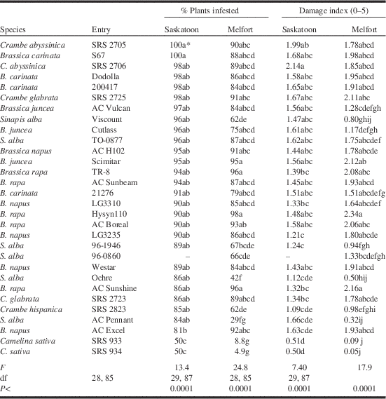
In a two-site field trial conducted in the year 2000, four replicates each of 28 (Saskatoon, Saskatchewan, Canada) or 29 (Melfort, Saskatchewan, Canada) entries, encompassing nine brassicaceous species including C. sativa, were seeded in four-row plots 6.1 m in length with 0.30 m row spacings and 200 seeds per row, using a randomised complete block design. The Saskatoon location was at the SRC farm, previously described, while the Melfort site was at the Melfort Research Farm of Agriculture and Agri-Food Canada (52°49'N, 104°35'W) (black chernozem soil zone with up to 10% organic matter). After swathing, 25 roots were randomly selected from the inner two rows of each plot and rated for presence (% infestation in 25 plants) and degree of damage using the 0–5 scale of Dosdall et al. (Reference Dosdall, Herbut and Cowle1994).
Diamondback moths
Two laboratory bioassays were undertaken to assess interactions between C. sativa and diamondback moth adults and larvae. Four plant entries, two commercial B. napus cultivars 46A65 and Q2, and two C. sativa accessions CN 101981 and CN 111334, were tested in both bioassays.
In the first bioassay, plants were grown individually in 13 cm diameter plastic pots in a soil-less mix of peat moss, vermiculite, sand, and plant nutrients. Seeded pots were placed in a walk-in growth chamber set at 20 °C day, 18 °C night, 16:8 hour light:dark cycle. Five weeks after seeding, plants were placed in either of two types of cages: choice test cages were wooden-based, wooden-framed cubes of dimensions 49×49×47 cm, with nylon screen sides, a clear Plexiglas® top, and a wooden door on one side for access. No-choice cages were also wooden framed, of dimensions 26×26×38 cm, with three screened sides and top, and a sliding Plexiglas® panel on one side for access. At the time of testing the plants had reached the bolting (B. napus) to bolting-early flowering (C. sativa) stages. One pot of each of the four plant lines was randomly arranged in each of four choice cages (16 pots in total in four cages), and one pot was placed in each no-choice cage, with four replicates (16 pots in total in 16 cages). Laboratory-reared fourth instar diamondback moth larvae were separated by sex (males have a noticeable white stripe on their dorsal surface (Liu and Tabashnik Reference Liu and Tabashnik1987)) and allowed to pupate and emerge as adults in cages in a growth room set at constant 25 °C, 18:6 hour light:dark conditions. Four female and four male newly emerged moths per plant were placed in choice test cages that contained one five-week-old plant of each of the four plant lines (32 moths and four plants per cage and four cage replicates in total), and four female and four male moths were placed in no-choice cages containing a single five-week-old plant (eight moths per cage with four cages of each plant entry, four replicates or 16 cages in total). Moths were allowed to oviposit for 48 hours and then were removed from the cages. The number of eggs laid per plant was recorded. Larvae were monitored after they emerged from eggs, and the pupal weights and time to pupation of up to 10 larvae per entry were determined.
The second bioassay was a larval feeding trial. Seeds of the same four plant lines, B. napus cultivars 46A65 and Q2, and C. sativa accessions CN 101981 and CN 111334, were planted in 13-cm pots in soil-less mix and grown in the greenhouse. Twenty-seven days after seeding, when they were in the late rosette stage, plants were placed in random arrangements of four pots, one plant of each entry, in the choice experiment (four plants per cage and four replicate cages), and singly (four entries and four replicates or 16 cages) in the no-choice experiment. Eight second-instar to third-instar diamondback moth larvae were placed on each plant in the trial and allowed to feed for 48 hours in a growth room set at 25 °C day, 22 °C night, 16:8 hour light:dark conditions. After this time the number of larvae and the feeding damage on each plant were noted.
Bertha armyworms
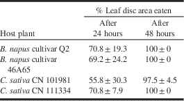
Two growth chamber bioassays were conducted to evaluate the interactions of C. sativa and bertha armyworms. In the first trial, a no-choice leaf disc feeding bioassay, 12 leaf discs of one of four plant lines were placed in each of four glass Petri dishes of 100 mm outside diameter, the bottom of which was covered with two circles of #3, 90 mm qualitative Whatman filter paper (Canadawide Scientific, Ottawa, Ontario, Canada) moistened with 5 mL of distilled water per dish. The same four cultivars as were evaluated in the diamondback moth bioassays were used: the B. napus cultivars Q2 and 46A65, and the C. sativa lines CN 101981 and CN 111334. Leaf discs were cut from rosette leaves using a #4 cork borer, and were about 8 mm in diameter or 50 mm2 in area. Care was taken to avoid sectioning across the leaf midrib. Ten second-instar bertha armyworms from a laboratory colony were placed in the Petri dishes containing leaf discs, and the lids were sealed with Parafilm® to prevent drying. Dishes were placed in a reach-in growth chamber set at 25 °C day, 22 °C night, 16:8 hour light:dark cycle. Larvae were allowed to feed for 24 and 48 hours, after which damage was visually evaluated as an average per cent leaf area eaten of the discs in each dish.
The second bertha armyworm bioassay consisted of choice and no-choice feeding tests using entire plants. Individual plants of B. napus cultivars Q2 and 46A65 and C. sativa lines CN 101981 and CN 111334 were grown in 13 cm diameter plastic pots as described for the diamondback moth bioassays. Five weeks after seeding, when plants were in pre-bolt stage, pots were placed in the same choice and no-choice cages as were used in the diamondback moth trials. One pot of each of the four plant lines was randomly arranged in each of four choice cages (16 pots in total in four cages), and one pot was placed in each no-choice cage, with four replicates (16 pots in total in 16 cages). Five laboratory-reared, second-instar bertha armyworms were weighed collectively, placed on each plant, and allowed to eat and move where they wished. After about 10 days, most larvae had reached the sixth-instar stage and searched out pupation sites, usually in the soil below the plants. In order to identify which larvae grew on which plant in the choice test cages, pots were moved at this time into individual cages. Prepupae were located (but not disturbed) in the soil around plants, allowed to pupate, and then weighed. The number of larvae that reached pupation, the prepupal development period, pupal weights, and biomass increase from five larvae to pupae were determined for armyworms on all four plant lines in both tests.
Leafhoppers
Field trials of Camelina sativa accessions
Field trials assessing aster yellows incidence in C. sativa lines were undertaken at the SRC Farm in 2009. On the basis of the results of a 2007 survey (Olivier et al. Reference Olivier, Séguin-Swartz, Galka and Olfert2011), six accessions of C. sativa exhibiting different levels of tolerance to aster yellows were selected. These accessions were SRS 933, SRS 934, CN 30487, and CN 30476, which had <10% incidence of aster yellows in 2007, and CN 101988 and CN 101897, which had the highest incidence of the disease (>20%) in the previous trial. The six accessions were seeded at 100 seeds per 3-m row, 0.30 m row spacings, with four rows and two replicates per accession, and each accession separated by two rows of barley, in a randomised complete block assay.
In mid-August, a visual estimation of aster yellows incidence in the six accessions was undertaken by counting the number of plants exhibiting typical symptoms of aster yellows in each row. At plant maturity, 20 plants were harvested randomly in each of the two middle rows of every accession and replicate. Approximately 1 g of stem, leaf, and root tissue was sampled from each of the 40 plants and stored at −20 °C in 2.0 mL Eppendorf tubes for polymerase chain reaction (PCR) testing. Because phytoplasmas are heterogeneously distributed within their host plants, plants for which at least one tissue sample tested positive for the presence of phytoplasma DNA were considered to be infected with phytoplasma.
Biodiversity and infectivity of leafhoppers
Leafhoppers were collected using 7.5×12.5 cm yellow sticky traps (Phero Tech Inc, Delta, British Columbia, Canada) placed 20 cm above the ground on a 27-cm wire holder (Natural Insect Control, Stevensville, Ontario, Canada). Twelve sticky traps, two per accession in each of two replicates, were set out within the rows and changed weekly from June until September. Sticky traps were stored in individual plastic bags at 4 °C until processing. Leafhoppers were carefully removed from each card using tweezers and identified using morphological characteristics and male genitalia. Species were identified using the keys of Beirne (Reference Beirne1956) and Hamilton (Reference Hamilton1998), with names cited according to Maw et al. (Reference Maw, Foottit, Hamilton and Scudder2000). After identification, leafhopper specimens belonging to species known to be phytoplasma vectors or potential vectors, as listed in Weintraub and Beanland (Reference Weintraub and Beanland2006) and Olivier et al. (Reference Olivier, Séguin-Swartz, Gugel and Gossen2009b), were individually PCR-tested, while other species were tested in groups of five or 10 specimens, depending on their size.
Polymerase chain reaction analysis and phytoplasma strain identification
Leaf tissues and leafhopper specimens were placed in 2.0-mL Eppendorf tubes. DNA extraction and purification was performed using the method described by Daire et al. (Reference Daire, Clair, Reinert and Boudon-Padieu1997). DNA extracts were amplified using universal primers for the 16Sr gene and separated on electrophoresis gels according to the methods described by Olivier et al. (Reference Olivier, Galka and Séguin-Swartz2010). The 1200 base-pair bands of phytoplasma DNA contained in leaves and leafhoppers were extracted from the agarose gel and purified using a Qia-Quick Gel Extraction Kit (Qiagen Canada, Mississauga, Ontario, Canada). Sequencing was performed at the Plant Biotechnology Institute (National Research Council, Saskatoon, Saskatchewan, Canada) and sequences were trimmed, compared, and assembled into consensuses using the Sdaten package (Bonfield and Whitwham Reference Bonfield and Whitwham2010), then compared with sequences recorded in GenBank using the BLAST program (http://www.ncbi.nlm.nih.gov).
Data analyses
In both laboratory and field feeding bioassays of flea beetles, tissue feeding damage data were analysed with plant phenotype as a fixed effect and arena or replicate as a random effect using analysis of variance (ANOVA) or PROC MIXED procedures (SAS Institute 2005). When significant differences were found between hosts in feeding levels at P⩽0.05, Tukey’s studentised range test was used to distinguish among means (Steel and Torrie Reference Steel and Torrie1980). Bertha armyworm and diamondback moth data were similarly analysed. Root maggot damage ratings were analysed for each experiment using the PROC MIXED procedure of SAS (SAS Institute 2005), with treatments as fixed effects considered significant at P⩽0.05. Pearson’s correlation coefficients for measured variables and Spearman’s rank correlation coefficients for damage index relationships were determined to estimate measures of association between parameters (Littel et al. Reference Littel, Milliken, Stoup and Wolfinger1996) in root maggot bioassays. T-tests (SAS Institute 2005) were used to compare per cent of aster yellows infection in C. sativa accessions and leafhopper species and numbers. In field assays, number of leafhoppers and aster yellows incidence between accessions were compared, using ANOVA procedures and regression analysis and best-fit lines from Microsoft Excel 2010 (Microsoft Corporation, Redmond, Washington, United States of America).
Results
Flea beetles
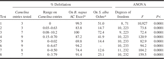
In total, 49 C. sativa entries were tested in eight laboratory bioassays. Most entries were tested only once, but all trials included a B. napus susceptible control and most had a moderately resistant S. alba mustard control. The results are summarised in Table 1. All of the C. sativa accessions were fed upon significantly less than both the susceptible B. napus entry AC Excel and the moderately resistant S. alba mustard entry Ochre. In only one trial, that with the greatest amount of defoliation to the two controls, did a C. sativa entry have defoliation >10% (Table 1).
Summary of laboratory bioassays testing levels of flea beetle Phyllotreta cruciferae feeding on various entries of Camelina sativa and two control entries of Brassica napus and Sinapis alba.
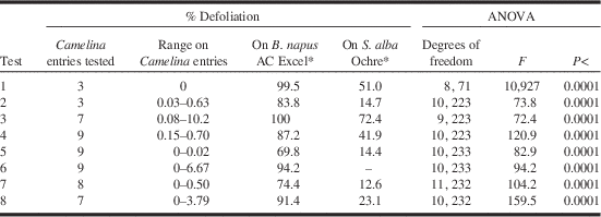
* B. napus cultivar AC Excel is the susceptible control and S. alba cultivar Ochre is the resistant control.
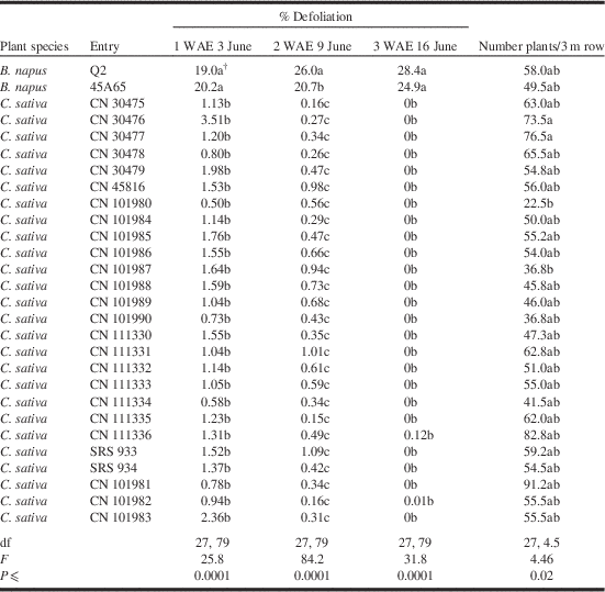
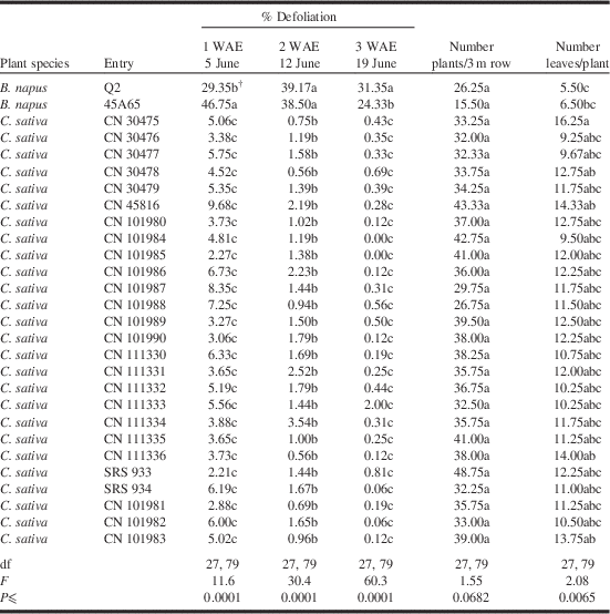
In the field trials flea beetle feeding levels varied with year and, therefore, data were not combined over years. However, the pattern of flea beetle feeding was similar in both years, and duplicated laboratory results. Feeding damage to the B. napus canola entries was high, surpassing the nominal economic threshold of 25% leaf area eaten in both years (Tables 2, 3). On the other hand, all 26 C. sativa entries had feeding levels significantly lower than did either of the B. napus entries, decreasing to near zero on the third sampling date in both years (Tables 2, 3).
Defoliation by flea beetles Phyllotreta species on youngest tissues one, two, and three weeks after seedling emergence (WAE) and plant densities three weeks after emergence of Brassica napus canola and Camelina sativa entries grown in the field at Saskatoon, Saskatchewan, Canada, in 2008.Footnote *

* Mean of damage to cotyledons one week after emergence, to cotyledons and first true leaf two weeks after emergence, and to cotyledons, first and second true leaves three weeks after emergence.
† Means within a column followed by the same letter are not significantly different from each other as determined by PROC MIXED or ANOVA at P⩽0.05 (means separation by Tukey’s studentised range test).
Defoliation by flea beetles Phyllotreta species on youngest tissues at one, two, and three weeks (WAE) after seedling emergence, plant densities, and number of leaves per plant rated three weeks after emergence of Brassica napus and Camelina sativa entries grown in the field at Saskatoon, Saskatchewan, Canada, in 2009.Footnote *
* Mean of damage to cotyledons one week after emergence, cotyledons and first true leaf two weeks after emergence, and cotyledons, first and second true leaves three weeks after emergence.
† Means within a column followed by the same letter are not significantly different from each other as determined by PROC MIXED or ANOVA at P⩽0.05 (means separation by Tukey’s studentised range test).
Measured agronomic characteristics did not appear to play a part in flea beetle preference to canola over C. sativa. Plant densities differed among entries but not between the two plant species in either year (Tables 2, 3). Likewise, plants of both species grew at relatively the same rate in 2008, as evidenced by similar growth stages among entries in that year (data not presented). Plant growth rate varied with entries in 2009, with the B. napus entry Q2 having fewer leaves than five of the C. sativa entries (Table 3). All B. napus plots were noted to have poor stand emergence and slower initial growth in 2009, while most C. sativa entries exhibited excellent emergence and growth.
Although formal measurements were not taken, visual observations of the field trial found very few flea beetles in the C. sativa plots late in the season, while the newly eclosed summer generation adults were commonly found feeding on B. napus plants in the trial.
Root maggots
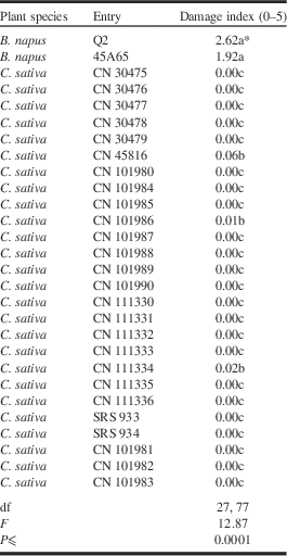
As the 2009 season progressed, moisture levels increased, but temperatures continued to be cool. These conditions were conducive to development of crucifer-feeding root maggot Delia species populations, and severe maggot injury was seen in rutabaga trials in the vicinity of the C. sativa trial. However, maggot feeding pressure in the C. sativa trial was not heavy, with 10–50% of the B. napus roots exhibiting injury. The damage index of 1.9–2.6 found on B. napus plants, indicating damage to 10–25% of the surface area of their roots, was in stark contrast to that seen in the C. sativa accessions, which were virtually immune to root maggot feeding. Only three of the 26 C. sativa entries exhibited any feeding damage at all, and the damage index on these three accessions averaged 0.06 on the 0–5 damage index scale (Table 4).
Root damage on a scale of 0 (root undamaged) to 5 (root severely damaged or completely severed) to two Brassica napus canola cultivars and 26 lines of Camelina sativa subjected to attack by root maggots, Delia species, in the field at Saskatoon, Saskatchewan, Canada, in 2009.
* Means within a column followed by the same letter are not significantly different from each other as determined by PROC MIXED or ANOVA at P⩽0.05 (means separation by Tukey’s studentised range test).
In the trial assessing root maggot injury to entries of nine brassicaceous species conducted at two sites in 2000, infestation and damage rates were significantly lower in the two C. sativa entries than in most of the 28 Brassica, Sinapis, and Crambe entries at both locations (Table 5).
Field bioassays measuring infestation rate of and damage to cruciferous plants infested by root maggots, Delia species, in the field at Saskatoon and Melfort, Saskatchewan, Canada, 2000.
* Means within a column followed by the same letter are not significantly different from each other as determined by PROC MIXED or ANOVA at P⩽0.05 (means separation by Tukey’s studentised range test).
Diamondback moths
When diamondback moths were given a choice of host plant on which to oviposit, they laid significantly more eggs on the two B. napus entries than on the C. sativa entries (Fig. 1). However, when moths were placed in the no-choice test with only one plant per cage, the numbers of eggs laid on the plants were low and did not vary with plant entry.
Diamondback moth eggs (mean+SE) laid over 48 hours by four female moths per plant on four entries of crucifers (left), and combined over crucifer species in choice and no-choice laboratory bioassays (right). Different letters over columns within a test indicate differences in oviposition levels, P⩽0.05.
When diamondback moth larval movement was monitored, larvae did not remain on C. sativa leaves when placed there. In feeding bioassays with a choice of hosts, 97–100% of the larvae moved off C. sativa plants when placed on them (Fig. 2). Some moved to B. napus plants, while others disappeared. On the other hand, 62–75% of the larvae placed on B. napus leaves stayed where they were placed (Fig 2). Results were similar in the no-choice tests. About 88% of the larvae originally placed on B. napus leaves stayed on the plants on which they were placed, while virtually all of the larvae placed on C. sativa plants moved off the plants and were not recovered. This held true for all four entries and over both species (Fig. 2).
Diamondback moth larval distribution 48 hours after placing eight larvae on each plant on two Brassica napus and two Camelina sativa plants in growth chamber bioassays (left), and combined over crucifer species in choice and no-choice laboratory bioassays (right). Different letters over columns within a test indicate differences in larval number per location, P⩽0.05.
Avoidance of C. sativa was also evident in the amount of feeding injury that diamondback moth larvae inflicted on plants in the entries. In both choice and no-choice trials, C. sativa plants sustained 4–13% of the damage sustained by B. napus plants (Fig. 3).
Diamondback moth larval injury to plants at the late rosette stage 48 hours after placing eight larvae per plant on four entries of crucifers (left), and combined over crucifer species in choice and no-choice laboratory bioassays (right). Different letters over columns within a test indicate differences in feeding levels, P⩽0.05.
While many larvae pupated near the B. napus entries and 10 pupae were found per plant on each cultivar with ease in the choice trial, only 5.5±3.3 and 3.0±0.8 larvae reached pupation per plant on C. sativa lines CN 111334 and CN 101981, respectively. Similarly, while 10.0 and 9.0±2.0 pupae were found on cultivars Q2 and 45A65 plants in the no-choice test, respectively, the corresponding numbers of pupae found on C. sativa cultivars CN 111334 and CN 101981 plants were 2.5±4.2 and 1.0±0.
Developmental period varied among entries in choice tests; larvae on both C. sativa accessions took longer to reach pupation than larvae on B. napus cultivar Q2 (Fig. 4). In no-choice tests, low numbers of pupae on both C. sativa entries and high variation within the C. sativa entry CN 111334 rendered this trend not significant (P<0.06). When combined over species, the trend towards increased larval developmental time when feeding on C. sativa was not significant (P⩽0.09 and 0.06 in choice and no-choice tests, respectively) (Fig. 4).
Diamondback moth developmental period from second instar to pupation when reared on four entries of crucifers (left), and combined over crucifer species in choice and no-choice laboratory bioassays (right). Different letters over columns within a test indicate differences in developmental times among entries or between crucifer species, P⩽0.05.
Despite differences in pupal number and developmental time among entries, there were no differences in pupal weights among plant entries or species in either choice or no-choice tests (Fig. 5).
Diamondback moth pupal weights when reared on four entries of crucifers (left), and combined over crucifer species in choice and no-choice laboratory bioassays (right). There were no differences in pupal weights among plant entries or between crucifer species in either test, P⩾0.05.
Although not formally assayed, the green peach aphid Myzus persicae (Sulzer) (Hemiptera: Aphididae), a generalist feeder, was noticed on plants of both species in the greenhouse before the setup of the diamondback moth larval feeding trial. Aphids were brushed off the plants before these were used in the bioassays, and on three occasions during the bioassays. There did not appear to be any difference in aphid colony growth among the four plant entries, with the two C. sativa lines supporting as many aphids as the two B. napus lines. After the third elimination of aphids in the bioassay, their numbers were too low to be noticeable.
Bertha armyworms
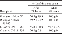
Under heavy feeding pressure, no differences were found in feeding levels of bertha armyworm larvae on four plant entries in the no-choice Petri dish leaf disc bioassay (Table 6). Likewise, when larvae were placed on entire plants, they did not move to any noticeable extent on the plants, and heavy feeding levels were seen on all entries. Indeed, in the no-choice trial, two CN 101981 plants were so severely defoliated that larval development may have been affected by lack of food material.
Mean defoliation (% leaf area eaten±standard deviation of the mean) of 12 8-mm diameter leaf discs in two Brassica napus canola cultivars and two lines of Camelina sativa by 10 bertha armyworm second instars in a no-choice Petri dish trial.
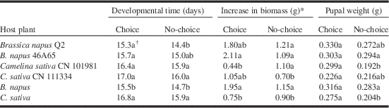
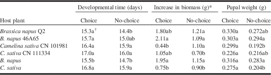
Bertha armyworm developmental time from placement of the larvae on leaves as second instars to pupation was not different among the plant entries in the choice trial (Table 7). However, when data were combined over species, bertha armyworms took more than a day longer to develop to pupae on C. sativa than on B. napus plants. Similarly, in the no-choice trial, larvae on B. napus cultivar Q2 developed faster than did larvae on the two C. sativa accessions, and when data were combined over species, larvae feeding on B. napus plants developed into pupae over one day sooner than did those on C. sativa plants (Table 7). The increase in biomass was 2.6 and 1.3 times greater for bertha armyworms feeding on B. napus plants than on those feeding on C. sativa accessions in choice and no-choice trials, respectively (Table 7).
Developmental period, increase in biomass of bertha armyworms from second instar to pupation, and weights of bertha armyworm pupae extracted from soil surrounding four plant entries in growth chamber choice and no choice feeding bioassays.
* Average weight of five pupae minus average weight of five larvae, in grams.
† Means within a column followed by the same letter are not significantly different from each other as determined by analysis of variance at P⩽0.05.
When pupal numbers were averaged over both species in the choice trial, fewer bertha armyworm pupae emerged from pots containing C. sativa plants than from pots containing B. napus plants (2.75±0.87 and 6.25±0.65 pupae, respectively, out of 10 larvae originally placed per two plants). However, because some larvae were seeking pupation sites at the time the plants were separated, it was difficult to determine with complete certainty from which plant in the choice trial that pupae originated. In the no-choice trial, equally high proportions of pupae were found in pots of either plant species (4.50±0.41 and 4.12±0.25 pupae from C. sativa and B. napus, respectively, out of five larvae originally placed per plant). No significant differences were found in the weights of pupae found among entries in the choice test (Table 7). In the no-choice tests, pupal weights from cages containing plants of C. sativa accession CN 101981 were lower than from cages containing B. napus cultivar 46A65 plants (Table 7). Similarly, when data were combined over plant species, heavier pupae were found in the no-choice cages containing B. napus plants than in the cages containing C. sativa plants (Table 7).
Leafhoppers
Phytoplasma strain identification in plant and leafhoppers
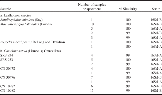
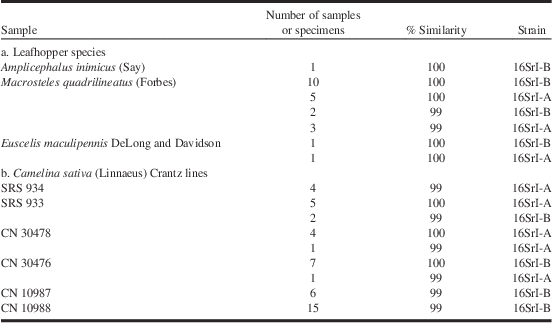
Polymerase chain reaction tests were positive for aster yellows presence in 96 plant samples and 97 leafhopper samples. Forty-five PCR products obtained from plant samples and 23 PCR products from leafhopper samples were sequenced. All sequences showed 100% or 99% nucleotide sequence identity to aster yellows phytoplasma subgroup 16SrI-A (Genbank Accession NR_074759) or aster yellows phytoplasma subgroup 16SrI-B (Genbank Accession NR_074811) (Table 8). The distribution of the strains in the accessions and in the leafhoppers appeared random.
Per cent similarity between sequences of phytoplasma strains found in (a) leafhopper specimens or (b) samples of Camelina sativa, and strains of 16SrI-A (accession number NR_074759) and 16SrI-B (accession number NR_ 074811).
Aster yellows incidence in Camelina sativa accessions
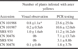
Aster yellows incidence differed significantly among accessions in both visual and PCR-based analyses in 2009 (Table 9). Accession CN 101988 had a significantly greater number of C. sativa plants infested with aster yellows than SRS 934 and CN 30478 in both assessments. Also, based on both assessments, there were no significant differences in levels of infectivity between accessions SRS 933 and CN 1901987. Polymerase chain reaction assessment revealed a higher number of aster yellows-infected plants in all accessions as compared with visual observations. This suggests the presence of a high number of aster yellows-infected plants that do not express visual symptoms. In 2010, aster yellows incidence was very low over the entire season, and only one aster yellows-infected plant (in accession CN 101988) was detected using PCR tests (data not shown).
Average (±standard deviation of the mean) aster yellows incidence estimated using visual observation and PCR tests in Camelina sativa accessions grown in the field at Saskatoon, Saskatchewan, Canada, in 2009.
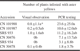
PCR, polymerase chain reaction.
* Means within a column followed by the same letter are not significantly different from each other as determined by analysis of variance at P⩽0.05.
Leafhopper biodiversity in Camelina sativa and aster yellows incidence
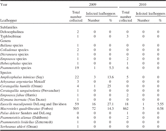
In total, 1539 leafhoppers were collected from the sticky traps, 632 and 907 over the 2009 and 2010 summer seasons, respectively. Of these, 1489 specimens were identified to species, 34 to genus and six to subfamily level (Table 10). Three species comprised 92% of all leafhoppers identified: aster leafhopper, M. quadrilineatus, was by far the most abundant leafhopper species in the C. sativa plots, followed by Euscelis maculipennis DeLong and Davidson (Hemiptera: Cicadellidae) and Amplicephalus inimicus Say (Hemiptera: Cicadellidae). In 2009, numbers of M. quadrilineatus peaked on 26 June with 287 specimens caught, remained high until 24 July with an average of 20 specimens caught per sampling date, and increased again from 6 August onwards to reach a total of 57 specimens caught on 27 August, just before harvest (Fig. 6). In 2010, numbers of M. quadrilineatus peaked on 8 July with a total of 303 specimens collected and, except for a slight drop on 19 August, numbers remained high with an average of 97 specimens collected per sampling date (Fig. 6). Numbers of species other than M. quadrilineatus, E. maculipennis, or A. inimicus collected from the C. sativa nursery were very low, averaging two and one specimens caught in 2009 and 2010, respectively (Fig. 6).
Number of aster yellows infected and uninfected leafhoppers collected from sticky traps placed in a Camelina sativa nursery per sampling date, Saskatoon, Saskatchewan, Canada, 2009 and 2010.
Subfamilies, genera, and species of leafhoppers and number and percent infected with aster yellows phytoplasma when collected on yellow sticky traps in a Camelina sativa nursery at Saskatoon, Saskatchewan, Canada, from June to August 2009 and 2010.
While significant differences were found between the average numbers of leafhoppers trapped among accessions of C. sativa in 2009 and 2010 (Fig. 7), there was no significant difference in the number of A. inimicus and E. maculipennis collected in the accessions in either 2009 or 2010. However, in both years, significantly fewer specimens of M. quadrilineatus were trapped in accessions SRS 933 and SRS 934 as compared with accessions CN 101987 and CN 101988.
Average number of leafhoppers Amplicephalus inimicus, Euscelis macilupennis, and Macrosteles quadrilineatus trapped on sticky traps per accession of Camelina sativa per year at Saskatoon, Saskatchewan, Canada, from June to September 2009 and 2010. Columns representing M. quadrilineatus with the same letter are not significantly different.
The greatest numbers of leafhoppers infected with aster yellows were M. quadrilineatus (72) and E. maculipennis (16). Proportionately, however, E. maculipennis and C. humilis leafhoppers had the highest infectivity rates (Table 10). There were significantly more aster yellows-infected M. quadrilineatus in 2009 (n=72) than in 2010 (n=5). Regression analysis of the number of leafhoppers sampled in 2009 at the peak of the season (June 25) plotted against aster yellows incidence in the different accessions showed that the best fit of the data is a logarithmic regression (R 2=0.94) (Fig. 8). No regression analysis was done in 2010 due to the very low incidence of aster yellows.
Correlation between the aster yellows incidence observed in accessions of Camelina sativa and the total number of Macrosteles quadrilineatus trapped in each accession on sticky traps harvested in the field at Saskatoon, Saskatchewan, Canada on 25 June 2009.
Discussion
This investigation determined the host suitability of several C. sativa entries for five insect pests of canola in numerous laboratory and field experiments. Our results revealed that insect responses to C. sativa depend on the host specificity or dietary specialisation of each insect studied.
This is the first study to examine systematically the levels of flea beetle feeding injury to C. sativa in field trials on the Canadian prairies, and substantiates the results of other observations. Decreased flea beetle feeding injury on C. sativa as compared with B. napus entries has been described previously, both anecdotally (Plessers et al. Reference Plessers, McGregor, Carson and Nakoneshny1962; Gugel and Falk Reference Gugel and Falk2006) and in more thorough investigations in the laboratory (Pachagounder et al. Reference Pachagounder, Lamb and Bodnaryk1998; Henderson et al. Reference Henderson, Hallett and Soroka2004; Soroka and Grenkow Reference Soroka and Grenkow2013). Pachagounder et al. (Reference Pachagounder, Lamb and Bodnaryk1998) and Soroka and Grenkow (Reference Soroka and Grenkow2013) found that all C. sativa accessions that were tested in laboratory bioassays had virtually no feeding damage from P. cruciferae compared with damage on B. napus cultivar Westar. Even though C. sativa was less preferred in terms of feeding, Henderson et al. (Reference Henderson, Hallett and Soroka2004) found similar patterns of flea beetle time spent in chemosensory, nonchemosensory, and rest behaviours on cotyledons of C. sativa and B. napus. This suggests a lack of stimulatory chemical cues to initiate feeding as opposed to repellence in C. sativa towards P. cruciferae. Glucosinolates, secondary plant alkaloids, are thought to play a role in attraction of insects such as flea beetles to the Brassicacae (Chew Reference Chew1988; Hopkins et al. Reference Hopkins, van Dam and van Loon2009; Soroka and Grenkow Reference Soroka and Grenkow2013). Camelina sativa contains three relatively unique sulfinyl glucosinolates (Berhow et al. Reference Berhow, Polat, Glinski, Glensk, Vaughn and Isbell2013) but very low levels of glucosinolates typically found in crucifer field crops (Finch Reference Finch1978; Schuster and Friedt Reference Schuster and Friedt1998), including indole and hydroxybutyl glucosinolates that are most common in B. napus (Soroka and Grenkow Reference Soroka and Grenkow2013). Flea beetles may not encounter sufficient chemosensory stimuli to initiate and maintain feeding on C. sativa.
The excellent emergence and growth of the C. sativa entries in the field in the cold, dry spring of 2009, compared with the poor stand emergence and slow initial growth of B. napus plants, is a reflection of the drought and cold stress tolerance of seedlings of C. sativa (Marinitch Reference Marinitch1954 cited in Plessers et al. Reference Plessers, McGregor, Carson and Nakoneshny1962; Gugel and Falk Reference Gugel and Falk2006). The very low levels of feeding on C. sativa seedlings in the field in both 2008 and 2009 substantiates laboratory results in this and other studies (Pachagounder et al. Reference Pachagounder, Lamb and Bodnaryk1998, Soroka and Grenkow Reference Soroka and Grenkow2013), and is the more remarkable given the lack of vigour of the B. napus lines in the latter year. The fact that flea beetle feeding on C. sativa entries had virtually ceased by 21 days after seedling emergence suggests that what little attraction C. sativa initially held disappeared as the plants grew. The reasons for this are unclear, but are more likely related to chemical rather than phenological attributes of the species.
Many genera of Brassicaceae have been recorded as hosts of crucifer-feeding Delia species, including Barbarea Aiton, Conringia Heister ex Fabricius, Hesperis Linnaeus, Lepidium Linnaeus, Sinapis Linneaus, Sisymbrium Linnaeus, and Thlaspi Linnaeus (Brassicaceae) (Griffiths Reference Griffiths1993). Camelina sativa has not previously been recorded as a host of Delia species maggots, and our findings of little root maggot feeding on the crop suggest that it is not a preferred host. Finch (Reference Finch1978) found that female cabbage maggot flies, D. radicum (L.), do not oviposit on C. sativa plants, possibly because of an absence of volatile glucosinolates in the plants. Root maggot flies require a complex set of chemical and physical recognition cues in order to select an oviposition host (Hopkins et al. Reference Hopkins, Birch, Griffiths, Baur, Stadler and McKinley1997), and it may be that C. sativa plants either do not have positive cues or else have deterrent factors that prevent Delia oviposition and/or larval development on them. Our results from Saskatoon in Reference Maw, Foottit, Hamilton and Scudder2000 show that, under high infestation rates in small plots surrounded by preferred hosts, infestation of Delia species on C. sativa can occur. However, even under such conditions feeding injury levels by the maggots were very low, and it is likely that oviposition by female Delia and feeding by root maggots in larger scale plantings of Camelina would not be of economic importance.
In the diamondback moth oviposition study, our choice test results indicated that female moths exhibited a strong ovipositional avoidance of C. sativa plants. The low numbers of eggs on plants of both species in the no-choice trial may have been a reflection of the small size of the no-choice cages. Chemical cues mediate host plant selection in diamondback moth (Pivnick et al. Reference Pivnick, Jarvis and Slater1994). Oviposition by diamondback moth females depends largely on the presence of stimulatory compounds in their hosts, rather than deterrent ones (Renwick and Radke Reference Renwick and Radke1990); intact indole glucosinolates and, to a lesser extent, aliphatic glucosinolates from damaged plants are crucial in initiating oviposition by diamondback moth females (Sun et al. Reference Sun, Sønderby, Halkier, Jander and de Vos2009), and both types of glucosinolates promote feeding by diamondback moth larvae (Muller et al. Reference Muller, de Vos, Sun, Sonderby and Halkier2010). Camelina sativa, with its low level of glucosinolates, may not be recognised as a potential host by the moth. Results from oviposition trials of Deng et al. (Reference Deng, Yun, Zhang, Xu and Cai2004), testing oviposition of diamondback moths to C. sativa and cabbage (B. oleracea Linnaeus), contradict this hypothesis. These researchers found only about 1.5 times the number of eggs on cabbage than on C. sativa after two days, a much lower proportion than the 6.5 times greater proportion of eggs on B. napus than on C. sativa in our oviposition choice study. In a trial comparing oviposition of diamondback moths in five Brassica species, Sarfraz et al. (Reference Sarfraz, Dosdall and Keddie2007) found three to eight times more eggs were laid on B. napus canola cultivars than on a cultivar of cabbage. The disparity in the oviposition results of our investigation and those of Deng et al. (Reference Deng, Yun, Zhang, Xu and Cai2004) may be a function of lower ovipositional response of the moths to cabbage over B. napus rather than no difference in response to cabbage and C. sativa.
We found antixenosis effects of C. sativa on diamondback moth larvae and pupae. Larvae did not remain on C. sativa plants when placed on them, and C. sativa plants sustained much less damage than B. napus plants. The larval development period was shorter on one B. napus cultivar than on the two C. sativa entries in the choice test, and had more larvae reached pupation on C. sativa, significant difference may have been found in the no-choice experiment as well. These results were similar to those of Deng et al. (Reference Deng, Yun, Zhang, Xu and Cai2004), who found that diamondback moth larval feeding on cabbage was much higher than on C. sativa. Deng et al. (Reference Deng, Yun, Zhang, Xu and Cai2004) found that first instar larvae had a 79% mortality rate after three days on C. sativa, while third instars did not feed on C. sativa when given a choice with cabbage, and in no-choice trials consumed only a small amount of C. sativa leaf area in comparison to cabbage. Unlike our results, in the study of Deng et al. (Reference Deng, Yun, Zhang, Xu and Cai2004) pupal weights were decreased when larvae fed on C. sativa than on cabbage. This may be a factor of the fitness of diamondback moth larvae on cabbage versus B. napus, or the B. napus entries used (Sarfraz et al. Reference Sarfraz, Dosdall and Keddie2007), or a function of the low number of pupae that developed on C. sativa in our trials. While further research must be undertaken to discover the specific mechanisms for antixenosis and antibiosis of C. sativa towards diamondback moth, it is probable that C. sativa will not support damaging populations of diamondback moths if grown on a large scale on the prairies.
In our current study, larvae of the generalist bertha armyworm fed equally voraciously on B. napus and C. sativa in a no-choice feeding trial, and did not move readily off C. sativa when placed there in a choice trial, suggesting that the crop exhibits little feeding antixenosis towards the insect. The increase in developmental time to pupation in larvae feeding on C. sativa as seen in both choice and no-choice trials, the decrease in biomass of larvae feeding on C. sativa in both choice and no-choice trials, and the decrease in weight of pupae that had been reared on C. sativa in the no-choice trial, however, suggest the presences of an antibiosis factor in C. sativa towards bertha armyworm. The fact that lower numbers of pupae were found in pots containing C. sativa than B. napus in the choice trial (antixenosis), coupled with the lack of differences in pupal weights between the two hosts in the choice trial (no antibiosis) contradicts the results of other bioassays, but can be explained if larvae reared on C. sativa chose to pupate away from the plant on which they had fed. Unlike other noctuid species such as corn earworm Helicoverpa zea (Boddie) (Lepidoptera: Noctuidae), that undergo a pre-pupal wandering phase during the last instar (Roach and Hopkins, Reference Roach and Hopkins1979), last instar bertha armyworms are not noted for substantial movement away from a preferred food source. Differences in soil microclimate under the two plant species could have triggered the movement away from C. sativa plants seen in our study.
The identity of the specific mechanism(s) responsible for antibiosis in C. sativa towards bertha armyworm is not known. Defensive secondary metabolites or transcriptional responses towards insects have not been identified in the crop. Whether these factors are sufficiently strong to prevent bertha armyworm from reaching economic status on the crop remains to be determined.
Aster yellows disease is caused by the aster yellows phytoplasma, which encompasses 11 subgroups (Zhao et al. Reference Zhao, Wei, Davis and Lee2010). Phytoplasma strains are considered novel if their 16S rRNA gene sequence has less than 97.5% similarity to that of any previously described phytoplasma species (IRPCM Phytoplasma/Spiroplasma Working Team – Phytoplasma Taxonomy Group 2004). In the current study, phytoplasma DNA for which 16Sr sequences were 99–100% identical to aster yellows strains 16SrI-A or 16SrI-B were identified in plants of C. sativa and in leafhoppers trapped in the C. sativa nursery in the field. The aster yellows strains 16SrI-A and/or -B were identified in C. sativa in 2001 (Khadhair et al. Reference Khadhair, Tewari, Howard and Paul2001), 2007 (Séguin-Swartz et al. Reference Séguin-Swartz, Eynck, Gugel, Strelkov, Olivier and Li2009) and again in 2009 and 2010 in this study. The same strains were detected in three leafhopper species –M. quadrilineatus, E. maculipennis and A. inimicus. Macrosteles quadrilineatus is an important vector of several phytoplasma strains, 16SrI-A and -B included (Weintraub and Beanland Reference Weintraub and Beanland2006), and is considered to be the main vector of aster yellows in oilseed crops on the Canadian prairies (Olivier et al. Reference Olivier, Lowery and Stobbs2009a, Reference Olivier, Séguin-Swartz, Galka and Olfert2011). Eucelis maculipennis is known to vector strain 16SrI-B (Lee et al. Reference Lee, Gundersen-Rindal, Davis, Bottner, Marcone and Seemüller2004) and to be infected with 16SrI-A (Olivier et al. Reference Olivier, Séguin-Swartz, Galka and Olfert2011). Amplicephalus inimicus is a vector of aster yellows phytoplasma (Weintraub and Beanland Reference Weintraub and Beanland2006; Olivier et al. Reference Olivier, Séguin-Swartz, Gugel and Gossen2009b). Among the species present in lower numbers in our study, A. argentarius (n=3) is the only known aster yellows vector (Olivier et al. Reference Olivier, Lowery and Stobbs2009a). Ceratogallia humilis (n=4) has been found infected with aster yellows phytoplasma in other studies, but is not known to be a vector of the phytoplasma. Macrosteles quadrilineatus (n=1367) was by far the most abundant leafhopper species, comprising 91% of the leafhopper population found in the C. sativa nursery during both years. This is similar to results of a preliminary study where the total number of M. quadrilineatus trapped during the growing season in 2007 at Saskatoon was 10 times higher than the number of all the other leafhopper species combined (C.O., personal observation). The abundance of this leafhopper has been related to its high numbers during migration (Valk and Stevenson Reference Valk and Stevenson1994) and its polyphagous habit, which allows it to feed and reproduce on many plant species (Kunkel Reference Kunkel1926; Whitcomb and Tully Reference Whitcomb and Tully1979). Little information is known about leafhoppers that feed on C. sativa (Gavloski et al. Reference Gavloski, Cárcamo and Dosdall2011) and, to our knowledge, no report has cited C. sativa as a host plant for M. quadrilineatus. However, M. quadrilineatus was observed in high numbers in C. sativa plots in 2007, 2009, and 2010, and nymphs were trapped on sticky cards in 2009 and 2010 (data not shown), suggesting that M. quadrilineatus can feed and reproduce on C. sativa. Species other than M. quadrilineatus that were collected from the C. sativa nursery are principally forb or graminoid feeders (Beirne Reference Beirne1956). However, a few unidentified nymphs were caught on sticky traps, suggesting that other species of leafhoppers might be able to use C. sativa as a plant host.
The existence of genetic diversity in C. sativa for resistance to aster yellows disease has been previously suggested (Séguin-Swartz et al. Reference Séguin-Swartz, Eynck, Gugel, Strelkov, Olivier and Li2009). In 2007, a preliminary survey for aster yellows found that C. sativa accession SRS 934 showed the lowest incidence of aster yellows when compared with other accessions based on visual survey only (C.O., unpublished data). In the current study, based on visual survey and PCR tests, we confirmed that accessions SRS 934 and CN 30478 had significantly lower incidences of aster yellows compared with other lines and that accession CN 101988 showed the highest incidence of aster yellows. Interspecific and intraspecific variations in susceptibility to phytoplasma diseases have been reported for decades, with most of the data obtained from perennial plants (Seemüller and Harries Reference Seemüller and Harries2010), but also from rice, carrots, and pasture legumes (Weintraub and Wilson Reference Weintraub and Wilson2010). To our knowledge, no line or cultivar of Brassicaceae has been reported to be resistant or tolerant to aster yellows. Also, few breeding programmes have been carried out that result in information on inherited resistance (Seemüller and Harries Reference Seemüller and Harries2010). The accessions SRS934 and CN 30478 have been integrated into the C. sativa breeding programme at Saskatoon Research Centre (Saskatchewan, Canada) for aster yellow resistance development.
Resistance to phytoplasma disease can be due to true resistance towards the phytoplasma or to a deterrent/attractant effect from the tolerant/susceptible plants on the vectors. Feeding preference has been suggested in the resistance of coconut palms to coconut lethal yellowing, antixenosis has been demonstrated in the resistance of pear-to-pear decline, and direct resistance to the causal phytoplasma was suggested for apple proliferation (Seemüller and Harries Reference Seemüller and Harries2010). In both years of the current study, we found that significantly fewer specimens of M. quadrilineatus were collected from accessions SRS 933 and SRS 934 compared with the other accessions, and accessions CN 101987 and CN 101988 recorded higher numbers of M. quadrilineatus. As well, in 2009 the number of M. quadrilineatus during the population peak was strongly correlated with incidence of aster yellows in the different accessions. The population peak consists mostly of migrant leafhoppers and nymphs and adults that hatch from overwintering eggs (Olivier et al. Reference Olivier, Lowery and Stobbs2009a; Gavloski et al. Reference Gavloski, Cárcamo and Dosdall2011). These results suggest that incidence of aster yellows in C. sativa is correlated with the number of leafhoppers and therefore to the feeding preference of M. quadrilineatus. Whether the feeding preference is based on antixenosis from tolerant lines or attraction from susceptible lines is not known.
This study considered the individual interaction of C. sativa and five insect pests common to canola on the Canadian prairies, and found that the crop had antixenotic and/or antibiotic properties towards most of them. Brassicaceae specialist feeders were the most strongly affected by the crop, with bertha armyworms and leafhoppers (and, anecdotally, green peach aphids) less affected. While the feeding response to C. sativa of other canola insect pests such as grasshoppers, cutworms, and Lygus Hahn (Hemiptera: Miridae) species has not been well studied, if C. sativa becomes a commonly grown oilseed crop on the Canadian prairies, it will likely suffer little damage from the current key insect pests of canola.
Acknowledgements
Funding for part of this research was provided by the Canadian Food Inspection Agency through a grant to Ginette Séguin-Swartz. We thank Robert Bodnaryk of Winnipeg Research Station, Agriculture and Agri-Food Canada, and Richard Gugel, Plant Gene Resources Canada, for providing seed of the C. sativa accessions used in this investigation. Our heartfelt thanks to our summer students for their cheerful assistance in carrying out this investigation.