Introduction
All insects harbour rich and complex facultative or obligate ecto- and/or endo-microbial consortia (Rajagopal, Reference Rajagopal2009; Rani et al., Reference Rani, Sharma, Rajagopal, Adak and Bhatnagar2009). In disease vectors, the microbes are thought to produce various growth factors or else bioactive compounds, which play a role in the growth, development, digestion, modulation of immune response, vector competence and survival of disease pathogens in the insect (Chernysh et al., Reference Chernysh, Kim, Bekker, Pleskach, Anikin, Platonov and Bulet2002; Dillon and Dillon, Reference Dillon and Dillon2004; Zhang and Brune, Reference Zhang and Brune2004; Rajagopal, Reference Rajagopal2009; Rani et al., Reference Rani, Sharma, Rajagopal, Adak and Bhatnagar2009).
Chemical control of disease-vectoring mosquito species has resulted in resistance development and necessitated more innovative strategies, such as the manipulation of beneficial gut microbes in the mosquito (Poopathi and Tyagi, Reference Poopathi and Tyagi2006; Rani et al., Reference Rani, Sharma, Rajagopal, Adak and Bhatnagar2009). The judicious exploitation or regulation of these microbes might provide an avenue to limit the success of the insect's ability to vector diseases (Lehane et al., Reference Lehane, Wu and Lehane1997; Dillon et al., Reference Dillon, Vennard, Buckling and Charnley2005; Rani et al., Reference Rani, Sharma, Rajagopal, Adak and Bhatnagar2009). However, very little information is available on the resident microbial guilds in the mosquito midgut and the role that they play in the growth and development of their host (Straif et al., Reference Straif, Mbogo, Toure, Walker, Laufman, Toure and Beier1998; Rani et al., Reference Rani, Sharma, Rajagopal, Adak and Bhatnagar2009).
Culex vishnui occurs in tropical and subtropical countries, where it breeds exclusively in paddy fields. It is the vector of Japanese encephalitis in India and other tropical countries (Hati, Reference Hati1986). We isolated and characterized the predominant beneficial bacteria from the midgut of C. vishnui larvae and conducted experiments aimed at elucidating their importance to the mosquito's survival.
Materials and methods
A Culex vishnui culture was maintained on honey-soaked cotton pads at 28 ± 2°C and 80 ± 5% relative humidity in cages (60 cm3) in the Medical Entomology Research Unit of Burdwan University, West Bengal, India. A second culture was raised from 17 second and 9 third instars collected from the paddy fields of the Botany Department, Burdwan University, and was maintained in 2 l beakers filled halfway with paddy field water or tap water.
Two batches of five larvae each (three third instars and two fourth instars), one from the paddy field and the other from a laboratory culture, were obtained for the experiments. The larvae were sterilized with 90% ethanol for 3 min, washed thoroughly with sterile distilled water and their midguts dissected out.
Each midgut was crushed separately on a sterile slide with 250 μl sterile distilled water, and the gut extracts (100 μl each) were mixed separately with 100 ml nutrient agar (NA) medium (peptone–beef extract–NaCl–agar at 5:3:3:18 g/l) at pH 7, plated on five Petri plates and incubated in a biochemical oxygen demand incubator at 30 ± 0.1 °C for 24 h. The bacteria were isolated at monthly intervals for 12 months. The most prevalent colonies (n = 461) developed from the gut triturate of different insects were then maintained on NA slants at 4 ± 0.1 °C in a refrigerator. The size, shape, elevation, margin, colour, opacity and consistency of the colonies, and the morphology (under × 100 phase-contrast microscope) of the bacteria were recorded from 24 h cultures on NA plates. The bacteria (n = 106) from all the midguts, which produced apparently identical colonies and occurred throughout the year, were selected for further studies and identified as Pseudomonas fluorescens (Ps).
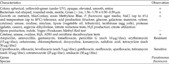
The bacteria were sorted into four strains (Ps1, Ps2, Ps3 and Ps4) based on their cultural, morphological and phenotypic characters. Cultural characteristics (shape, size, colour, margin and opacity of the colonies on NA plates), morphology (shape and size of vegetative cells and spores, if any) and motility of the strains (Ps1–Ps4) were recorded under 100 × of a phase-contrast microscope following standard microbiological methods (Smibert and Krieg, Reference Smibert, Krieg, Gerhardt, Murray, Wood and Krieg1995; Lacey, Reference Lacey1997). Gram staining, NaCl (1–10%) tolerance, hydrolysis of different substrates (starch, protein, lipid and lecithin) and sensitivity (judged by inhibition zone formation) to different antibiotics (Table 1) were also investigated. The potent bacterium strain (Ps1), which supported the growth of C. vishnui, was phenotyped following Palleroni (Reference Palleroni and Krieg1984).
Phenotypic characterization of the Pseudomonas fluorescens (Ps1) isolate

ADH, arginine dihydrolase.
To assess the beneficial effects of the prevalent bacteria (namely Ps1, Ps2, Ps3 and Ps4) on the larvae, 30 third instar C. vishnui were released into 2 l glass beakers filled halfway with sterile tap water mixed separately with a 5 ml suspension (106 bacteria/ml) of each isolate and maintained in cages. The potent bacterium, Ps1, was selected that supported normal development.
To cure experimental mosquito larvae of Ps1, third instar C. vishnui were fed for 24 h on a 10% sterile sucrose solution containing a mixture of the antibiotics to which the strain was sensitive (Table 1). Untreated (wild) and antibiotic-treated (cured) larvae (30 larvae/l water) were used to assess the role of Ps1, at 5 ml inoculum (106 bacteria/ml)/l water, on larval development, in beakers containing unsterile or sterile paddy field water, tap water or distilled water. Survival of the larvae was checked at 12 h intervals.
The data on assessment of P. fluorescens were analysed by SPSS 11.0 SW for standard error calculation.
Results and Discussion
Pseudomonas spp. are regular inhabitants of different mosquito species from diverse habitats (Takahiro et al., Reference Takahiro, Yoshitomo, Naruo, Masakazu and Takema2006; Rajagopal, Reference Rajagopal2009; Rani et al., Reference Rani, Sharma, Rajagopal, Adak and Bhatnagar2009). Among the four P. fluorescens strains (Ps1, 2, 3 and 4) present in the third and fourth instars of C. vishnui throughout the year, only the Ps1 strain supported the development of the mosquito's larval stages.
This strain formed yellowish-green, opaque, elevated, entire and smooth colonies (Table 1). They were aerobic, non-spore-forming and Gram-negative rods, tolerated 10% NaCl and could grow at temperatures of up to 40 ± 0.1°C (Table 1). It was negative for indole, methyl red and Voges–Proskauer tests, and positive for catalase, urease, oxidase, H2S, arginine dihydrolase and ornithine decarboxylase tests (Table 1).
Its phenotypic characteristics indicated that the bacterium was P. fluorescens (Palleroni, Reference Palleroni and Krieg1984). Table 1 shows the sensitivity of Ps1 to common antibiotics. The data suggest that Pseudomonas spp. harbour several antibiotic-resistant genes, which impart resistance to different antibiotics, including pollutants from anthropogenic activities (Palleroni, Reference Palleroni and Krieg1984).
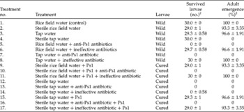
In all experimental conditions, C. vishnui larvae died when fed with antibiotics that were active against Ps1 (Table 2). In contrast, 93.3–96.6% of larvae cured of Ps1 survived to adult emergence in Ps1-treated sterile rice field water or tap water. When no Ps1 was introduced into Ps1-cured larvae, or the Ps1 was introduced along with anti-Ps1 antibiotics (Table 2), none of the larvae survived.
Effect of Pseudomonas fluorescens (Ps1) on development of the third instar Culex vishnui larvae1

1 For each test, 30 third instar larvae and three replications were used.
2 Data are means of three replications ± SE. Data for the strains Ps2, Ps3 and Ps4 are not shown, as these had no effect on the development of C. vishnui.
All cured larvae survived in unsterile water or rice field water treated with antibiotics that were ineffective against Ps1 (Table 2), which indicates that the larvae acquired the Ps1 from water in its natural habit. This observation is similar to that observed in the mosquito Anopheles gambiae, which acquires the gut bacteria Thorsellia anopheles from rice field soil (Rani et al., Reference Rani, Sharma, Rajagopal, Adak and Bhatnagar2009). The mortality of all the cured larvae in sterile water or in water containing Ps1 along with anti-Ps1 antibiotics proved that this strain of gut bacteria was vital to the development of the larvae.
Gut-inhabiting P. fluorescens of C. vishnui probably produces siderophore, hydrogen cyanide, antibiotics and other compounds that inhibit some bacteria, as well as growth factors. The bacteria also fix nitrogen, which supports the growth of the mosquito (Perry et al., Reference Perry, Balbo, Jones, Fetherston and DeMoll1999; Quadri and Ratledge, Reference Quadri, Ratledge, Cole, Eisenach, McMurray and Jacobs2005; Rajagopal, Reference Rajagopal2009). It has also been reported that several other gut bacteria, such as Acetobacter, Pseudomonas, Klebsiella, Gluconobacter and Enterobacter spp. inhabiting the insect orders Isoptera, Homoptera, Heteroptera, Coleoptera, Hymenoptera and Diptera, also affect larval growth. The bacteria influence digestion and growth factor production, and protect their host from harmful microbes by producing anti-microbial products and modulating their immune response (Baumann et al., Reference Baumann, Baumann, Lai, Rouhbakhsh, Moran and Clark1995; Takahiro et al., Reference Takahiro, Yoshitomo, Naruo, Masakazu and Takema2006; Rajagopal, Reference Rajagopal2009; Rani et al., Reference Rani, Sharma, Rajagopal, Adak and Bhatnagar2009).
Evidently, since C. vishnui is dependent on Ps1 for its survival, eliminating this strain of bacterium from mosquito breeding areas could be an innovative and effective means of managing the encephalitis problem in tropical rice-growing countries. This could be achieved by treating rice fields with leaves of anti-pseudomonal plants, such as tamarind Tamarindus indica (Muthu et al., Reference Muthu, Nandakumar and Rao2005), or by using anti-Ps1 antibiotics.
In the future, Ps1 could be genetically engineered to express genes that confer entomopathogenicity, which would suppress its host, or even with the anti-pathogenic or anti-parasitic genes, to eliminate the ability of the mosquito to support disease-causing agents (Poopathi and Tyagi, Reference Poopathi and Tyagi2006; Rani et al., Reference Rani, Sharma, Rajagopal, Adak and Bhatnagar2009).