The recently published 2013 Global Burden of Disease study reports that there are at least 32.9 million persons currently affected by rheumatic heart disease. 1 Secondary prophylaxis with penicillin prevents disease progression and can even lead to regression in patients with known acute rheumatic fever or mild clinical rheumatic heart disease.Reference Tompkins, Boxerbaum and Liebman 2 In endemic areas, many patients present late with irreversible valve damage and life-threatening complications, whereas almost none recall a history of rheumatic fever.Reference Okello, Wanzhu and Musoke 3 Thus, identification of patients with only latent rheumatic heart disease (mild valve disease without symptoms) who would benefit from secondary prophylaxis in childhood presents a unique opportunity to prevent progression of disease in these regions. There are several recent echocardiographic studies using World Heart Federation guidelinesReference Remenyi, Wilson and Steer 4 that have found the prevalence of latent rheumatic heart disease in endemic regions to be significant, as high as 40/1000;Reference Beaton, Aliku and Okello 5 – Reference Engel, Haileamlak and Zuhlke 10 however, studies that include longitudinal follow-up and complementary assessment, including other echocardiography measurements and non-echocardiography covariates, are needed to determine which of these children are at higher risk for developing clinically significant valve disease.Reference Zuhlke and Mayosi 11 , Reference Saxena, Zuhlke and Wilson 12
There is little known about the pathophysiology of latent rheumatic heart disease, but two studies confirm that it is more likely to be present in rheumatic heart disease-endemic regions.Reference Roberts, Maguire and Brown 13 , Reference Clark, Krishnan, McCarter, Scheel, Sable and Beaton 14 Even though advanced rheumatic heart disease is not thought to involve the myocardium, there is evidence that the initial episode of carditis does involve the myocardium,Reference Ozdemir, Oguz, Atmaca, Sanli, Yildirim and Olgunturk 15 and it is possible that subtle myocardial changes could persist or recur with repeated exposure to streptococcal infection in endemic regions. Echocardiographic identification of these subtle myocardial abnormalities has the potential to facilitate identification of patients predisposed to developing more significant rheumatic carditis, potentially before onset of clinical valve disease.
Over the last decade, echocardiographic measurement of myocardial mechanics has become effective in many disease processes. Myocardial strain, a measurement of shortening and lengthening of muscle fibres, is a more sensitive indicator of cardiac performance than traditional measures of ejection fraction and shortening fraction.Reference Gorcsan and Tanaka 16 Longitudinal strain is more sensitive to early, subtle myocardial injury than other strain measurements.Reference Mor-Avi, Lang and Badano 17 Strain assessment analyses myocardial motion by tracking natural acoustic markers, or speckles, within the myocardium.Reference Geyer, Caracciolo and Abe 18 Multiple studies of patients with isolated valvular diseaseReference Tayyareci, Yildirimturk, Yurdakul and Aytekin 19 – Reference Sengupta, Amaki and Bansal 21 or systemic diseaseReference Gaur, Waloff, Schiller, Sable and Frank 22 – Reference Poterucha, Kutty, Lindquist, Li and Eidem 27 have shown abnormalities in strain that are undetectable by traditional measures.
We recently reported on a cohort of children who underwent prospective screening for rheumatic heart disease in Gulu, Northern Uganda, using 2012 World Heart Federation criteria.Reference Beaton, Lu and Aliku 6 The prevalence of definite and borderline rheumatic heart disease in that cohort was 1 and 2.9%, respectively. To test the hypothesis that abnormalities in myocardial strain may be present in children with latent rheumatic heart disease, children from this cohort, an earlier cohort of children screened in 2010,Reference Beaton, Okello, Lwabi, Mondo, McCarter and Sable 28 and additional control subjects underwent electrocardiogram-gated echocardiograms with subsequent offline analysis of traditional measures of cardiac systolic function as well as myocardial strain.
Materials and methods
Patients
Approval for this prospective case–control study was obtained from the Institutional Review Board at Children’s National Medical Center, Makerere University School of Medicine, and the Ugandan National Council of Science and Technology. Electrocardiogram gating, not routinely used in our Ugandan studies, was added to echocardiography during regularly scheduled follow-up visits of children identified to have definite or borderline rheumatic heart disease from previous studies.Reference Beaton, Aliku and Okello 5 , Reference Beaton, Lu and Aliku 6 , Reference Beaton, Okello, Lwabi, Mondo, McCarter and Sable 28 Similarly, normal studies were obtained from the addition of electrocardiogram gating to concurrent school-based screening. Permission for offline review of echocardiograms obtained for clinical follow-up and research purposes was covered by the standard informed consent for both patient populations. More detailed demographic information was not available, and blood analyses to determine nutritional and HIV statuses were not included in this study.
Conventional two-dimensional echocardiography
All two-dimensional transthoracic echocardiograms were performed by a single operator (M.P.) with a standardised protocol using a commercial echocardiographic system (General Electric VIVID Q; General Electric, Milwaukee, Wisconsin, United States of America). A limited protocol included parasternal long-axis, short-axis, and apical four-chamber black and white and color Doppler images as well as spectral Doppler images of the mitral and aortic valves from the apical four-chamber view; in addition, two heartbeat cine-loops were recorded with electrocardiogram gating. Conventional echocardiographic parameters were obtained by a single reviewer (H.R.), including ejection fraction, shortening fraction, and left atrial, aortic root, and left ventricular chamber size. Ejection fraction was calculated by the single-plane method of disks in the apical four-chamber view, and shortening fraction was calculated by standard left ventricular two-dimensional measurements at the level of the papillary muscles.Reference Lopez, Colan and Frommelt 29 In addition, a cardiologist with expertise in rheumatic heart disease (C.S.) graded the presence and qualitative assessment of mitral regurgitation, mitral stenosis, and aortic regurgitation as well as classified the echocardiograms according to 2012 World Heart Federation criteria into the following – normal, borderline rheumatic heart disease, definite rheumatic heart disease, or other.Reference Remenyi, Wilson and Steer 4
Myocardial deformation analysis
Echocardiograms were imported into a picture archival and communication system network (Philips Xcelera; Philips, Best, Holland) in DICOM format and were analysed offline using Tomtec Cardiac Performance Analysis (Tomtec Imaging Solutions, Unterschleissheim, Germany). Analysis was performed on images with an average viewed frame rate of 70 Hz. Endocardial tracings of the left ventricle were performed in apical four-chamber views for each subject, and subjects were excluded from the analysis if the entirety of the left ventricle was not captured. Apical four-chamber views with good visualisation of the left ventricular endocardium were used to obtain global peak systolic longitudinal strain. In all, three separate measurements for global longitudinal strain were recorded on the same two-beat clip and averaged for each patient by a single reviewer (H.R.). A second reviewer (C.S.) repeated this process for studies that had >20% variation in the three strain measurements. Both reviewers were blinded to other findings when performing strain assessment on the apical four-chamber view. Strain analysis was performed 4–12 weeks after conventional assessment and rheumatic heart disease grading.
Statistical analysis
Subjects with rheumatic heart disease were classified into groups on the basis of (1) the presence of any rheumatic heart disease, (2) the presence of definite rheumatic heart disease, and (3) the presence of borderline rheumatic heart disease. Control subjects were sub-classified into two groups – those with mitral regurgitation, but not meeting criteria for rheumatic heart disease, and those with no mitral regurgitation. Strain was categorised as normal or abnormal (<5th percentile for age) using a cut-off absolute number <18.4 for 5- to 9-year-olds, 19.2 for 10- to 14-year-olds, and 19.9 for 15- to 19-year-olds.Reference Marcus, Mavinkurve-Groothuis and Barends 30 Fisher’s exact test was used to compare the presence or absence of abnormal strain between groups as a categorical variable. Continuous data were not normally distributed. Therefore, strain values were compared between groups as a continuous variable using the two-tailed Mann–Whitney U-test.Reference Fay and Proschan 31 Sample size calculations were performed on an anticipated recruitment ratio of four control subjects for one rheumatic heart disease subject on the basis of the availability of children who return for screening. Sample sizes of 156, 100, and 68 control and 39, 25, and 17 rheumatic heart disease subjects provided 80% power (with 0.05 type 1 error rate) to detect 10, 12.5, and 15% differences in strain, respectively.
Results
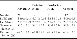
Echocardiography was performed on 148 children. Of these, 139 had apical four-chamber images suitable for strain analysis. There were 14 patients with definite rheumatic heart disease and 13 with borderline rheumatic heart disease. The remaining 112 subjects made up the control group, and included 35 subjects with trivial or mild mitral regurgitation that did not meet World Heart Federation criteria for rheumatic heart disease. There was >20% variation in initial strain measurements in 14 subjects – 12 control subjects, one subject with definite rheumatic heart disease, and one subject with borderline rheumatic heart disease. Demographic variables are shown in Table 1. Subjects with any rheumatic heart disease and with borderline rheumatic heart disease were older than control subjects. No differences were found when comparing demographic variables within the two control subgroups. No rheumatic heart disease or control subjects had left ventricular systolic or diastolic diameter >95th percentile or ejection or shortening fraction <5th percentile. There were no significant differences in these measurements between groups (Table 2).
Demographic variables.

RHD=rheumatic heart disease
* p=0.05 for age (any RHD versus control)
** p=0.006 for age (borderline RHD versus control)
Standard echocardiographic measurements.

LVED=left ventricle end-diastolic dimension; LVES=left ventricle end-systolic dimension; RHD=rheumatic heart disease
p=NS for all comparisons
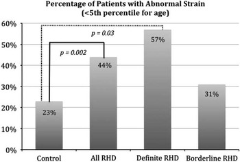
Categorical results for global longitudinal strain are shown in Figure 1. Strain was lower (absolute value) than the 5th percentile in 12 of 27 (44%) subjects with any rheumatic heart disease (p=0.002 versus controls) and in 8 of 14 (57%) subjects with definite rheumatic heart disease (p=0.03 versus controls). There was no difference when comparing borderline rheumatic heart disease with controls or when comparing definite rheumatic heart disease with borderline rheumatic heart disease. The number of subjects in each control subgroup with strain below the 5th percentile was not different – 20% of subjects with mitral regurgitation who did not meet criteria for rheumatic heart disease versus 27% of subjects with no mitral regurgitation.
Categorical comparison of percentage of patients with abnormal strain (<5th percentile for age). RHD=rheumatic heart disease.

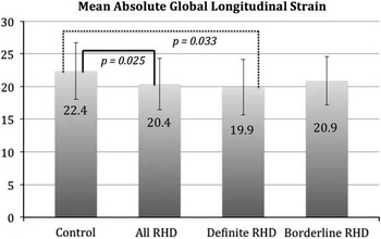
Figure 2 shows absolute mean strain values for all groups. The mean absolute strain values were significantly lower when comparing subjects with any rheumatic heart disease with controls (20.4±3.95 versus 22.4±4.35, p=0.025) and subjects with definite rheumatic heart disease with controls (19.9±4.25 versus 22.4±4.35, p=0.033). There was no difference when comparing definite rheumatic heart disease with borderline rheumatic heart disease subjects or borderline rheumatic heart disease subjects with control subjects.
Mean absolute global longitudinal strain comparison between groups using the Mann–Whitney U-test. RHD=rheumatic heart disease.

Discussion
This is the first study to assess myocardial strain in a paediatric cohort with rheumatic heart disease. Our results from an endemic region in Africa show that global longitudinal strain is abnormal in rheumatic heart disease as compared with a control indigenous population. Strain below the 5th percentile is more likely to be present in subjects with definite rheumatic heart disease. Mean longitudinal strain is statistically significantly lower in subjects with definite rheumatic heart disease. Although not statistically significant, when assessed separately, the borderline rheumatic heart disease subgroup, with minimal valve changes, tended to have a higher percentage of patients with lower strain. It may be that strain imaging identifies myocardial changes more characteristic of early or latent disease. Our cross-sectional study provides a starting point for further investigations that should include longitudinal follow-up to determine the utility of strain in assessing latent rheumatic heart disease.
These differences are unlikely to be the result of other factors. Our subjects with rheumatic heart disease were older than control subjects; this is consistent with other studies as well.Reference Beaton, Okello, Lwabi, Mondo, McCarter and Sable 28 , Reference Paar, Berrios and Rose 32 The differences in global longitudinal strain, however, cannot be accounted for by this finding, given that the absolute longitudinal strain values actually get higher with age.Reference Marcus, Mavinkurve-Groothuis and Barends 30 We may have seen a more exaggerated difference if we had used age-matched control subjects. Ejection fraction was within normal limits in all subjects, and there were no differences in ejection fraction or shortening fraction between groups. Conversely, 57% of subjects with definite rheumatic heart disease had global longitudinal strain below the normal range. Presence of non-pathological mitral regurgitation – seen in 35 of 112 of control subjects – was not significant for abnormal strain, providing some evidence against increased preload from mitral regurgitation playing a significant role in our strain results.
Although rheumatic heart disease appears to be primarily a disease of the endocardium and valves with minimal myocardial involvement,Reference Mishra, Mohanty, Mishra and Rath 33 , Reference Tandon 34 this is not the first study to suggest significant early myocardial involvement. Ozdemir et al.Reference Ozdemir, Oguz, Atmaca, Sanli, Yildirim and Olgunturk 15 , reported an increase in cardiac troponin in patients with acute rheumatic carditis, speculating that there may be some myocardial damage in patients with active carditis. A previous study looking at myocardial strain in adults with rheumatic mitral stenosis before and after mitral balloon valvulplasty Reference Sengupta, Amaki and Bansal 21 demonstrated that global longitudinal strain was reduced at baseline, compared with controls, and improved after valvuloplasty. In contrast to our study of patients with lesser disease, these patients had impaired systolic function. An explanation for abnormal strain in mitral stenosis is restriction of the posterior basal myocardium by an abnormal mitral valve.Reference Gaasch and Folland 35 Although these abnormalities are unlikely to be present in our cohort, it is possible that minor changes could occur from more subtle abnormalities in mitral valve function.
There are several limitations to our study. Although subtle findings that could differentiate latent rheumatic heart disease from normal variants would be more useful in the borderline group, only subjects with definite rheumatic heart disease had significant differences in strain when compared with our control population. Although we met our sample size goal for detecting 12.5% difference in strain at 80% power, our study was underpowered to detect differences in strain in subgroups of rheumatic heart disease subjects. We were limited to subjects who were able to return for additional screening with electrocardiogram gating. Longitudinal follow-up of this cohort will be of critical importance to determine whether strain can predict which subjects develop more significant valvular disease.
Even though strain was significantly lower in subjects with latent rheumatic heart disease than in control subjects, it was unexpected that >5% of our control subjects had strain outside the normal range. This is likely due to both patient and technical factors. No normative paediatric strain data are available from Africa, but there could be regional differences in paediatric strain that may, in part, be influenced by malnutrition and other chronic diseases.Reference Sgambat, Frank, Ellini, Sable and Moudgil 23 , Reference Sims, Frank and Cross 24 , Reference Hammoudi, Arangalage and Djebbar 36 Covariates, such as HIV and nutritional status would have strengthened this study but were not available to us for this cohort. Inclusion of blood analysis would have added important information but would have limited subject recruitment and the ability to work in schools. It is also possible that differences in echocardiography machines, strain analysis software, and type of images used for analysis could account for differences in strain values across all subjects. Our study used General Electric VIVID Q echocardiography machines and Tomtec software to analyse DICOM images, whereas the reference study used General Electric VIVID 7 echocardiography machines and General Electric Echo picture archival and communication system software to analyse native data images.Reference Marcus, Mavinkurve-Groothuis and Barends 30
In conclusion, our study shows that global longitudinal strain, a sensitive marker of early subtle myocardial injury, is decreased in a significant number of subjects with definite latent rheumatic heart disease. The mechanism of this is unknown, but it does not appear to be related to other more global measures of systolic function or increased preload from mild mitral regurgitation. Our findings raise the possibility of recurrent subtle myocardial inflammation being present in latent rheumatic heart disease. Larger studies with a longer-term follow-up and collection of covariates are required to determine whether there is a potential role for strain to help better understand the significance of latent rheumatic heart disease in the pathophysiology of rheumatic heart disease in endemic regions and to differentiate which patients with latent rheumatic heart disease will develop progressive disease.
Acknowledgements
None.
Authors’ Contributions: A.B.: concept/design, data analysis/interpretation, drafting of article, critical revision of article, statistics. H.R.: concept/design, data analysis/interpretation, approval of article. M.P.: concept/design, critical revision of article, approval of article. L.G.: critical revision of article, approval of article. T.A.: concept/design, critical revision of article, approval of article. P.L.: critical revision of article, approval of article. G.E.: critical revision of article, approval of article. C.S.: concept/design, data analysis/interpretation, drafting of article, critical revision of article, statistics.
Financial Support
This project was supported by Award Nos UL1TR000075 and KL2TR000076 from the NIH National Center for Advancing Translational Sciences. Its contents are solely the responsibility of the authors and do not necessarily represent the official views of the National Center for Advancing Translational Sciences or the National Institutes of Health. This study was also funded in part by grants from General Electric and Edwards Life Sciences.
Conflicts of Interest
None.
Ethical Standards
The authors assert that all procedures contributing to this work comply with the ethical standards of the national guidelines on human experimentation of the United States of America and Uganda and with the Helsinki Declaration of 1975, as revised in 2008. This work has been approved by the Institutional Review Boards of Children’s National Medical Center, Makerere University School of Medicine, and the Ugandan National Council of Science and Technology.