Introduction
Alveolar echinococcosis in humans is a parasitic disease caused by Echinococcus multilocularis (Cyclophyllidea: Taeniidae). Human infection occurs through accidental ingestion of parasite eggs excreted in the faeces of carnivorous definitive hosts such as foxes and dogs (Deplazes & Eckert, Reference Deplazes and Eckert2001). Detection of adult worms in the intestinal duct via necropsy is the definitive diagnostic method for determining Echinococcus infections in definitive hosts (Conraths & Deplazes, Reference Conraths and Deplazes2015). However, performing necropsy examinations on wild animals requires collecting their carcasses, which is difficult, and safety equipment must be used during the examination to prevent human infection. Thus, coprological examinations are alternatively performed to detect the parasite eggs, antigens and DNA in the host faeces.
Among these coprological methods, worm-specific copro-DNA examination based on alkaline extraction achieved 89% sensitivity and complete specificity in fox faeces (Dinkel et al., Reference Dinkel, von Nickisch-Rosenegk, Bilger, Merli, Lucius and Romig1998), and its modified technique achieved successful detection in both the prepatent and patent periods in faeces of experimentally infected dogs (Irie et al., Reference Irie, Ito, Kouguchi, Yamano, Uraguchi, Yagi and Nonaka2017). However, the dogs in that study were fed commercial food, and minimal undigested residue derived from this food was found in their faeces. Therefore, the faecal characteristics may have differed from the faeces of wild definitive hosts; thus, the test's reliability must be evaluated before using it to survey wild animals.
We preliminarily performed copro-DNA examinations using rectal faeces from red foxes via the method described in our previous report (Irie et al., Reference Irie, Ito, Kouguchi, Yamano, Uraguchi, Yagi and Nonaka2017), in which foxes were diagnosed with E. multilocularis infection upon necropsy. However, the copro-DNA examination results yielded several false-negatives. One reason for this may have been that the substantial amount of faecal material used was smaller than the measured weight because the red fox faeces contained undigested vegetable and animal residue. Increasing the amount of faeces used for copro-DNA extraction should improve sensitivity; therefore, the amount tested was changed from 1.0 g to 3.0 g in this study. Another reason for the false-negative results was that some faeces indicated a strong acidic pH when diluted in water (the lowest was pH = 3); therefore, extracting the copro-DNA from these faeces may have failed because alkalinity could not be achieved in the lysis step. Thus, in the present technique, a buffered solution was used to neutralize the faeces before copro-DNA extraction. In this study, we evaluated the reliability of the modified copro-DNA examination for detecting E. multilocularis infections in red foxes by comparing the results with those obtained via necropsy examination.
Materials and methods
Collection of small intestine and rectal faeces from red foxes
Red foxes that had been found as roadkill in Sapporo City, Hokkaido, Japan, were collected by city government workers and stored at −80°C for several days. The carcasses were then transported to the Hokkaido Institute of Public Health. From November 2015 to March 2019, whole small intestines and rectal faeces were collected from 258 foxes that were not completely decomposed. Necropsy and sample collection were performed in a biological safety room for Echinococcus infection in the Hokkaido Institute of Public Health.
Worm examination by the modified sedimentation and counting technique (SCT)
The collected intestines were immediately subjected to mucosal scraping and sedimentation. The small intestines were incised longitudinally, and the mucosa was scraped. The scraped mucosa was well mixed in 0.9% sodium chloride (NaCl) solution and passed through a wire mesh (pore size: ~2 mm) into a glass bottle. NaCl solution was added up to 1 l, then the sample was sedimented for 30 min, followed by three changes of supernatant as described by Hofer et al. (Reference Hofer, Gloor, Müller, Mathis, Hegglin and Deplazes2000). The sediments were kept in 70% ethanol until the worm examination. The sediment fraction was examined under a stereomicroscope. Whole sediment was examined when the worm burden was fewer than 100. One-tenth or one-twentieth of the sediment was used in cases with more than 100 worms, and the approximate number was recorded.
Copro-DNA examination using modified alkaline-sodium dodecyl sulphate (SDS) extraction
Faecal samples were heated at 70°C for 12 h to inactivate the E. multilocularis eggs, then the samples were stored at −30°C until use. Faecal E. multilocularis DNA was extracted using 3.0 g of faeces via a modified alkaline-SDS extraction technique and adding a pre-wash treatment per a previously described method (Dinkel et al., Reference Dinkel, von Nickisch-Rosenegk, Bilger, Merli, Lucius and Romig1998; Irie et al., Reference Irie, Ito, Kouguchi, Yamano, Uraguchi, Yagi and Nonaka2017). Briefly, 3.0 g of rectal faeces were collected into a centrifuge tube and thoroughly mixed in 2 M Tris-hydrochloride (HCl) (pH 8.3), filled to 15 ml, then stored at 4°C overnight. After centrifugation at 1710 × g for 10 min, the supernatant was decanted and thoroughly mixed in 900 μl of 1 M potassium hydroxide and 50 μl of 2 M dithiothreitol, followed by adding 50 μl of 20% SDS. The mixture was then heated at 65°C for 30 min. After neutralization with 500 μl of solution (2 M Tris-HCl: 25% HCl = 3:1), the mixture was centrifuged at 1710 × g for 10 min. Phenol/chloroform/isoamyl alcohol (25:24:1) extraction was performed on 400 μl of supernatant. After centrifugation at 10,000 × g for 5 min, the supernatant layer was collected and purified using a Gel/polymerase chain reaction (PCR) purification kit (Nippon Genetics, Tokyo, Japan) using 135 μl of specimen and extracted with 200 μl of extraction buffer.
Echinococcus multilocularis-specific PCR was performed on the cytochrome c oxidase subunit I gene according to previous reports (Irie et al., Reference Irie, Ito, Kouguchi, Yamano, Uraguchi, Yagi and Nonaka2017). For PCR-negative samples, presence of PCR inhibitors was checked via repeated PCR using the extracted faecal DNA solution added with 10 fg of control DNA from an adult E. multilocularis worm in the reaction mixture (Dinkel et al., Reference Dinkel, Kern, Brinker, Oehme, Vaniscotte, Giraudoux, Mackenstedt and Romig2011). For PCR-positive samples that were SCT-negative, amplified DNA was directly sequenced and analysed to confirm whether it belonged to E. multilocularis.
For specimens demonstrating the presence of PCR inhibitors on addition-recovery tests using control DNA, the following examinations were conducted. For SCT-positive cases, PCR was repeated using ten- and 100-fold diluted extractions of faecal DNA. For SCT-negative cases, PCR was repeated by adding 100 fg, 1 pg and 10 pg of control DNA from adult worms into the reaction mixture.
For faeces from cases that were negative for both copro-DNA and PCR inhibitors, despite being SCT-positive, copro-DNA extractions were conducted two to five more times using the same methods (depending on how much of the faecal subsample remained).
Statistical analysis
A binomial test for calculating 95% confidence intervals (CIs) was conducted using Excel Statistics 2010 for Microsoft Excel 2010 for Windows (Social Survey Research Information Co., Ltd., Tokyo, Japan).
Results and discussion
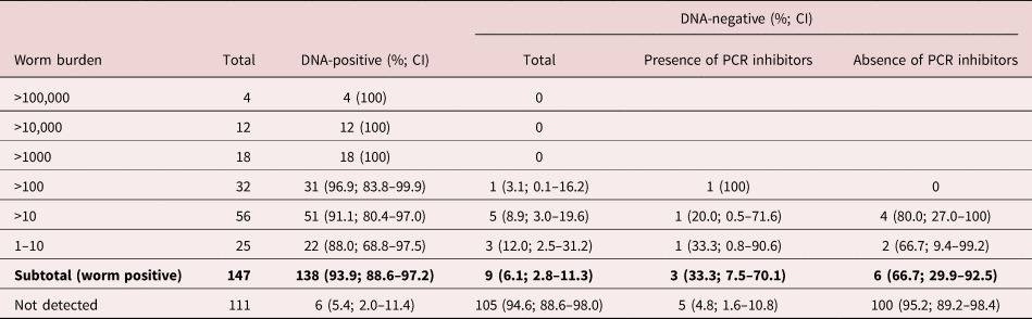
A total of 147 faecal specimens were positive for E. multilocularis worms by the modified SCT, and, of these, 138 (93.9%, CI: 88.6%–97.2%) were positive on the copro-DNA examination (supplementary table S1). The present copro-DNA detectability was increased depending on the worm burden and showed 100% sensitivity in cases with more than 1000 worms (table 1), as suggested in the reference method of Dinkel et al. (Reference Dinkel, von Nickisch-Rosenegk, Bilger, Merli, Lucius and Romig1998). Conversely, from 111 worm-negative specimens, six cases were copro-DNA-positive, and all were confirmed as E. multilocularis via sequencing analysis. The SCT described by Hofer et al. (Reference Hofer, Gloor, Müller, Mathis, Hegglin and Deplazes2000) has been widely used as a reference standard for evaluating the diagnostic method and epidemiological study of E. multilocularis infections in definitive host animals (Eckert et al., Reference Eckert, Deplazes, Craig, Gemmell, Gottstein, Heath, Jenkins, Kamiya, Lightowlers, Eckert, Gemmell, Meslin and Pawlowski2001; Tackmann et al., Reference Tackmann, Mattis and Conraths2006; Conraths & Deplazes, Reference Conraths and Deplazes2015; König et al., Reference König, Romig and Holzhofer2019); however, its sensitivity is suspected to be incomplete at approximately 83% (Isaksson et al., Reference Isaksson, Hagstrom and Armua-Fernandez2014; Maksimov et al., Reference Maksimov, Isaksson, Schares, Romig and Conraths2019). Therefore, the cases that were copro-DNA-positive but worm-negative in our study may have been due to worm loss in the sedimentation-decantation step and/or by an oversight on microscopy.
Number of specimens showing as positive/negative on Echinococcus multilocularis-specific copro-DNA examinations by worm burden strata as detected upon necropsy.

CI, 95% confidence intervals.
Increasing the test amount of the faeces processed for copro-DNA extraction may improve the probability of detecting faecal E. multilocularis DNA; however, it can also enrich the PCR inhibitors, which can cause false-negatives (Maksimov et al., Reference Maksimov, Schares, Press, Fröhlich, Basso, Herzig and Conraths2017). Therefore, we checked the remaining PCR inhibitors in the cases that were PCR-negative but SCT-positive as well as all SCT-negative cases. Among nine cases showing false-negative results on the copro-DNA examination, three showed the presence of PCR inhibitors, and the other six cases were suspected to be false-negatives due to an absence of E. multilocularis copro-DNA (table 1). Five of the 105 specimens (4.8%, CI: 1.6%–10.8%) that were negative for both intestinal worms and copro-DNA also demonstrated the presence of PCR inhibitors, suggesting that approximately 5% of red fox faeces retain these inhibitors after treatment with the present copro-DNA extraction method. Concerning PCR inhibitors, several faecal agents can inhibit Taq-polymerase activity on PCR (e.g. bile acids, haemoglobin derivatives and polysaccharides) (Kohn & Wayne, Reference Kohn and Wayne1997; Monteiro et al., Reference Monteiro, Bonnemaison, Vekris, Petry, Bonnet, Vidal, Cabrita and Mégraud1997, Reference Monteiro, Gras and Megraud2001). Several methods have been developed to effectively remove these inhibitors, including techniques that capture parasite DNA (e.g. a magnetic capture-based DNA extraction), but with slightly decreased sensitivity (Isaksson et al., Reference Isaksson, Hagstrom and Armua-Fernandez2014; Maas et al., Reference Maas, van Roon, Dam-Deisz, Opsteegh, Massolo, Deksne, Teunis and van der Giessen2016; Maksimov et al., Reference Maksimov, Isaksson, Schares, Romig and Conraths2019). Combining such techniques in our extraction methods to remove PCR inhibitors may help further improve the reliability of the present copro-DNA examination method. Additionally, PCR yielded successful results when the copro-DNA extracts contained more E. multilocularis DNA than PCR inhibitors, because increasing the amount of control DNA added to the copro-DNA extracts from SCT-negative and inhibitor-present cases yielded PCR-positive results (supplementary table S2). Thus, among the SCT-positive cases, PCR-inhibitors were only confirmed in cases with low worm burdens (table 1). For such SCT-positive and inhibitor-present cases (n = 3), using ten- and 100-fold dilutions of extracted DNA resulted in a positive PCR conversion in all cases. This may have occurred because the inhibitors were diluted to less than the amount needed to effectively inhibit Taq-polymerase activity in the PCR.
To overcome false-negative copro-DNA examinations caused by the absence of E. multilocularis DNA in the extracted solution, the amount of the faecal sample tested should be further increased. Faecal E. multilocularis eggs are heterogeneously distributed during the patent period (Nonaka et al., Reference Nonaka, Iida, Yagi, Ito, Ooi, Oku and Kamiya1996; Maksimov et al., Reference Maksimov, Schares, Press, Fröhlich, Basso, Herzig and Conraths2017); hence, parasite tissue in the faeces is also likely to be heterogeneously distributed. In addition, the amount of parasite tissue in the faeces is suspected to be small, especially in the prepatent period, as we suspected in our previous study on experimentally infected dogs (Irie et al., Reference Irie, Ito, Kouguchi, Yamano, Uraguchi, Yagi and Nonaka2017). However, the amount of faeces that can be added to a test tube is limited. Therefore, repeated extraction of copro-DNA from each faecal subsample may help improve the reliability of the copro-DNA examination for diagnosing E. multilocularis infections. In our evaluation of five cases that were negative for both copro-DNA and PCR inhibitors, despite being SCT-positive, two cases were PCR-positive in other DNA extraction trials (supplementary table S3).
Relatively fresh rectal faeces were used for the evaluations in this study. When adapting the copro-DNA examination for epidemiological studies, the specimens to be examined will likely be faeces collected in the field, which are affected by natural conditions (e.g. ultraviolet light) that may affect the copro-DNA. In addition, DNase in the field-collected faeces may damage any target DNA. Hence, further evaluations that consider the effects of nature are needed.
The copro-DNA examination protocol described herein, which was modified by increasing the sample amount and adding a neutralization step, is highly reliable for diagnosing E. multilocularis infection in red foxes. This method could be widely used as an epidemiological diagnostic tool for E. multilocularis infections in definitive hosts because it does not require special equipment and is easy to handle. This test could help monitor prevalence tendencies in already endemic areas. When used for detailed evaluations of endemicity (e.g. change in infection pressure or spread in non-endemic areas), the absence of PCR inhibitors should be confirmed, and multiple trials on faecal subsamples are recommended.
Supplementary material
To view supplementary material for this article, please visit https://doi.org/10.1017/S0022149X20000255
Financial support
This research did not receive any specific grant from funding agencies in the public, commercial, or not-for-profit sectors.
Conflicts of interest
None.
Ethical standards
This article does not contain any studies with human participants or animals performed by any of the authors.
Author contributions
Takao Irie designed the study, performed the material preparation, experiments and data collection and drafted the first version of the manuscript. Takuya Ito contributed to modifying the copro-DNA examination. Hirokazu Kouguchi supported the intestinal collection. Kohji Uraguchi performed the necropsies, supervised the study and reviewed the original draft. All authors read and approved the final manuscript.