INTRODUCTION
Tapeworm parasites of the genus Echinococcus Rudolphi, 1801 (Cestoda: Taeniidae) are causative agents of echinococcosis, which is one of the most important zoonoses worldwide. Larval stages (metacestodes) of Echinococcus species establish as cysts in the internal organs (mainly liver and lung) of intermediate hosts – frequently wild and domestic ungulates, but also humans – leading to severe health problems and even death. Adult parasites are found in the small intestines of wild and domestic canids and/or felids. Their effects on the health of these definitive hosts are less severe compared with those experienced by intermediate hosts (McManus and Thompson, Reference McManus and Thompson2003; Jenkins et al. Reference Jenkins, Romig and Thompson2005; Thompson, Reference Thompson2008). In order to understand the biology, life cycle/transmission, distribution and evolution of Echinococcus parasites, numerous studies have been conducted in recent times. Despite the major medical and veterinary importance of this genus, efforts to establish a widely-accepted taxonomy have not yet been successful. Based on existing data, 4 species have received general acceptance, namely E. granulosus, E. multilocularis, E. vogeli and E. oligarthrus. However, significant taxonomic uncertainties in the genus are yet to be resolved, particularly within E. granulosus. On the basis of considerable intraspecific variation, E. granulosus has been regarded as a collection of 10 different genotypes, which have been numbered consecutively G1–G10, reflecting the order in which they were described (Bowles et al. Reference Bowles, Blair and McManus1992, Reference Bowles, Blair and McManus1994; Scott et al. Reference Scott, Stefaniak, Pawlowski and McManus1997; Lavikainen et al. Reference Lavikainen, Lehtinen, Meri, Hirvelä-Koski and Meri2003). It has been proposed that some of these genotypes or groups of genotypes may deserve species status: G1–G3 as E. granulosus sensu stricto, G4 as E. equinus, G5 as E. ortleppi and G6–G10 as E. canadensis (Thompson and McManus, Reference Thompson and McManus2002; Nakao et al. Reference Nakao, McManus, Schantz, Craig and Ito2007; Thompson, Reference Thompson2008). Furthermore, a new species, E. shiquicus, has been described recently (Xiao et al. Reference Xiao, Qiu, Nakao, Li, Yang, Chen, Schantz, Craig and Ito2005), and it has been argued that E. felidis Ortlepp, 1937 should be reaccepted as a distinct species, rather than falling within E. granulosus sensu stricto (see Hüttner et al. Reference Hüttner, Nakao, Torsten, Siefert, Boomker, Dinkel, Sako, Mackenstedt, Romig and Ito2008). The existence of a human-specific genotype G9 of E. granulosus, as proposed by Scott et al. (Reference Scott, Stefaniak, Pawlowski and McManus1997), remains controversial. Analysis of mitochondrial sequence data revealed no significant differences between isolates from pigs (G7) and humans (G9) (Kedra et al. Reference Kedra, Swiderski, Tkach, Dubinsky, Pawlowski, Stefaniak and Pawlowski1999).
Although phylogenetic studies have contributed significantly toward a better understanding of the taxonomy of the genus Echinococcus, even the use of complete mitochondrial (mt) genome sequences (e.g. Nakao et al. Reference Nakao, McManus, Schantz, Craig and Ito2007) has failed to produce results in agreement with earlier phenotypic classifications (Williams and Sweatman, Reference Williams and Sweatman1963; Rausch, Reference Rausch1967). Specifically, the phylogeny of Echinococcus based on analyses of complete mtDNA sequence data has confirmed earlier studies using partial mtDNA sequences (Lavikainen et al. Reference Lavikainen, Lehtinen, Meri, Hirvelä-Koski and Meri2003, Reference Lavikainen, Lehtinen, Laaksonen, Ågren, Oksanen and Meri2006; Xiao et al. Reference Xiao, Qiu, Nakao, Li, Yang, Chen, Schantz, Craig and Ito2005; Thompson et al. Reference Thompson, Boxell, Ralston, Constantine, Hobbs, Shury and Olson2006). These indicated that different genotypes of E. granulosus do not form a monophyletic cluster, but include also E. multilocularis and E. shiquicus (see Nakao et al. Reference Nakao, McManus, Schantz, Craig and Ito2007), which are both phenotypically distinct from E. granulosus. Moreover, it has been demonstrated that the cervid genotypes of E. granulosus, G8 and G10, are not positioned on the phylogenetic tree as sister taxa; instead, the camel- and pig-specific genotypes G6/G7 were shown to form a clade with G10 (Moks et al. Reference Moks, Jõgisalu, Valdmann and Saarma2008). Taken together, these data indicate the need for additional studies to resolve the taxonomy of Echinococcus.
Contemporary phylogenetic hypotheses have been based largely on data derived from mitochondrial sequences, whereas an alternative source of phylogenetic characters from the nuclear genome has not received much attention. Although mtDNA provides useful genetic markers – it is, generally, haploid, multicopy, neutral and provides access to a set of genes with no recombination (Brown, Reference Brown, Nei and Koehn1983) and ‘rapid’ evolution (Brown et al. Reference Brown, George and Wilson1979) – it has some significant limitations. First, mtDNA is usually inherited through the maternal lineage (Giles et al. Reference Giles, Blanc, Cann and Wallace1980; Avise et al. Reference Avise, Arnold, Ball, Bermingham, Lamb, Neigel, Reeb and Saunders1987). This is presumably valid also for parasitic flatworms, although experiments have suggested that intraspecific paternal inheritance of mtDNA may occur across multiple generations in Schistosoma mansoni (Jannotti-Passos et al. Reference Jannotti-Passos, Souza, Parra and Simpson2001). Should female evolutionary history differ from that of the species, then mtDNA would not reflect the evolutionary history of the species as a whole. Furthermore, insertions of mitochondrial sequences into the nuclear genome and vice versa can occur (see Leister, Reference Leister2005). As genes in the mitochondrial genome are linked (Anderson et al. Reference Anderson, Bankier, Barrell, Debruijn, Coulson, Drouin, Eperon, Nierlich, Roe, Sanger, Schreier, Smith, Staden and Young1981; Le et al. Reference Le, Blair, Agatsuma, Humair, Campbell, Iwagami, Littlewood, Peacock, Johnston, Bartley, Rollinson, Herniou, Zarlenga and McManus2000), information from different mtDNA genes does not fulfil the criteria of statistical independence, which is a requirement if a phylogeny is to represent the evolutionary history of genomes and not just of single loci (Ballard and Rand, Reference Ballard and Rand2005). If the goal is to reconstruct a phylogeny that involves species and not just maternal lineages, the use of nuclear DNA is frequently recommended (for recent reviews dealing with the problems associated with using mtDNA data for phylogenetic analysis, see Funk and Omland, Reference Funk and Omland2003; Hurst and Jiggins, Reference Hurst and Jiggins2005; Ballard and Rand, Reference Ballard and Rand2005). Although nuclear genes show a number of favourable characteristics, one should be aware that paralogous copies of nuclear genes can occur in the genome and may lead to erroneous phylogenies. In the present study, the aim was to employ data from a range of nuclear genes to reconstruct the phylogeny of the genus Echinococcus and describe relevant properties of these genes.
MATERIALS AND METHODS
Parasite material
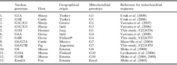
Samples of E. granulosus used in this study originated from various intermediate hosts from Eurasia (Italy, Greece, Turkey, Iraq, Finland and Estonia) and South-Central America (Argentina and Mexico) (Table 1). In addition, one adult specimen of E. multilocularis from Estonia (described by Moks et al. Reference Moks, Saarma and Valdmann2005) was used.
Details of samples used in this study

* The host imported to Finland, origin not documented.
DNA extraction, enzymatic amplification and sequencing
Total genomic DNA was isolated using the High Pure PCR Template Preparation Kit (Roche Diagnostics, Germany), following the manufacturer's protocols.
Partial sequences of the mitochondrial NADH dehydrogenase subunit 1 (nad1) gene were obtained for 3 previously uncharacterized samples to verify their respective mitochondrial genotypes (Table 1). Regions of nad1 were amplified and sequenced as described previously (Lavikainen et al. Reference Lavikainen, Lehtinen, Laaksonen, Ågren, Oksanen and Meri2006; Moks et al. Reference Moks, Jõgisalu, Saarma, Talvik, Järvis and Valdmann2006). Sequences were submitted to the GenBank database and are publicly available, with the Accession numbers being listed in Table 1. The mitochondrial sequences of other samples have been published previously, and the corresponding references can be found in Table 1. Thus, mitochondrial genotypes were known for all specimens used for further analyses.
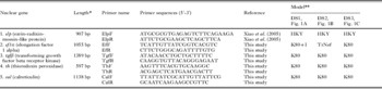
To amplify 4 individual nuclear genes elongation factor 1 alpha (ef1), transforming growth factor beta receptor kinase (tgf), thioredoxin peroxidase (th) and calreticulin (cal), we developed primers specific to Echinococcus (Table 2). For the nuclear gene coding for ezrin-radixin-moesin-like protein (elp), previously described primers were used (Xiao et al. Reference Xiao, Qiu, Nakao, Li, Yang, Chen, Schantz, Craig and Ito2005; Table 2). We sequenced the 5 nuclear genes, yielding an alignment of 5086 base-pairs (bp) in total (Table 2). Conditions for the polymerase chain reaction (PCR) for all nuclear gene fragments were as follows: 20–80 ng of purified genomic DNA and 5 pmol of primers were used in the PCRs, which were performed in a total volume of 20 μl containing 1×BD Advantage 2 PCR buffer, 1U BD Advantage 2 Polymerase mix (BD Biosciences, USA), 0·2 mm dNTP (Fermentas) and 1·5 mm MgCl2. Cycling was performed using a touchdown protocol: a 1 min-denaturing step at 95°C, followed by 10 cycles of 20 sec at 95°C, 30 sec at 59°C and 2 min at 68°C, but with the annealing temperature reduced by 0·5°C in each step (touchdown), followed by 27 cycles of 20 sec at 95°C, 30 sec at 55°C and 2 min at 68°C. The PCR was concluded with 5 min at 68°C. PCR products were purified with shrimp alkaline phosphatase/exonuclease I treatment (Fermentas). One unit of each enzyme was added to 10 μl of PCR reaction mix and incubated for 30 min at 37°C, followed by an inactivation at 80°C for 15 min.
Information about the 5 nuclear genes used in this study

* Sequence length of full nucleotide alignment (dataset 1) used in phylogenetic analysis. **Nucleotide substitution models, for a particular nuclear gene and dataset (DS), selected by Modeltest 3.7 according to the Bayesian Information Criterion.
DNA cycle sequencing was performed in a total volume of 10 μl using the Big Dye Terminator v.3.1 Cycle Sequencing Kit (Applied Biosystems), following the manufacturer's protocols. Initial denaturing was at 96°C for 1 min, followed by 27 cycles at 96°C for 10 sec (denaturation), 50°C for 15 sec (annealing) and 60°C for 4 min (extension). Sequences were resolved on the ABI3130xl automated DNA sequencer (Applied Biosystems). Both DNA strands were sequenced using the respective primers used in the primary amplification (Table 2). Consensus sequences were created using the Consed program (Gordon et al. Reference Gordon, Abajian and Green1998).
Sequences were aligned using Clustal W (Thompson et al. Reference Thompson, Higgins and Gibson1994) and checked and corrected in BioEdit (Hall, Reference Hall1999). All sequences were deposited in GenBank and are available under Accession numbers EU834886–EU834940.
Phylogenetic analysis
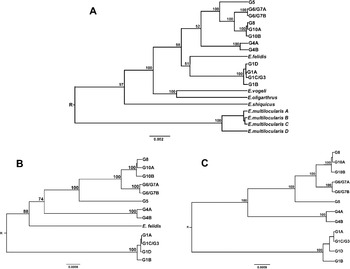
Phylogenies were constructed for 11 different sets of alignments (hereafter referred to as ‘datasets’): (1) a complete dataset of concatenated nucleotide sequences of 5 genes (5086 bp), each gene with its own best-fit nucleotide substitution model selected on the basis of Bayesian Information Criterion (BIC) scores from Modeltest 3.7 (Posada and Crandall, Reference Posada and Crandall1998). Concatenated nuclear sequences from this study were combined with all homologous sequences of Echinococcus spp. publicly available in GenBank: EF558359, EF558359, EF660053 (Hüttner et al. Reference Hüttner, Nakao, Torsten, Siefert, Boomker, Dinkel, Sako, Mackenstedt, Romig and Ito2008), AJ012663 (Brehm et al. Reference Brehm, Jensen, Frosch and Frosch1999), AB159141–AB159143 (Xiao et al. Reference Xiao, Qiu, Nakao, Li, Yang, Chen, Schantz, Craig and Ito2005) and AB306933–AB306940 (Nakao et al. unpublished observations). The resultant alignment included 20 taxa, comprising the entire genus Echinococcus; (2) a reduced dataset (13 taxa, only different genotypes of E. granulosus and E. felidis were included) of concatenated nucleotide sequences of 5 genes (5086 bp), each gene with its own best-fit nucleotide substitution model; (3) a reduced dataset (12 taxa, only different genotypes of E. granulosus were included) of concatenated nucleotide sequences of 5 genes (5086 bp), each gene with its own best-fit nucleotide substitution model; (4) a nucleotide dataset based on coding regions (3512 bp), each gene with its best-fit nucleotide substitution model; (5) a nucleotide dataset based on coding regions (3512 bp), each codon position with its best-fit nucleotide substitution model; (6) a complete dataset of concatenated amino acid sequences coded by 5 genes (1169 aa), using best-fit substitution model selected on the basis of BIC-criterion by ProtTest 1.4 (Abascal et al. Reference Abascal, Zardoya and Posada2005); (7) – (11) nucleotide sequences of each nuclear gene fragment separately, with its own best-fit nucleotide substitution model. All alignments are available from the authors upon request. Best-fit substitution models selected by Modeltest 3.7 are shown in Table 2 for datasets 1–3 (Fig. 1A–C), models for other datasets are not shown.
(A–C). Bayesian phylograms of the genus Echinococcus based on sequences of 5 nuclear genes. (A) Full set of taxa (20 taxa; dataset 1, see Materials and Methods section); (B) including only 13 taxa that belong to the E. granulosus/E. felidis clade according to (A) (dataset 2); (C) including taxa used in (B), but with E. felidis additionally removed (12 taxa; dataset 3). Posterior probability values are indicated above branches. R, root of the tree.

To determine whether the 5 different nuclear genes had significantly different signals, the incongruence-length difference (ILD) test (Farris et al. Reference Farris, Källersjö, Kluge and Bult1995) alias ‘partition homogeneity test’ implemented in PAUP*4.0b10 (Swofford, Reference Swofford1998) was used. The homogeneities between all 5 nuclear gene sequences were calculated in a pairwise manner with the branch-and-bound search option, using 1000 replicates.
Bayesian phylogenetic analysis was performed in BEAST 1.4.7 (Drummond and Rambaut, Reference Drummond and Rambaut2007) using the best-fit model of sequence evolution. Posterior distributions of parameters, including the tree, were estimated by Markov Chain Monte Carlo (MCMC) sampling. Following a discard burn-in of 300 000 MCMC steps, samples were drawn every 3000 steps over a total of 3 000 000 steps. Samples were verified for acceptable mixing and convergence to a stationary distribution using Tracer 1.4 (Rambaut and Drummond, Reference Rambaut and Drummond2007). A second phylogeny was calculated using MrBayes 3.1.2 (Ronquist and Huelsenbeck, Reference Ronquist and Huelsenbeck2003), with a best-fit model of sequence evolution (see Table 2). Searches were conducted with 4 simultaneous Markov Chains (3 heated and 1 cold chain) with over 2×10⁶ generations, sampling every 100 generations; burn-in trees were discarded, and a 50% majority rule consensus tree was calculated. This approach resulted in phylogenetic trees with identical topology and similar nodal support (data not shown) to that produced by the BEAST analysis. Given this similarity, results are only presented for the BEAST analysis, as it has the advantage of allowing the root of the phylogenetic tree to be identified from the data, without the need for an outgroup. Phylogenetic trees were displayed using TreeView 1.6.6 (Page, Reference Page1996) and FigTree 1.1.2 (http://tree.bio.ed.ac.uk/software/figtree).
Each gene was evaluated for the presence of paralogues using a BLAST-search against contigs assembled for E. multilocularis, provided via the Echinococcus Genome Project (http://www.sanger.ac.uk/Projects/Echinococcus). These contigs cover the vast majority of the genome (Dr M. Berriman, Echinococcus Genome Project Manager, personal communication).
RESULTS
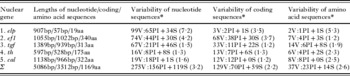
Phylogenetic analysis based on a concatenated nucleotide sequence alignment of 5 nuclear genes (5086 bp, dataset 1; see also Table 3 for sequence characteristics) indicated that E. multilocularis is a basal taxon in the Echinococcus genus and that all E. granulosus genotypes form a monophyletic clade (Fig. 1A). Moreover, the cervid genotypes, G8 and G10, were positioned as sister taxa and formed a single clade, whereas the camel-pig genotypes G6/G7 appeared as a sister taxon to G8/G10. Although the phylogenetic analysis based on the complete set of taxa yielded a tree with a well-resolved overall topology for Echinococcus (all major nodes received high posterior probability values), it fell short of providing reasonable support for the internal phylogeny of the E. granulosus/E. felidis cluster. Two nodes that describe phylogenetic relations between clades G1–G3/E. felidis and G4–G10 did receive posterior probability values of just slightly >50% (Fig. 1A). Attempts to resolve phylogenies with a wide range of taxa can produce nucleotide data matrices that contain homoplasic characters, i.e. distant taxa may share characters that are not common by descent, but have arisen due to convergence (Arendt and Reznik, Reference Arendt and Reznick2008). Homoplasy can lead to erroneous affinities between taxa and consequently result in a topology with low posterior probability values or even an erroneous topology (Naylor and Brown, Reference Naylor and Brown1998; Engstrom et al. Reference Engstrom, Shaffer and McCord2004). As it was not possible to resolve the phylogenetic relationships between clades G1–G3/E. felidis and G4–G10 by manipulating the gene sets included for analysis (data not shown), we hypothesized that homoplasy was most likely the reason why 2 nodes received only moderate posterior probability values when the full set of taxa was used (Fig. 1A). To test this hypothesis, we took a subset of the taxa whose relationship remained questionable and performed another analysis, which gave significantly better posterior probability values (Fig. 1B). When E. felidis was additionally removed, very high posterior probability values were obtained for clades G1–G3 and G4–G10 (Fig. 1C), confirming the topology of the phylogenetic tree constructed using the full set of taxa (see Fig. 1A). Based on the results of these 3 phylogenies, a summarized phylogeny of the genus Echinococcus is presented (Fig. 2B). Datasets 4–6 (i.e., coding regions and amino acid sequences) yielded phylogenetic trees with low nodal supports (data not shown). Datasets 7–11 (i.e., nucleotide alignments for each nuclear gene separately) yielded varying results (data not shown): dataset 7 (elp gene) provided a tree with a similar topology to that derived from dataset 1, with the notable difference that the positions of E. granulosus genotypes G1 and G4 were swapped. Dataset 8 (gene for ef1) also gave a different topology, namely, G4 grouped together with E. felidis to form a monophyletic clade, whereas the relative positions of G1 and G4 were in agreement with dataset 1. However, compared with dataset 1 (Fig. 1A), all of these differences were linked to lower posterior probability values and could thus be considered as less reliable. Alignments based on sequences of other genes (tgf, th and cal) yielded trees that were in agreement with dataset 1, albeit with moderate posterior probability values. Using the ILD test, no incongruence was found between the 5 nuclear genes. No paralogues were detected for any of the nuclear sequences.
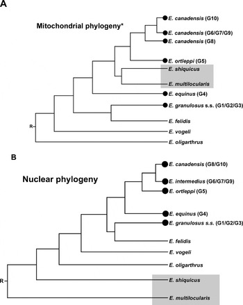
Comparison of phylogenies of the genus Echinococcus derived from mitochondrial and nuclear data. (A) Phylogenetic tree of the genus Echinococcus summarizing recent data based primarily on mitochondrial sequences. Relationships between G6–G10 are summarized from Nakao et al. (Reference Nakao, McManus, Schantz, Craig and Ito2007) and Moks et al. (Reference Moks, Jõgisalu, Valdmann and Saarma2008) ; the position of E. felidis is taken from Hüttner et al. (Reference Hüttner, Nakao, Torsten, Siefert, Boomker, Dinkel, Sako, Mackenstedt, Romig and Ito2008), who based their phylogeny on concatenated mitochondrial and nuclear data. (B) Phylogenetic tree of the genus Echinococcus from this study, based on 5 nuclear sequences. The position of E. multilocularis and E. shiquicus on both phylogenetic trees is indicated by a grey rectangle. Large dots represent the locations of E. granulosus genotypes on the trees.

Variability in the full nucleotide, coding and amino acid sequences of 5 nuclear genes

* Number of variable sites (V): parsimony informative sites (PI)+singletons (S), number of parsimony informative sites per 100 base-pairs of nucleotide sequences or 100 amino acid sequences are in parentheses.
Variation in nuclear genes
All nuclear genes were characterized in terms of the variability of their overall nucleotide sequences, coding sequences and inferred amino acid sequences. The main characteristics for each nuclear gene are presented in Table 3; here, we summarize these characteristics.
The fragment of the elp gene used in this study was composed of a long intron sequence and a relatively short protein-coding region. Although indels were found at the overall nucleotide level – one indel was 6 bp, two were 3 bp and three were 1 bp long – sequences could be aligned unambiguously. The nucleotide sequence of the elp gene fragment provided 65 parsimony informative sites for phylogenetic analysis, whereas its coding and amino acid sequences provided only 2 and 1, respectively.
The partial ef1 gene sequence was composed of 2, long coding sequences interrupted by a short intron. No indels were detected. The nucleotide sequence of the ef1 gene fragment provided 44 parsimony informative sites, whereas its coding and amino acid sequences provided 38 and 4 sites, respectively.
The partial tgf gene sequence was composed of 2 long coding sequences interrupted by a moderate sized intron. No indels were present. The nucleotide sequence of the tgf gene fragment provided 21 parsimony informative sites, whereas its coding and amino acid sequences provided 11 and 6 sites, respectively.
The partial th gene sequence was composed of 2 long coding sequences interrupted by a short intron. No indels were present. The nucleotide sequence of the th gene fragment provided 8 parsimony informative sites, whereas its coding and amino acid sequences provided 7 and 4 sites, respectively.
The partial cal gene sequence was composed of coding and intron sequences. No indels were found. The nucleotide sequence of the cal gene fragment provided 18 parsimony informative sites, whereas its coding and amino acid sequences provided 12 and 8 sites, respectively.
DISCUSSION
The taxonomy of the genus Echinococcus has remained controversial, despite numerous attempts to revise it (for a recent comprehensive review, see Thompson, Reference Thompson2008). Among the most difficult tasks has been to unravel relationships between E. granulosus and other species within the genus, and to explain the remarkable phenotypic and genetic variability between populations of E. granulosus from different host species and different geographical localities. Several of these populations exhibit distinct genetic and phenotypic characteristics that have prompted the distinction of separate populations into 10 genotypes within E. granulosus (note that the existence of genotype G9 is not well supported; see Introduction section). The most critical issues have been to determine whether these genotypes form a monophyletic entity and whether some of the genotypes can be recognized as separate species. Results from mitochondrial data have raised doubts about the monophyly of E. granulosus; the conclusion from a phylogeny based on the full mitochondrial genomic data was that E. granulosus genotypes do not form a monophyletic entity and are divided into 2 clusters, separated from each other by 2 other species of the genus, namely E. multilocularis and E. shiquicus (see Nakao et al. Reference Nakao, McManus, Schantz, Craig and Ito2007).
E. multilocularis, E. shiquicus, E. oligarthrus and E. vogeli
In the present study, we demonstrated that the phylogeny of Echinococcus inferred from nuclear genes (Figs 1A and 2B) differed significantly from the mitochondrial phylogeny (Fig. 2A). According to the nuclear phylogeny, E. multilocularis occupied a basal position in the genus with E. shiquicus positioned next to it. This result challenges the mitochondrial phylogeny, where both E. multilocularis and E. shiquicus were positioned between the clades G1–G3 and G4/G5–G10. The phylogenetic positions of E. vogeli and E. oligarthrus based on the nuclear data were also different from that of mitochondrial; whereas in the latter, the E. oligarthrus was shown as the basal taxon within the genus and E. vogeli was positioned next to it (cf. Nakao et al. Reference Nakao, McManus, Schantz, Craig and Ito2007), the nuclear phylogeny demonstrated that both of them were positioned as ingroup taxa between E. shiquicus and the E. granulosus/E. felidis clade.
E. granulosus sensu stricto (genotypes G1, G2, G3) and E. felidis
The results of this study, based on nuclear genes, are in agreement with previous mitochondrial data (Bowles et al. Reference Bowles, Blair and McManus1995; Thompson et al. Reference Thompson, Lymbery and Constantine1995; Obwaller et al. Reference Obwaller, Schneider, Walochnik, Gollackner, Deutz, Janitschke, Aspöck and Auer2004; Jenkins et al. Reference Jenkins, Romig and Thompson2005; Romig et al. Reference Romig, Dinkel and Mackenstedt2006; Nakao et al. Reference Nakao, McManus, Schantz, Craig and Ito2007; Busi et al. Reference Busi, Snabel, Varcasia, Garippa, Perrone, De Liberato and Amelio2007; Rinaldi et al. Reference Rinaldi, Maurelli, Capuano, Perugini, Veneziano and Cringoli2008) in placing E. granulosus genotypes G1, G2 and G3 as very closely related taxa. Moreover, these 3 genotypes have wide and overlapping geographical distributions and host spectra (Busi et al. Reference Busi, Snabel, Varcasia, Garippa, Perrone, De Liberato and Amelio2007; Varcasia et al. Reference Varcasia, Canu, Kogkos, Pipia, Scala, Garippa and Seimenis2007; Rinaldi et al. Reference Rinaldi, Maurelli, Capuano, Perugini, Veneziano and Cringoli2008). Thus, genotypes G1–G3 can be regarded as a single taxonomic unit: E. granulosus sensu stricto (see Nakao et al. Reference Nakao, McManus, Schantz, Craig and Ito2007; Thompson Reference Thompson2008).
Although E. felidis was positioned close to the genotypes G1–G3 in the study by Hüttner et al. (Reference Hüttner, Nakao, Torsten, Siefert, Boomker, Dinkel, Sako, Mackenstedt, Romig and Ito2008) and in the current study, its position in both cases was clearly as a sister taxon to G1–G3. Considering that contemporary samples (parasite eggs in lion faeces) yielded the same DNA sequences as obtained from a 40-year-old museum specimen of E. felidis (see Hüttner et al. Reference Hüttner, Nakao, Torsten, Siefert, Boomker, Dinkel, Sako, Mackenstedt, Romig and Ito2008), it is very likely that E. felidis is a distinct genetic entity. Based on all available data, it can be concluded that E. felidis shared a recent common ancestor with G1–G3, but has evolved into a separate phylogenetic entity and most likely also into a distinct species. However, further genetic and phenotypic studies of G1–G3 and E. felidis from a range of host species in Africa are required to test whether E. felidis deserves the status of a distinct species.
E. equinus (genotype G4) and E. ortleppi (genotype G5)
Extensive mitochondrial (Le et al. Reference Le, Pearson, Blair, Dai, Zhang and McManus2002; Nakao et al. Reference Nakao, McManus, Schantz, Craig and Ito2007) and nuclear analyses (this study) all place E. granulosus genotypes G4 and G5 on the phylogenetic tree as taxa separate from others and also from each other. Williams and Sweatman (Reference Williams and Sweatman1963) originally proposed that E. granulosus of horse origin (nowadays called genotype G4) can be regarded as the subspecies E. g. equinus. However, while subspecies can potentially interbreed, no evidence of any segregating mechanism between the sympatric E. g. equinus and E. g. granulosus has been identified that could explain their separation. Thus, the subspecies status of E. g. equinus was rejected (Rausch, Reference Rausch1967). More recent investigations of E. granulosus have shown that the parasite of horse origin is biologically and genetically distinct from that of sheep origin. Considering all of the data, Thompson and McManus (Reference Thompson and McManus2002) proposed that the equine genotype G4 should be regarded as a distinct species, E. equinus. Genotype G5, which occurs in Europe, Asia, Africa and South America, appears to be adapted to cattle as its principal intermediate host. Adult stages of the cattle isolate were originally described as E. ortleppi by Lopez-Neyra and Soler Planas (Reference López-Neyra and Soler Planas1943). Besides clear genetic differentiation, it is also phenotypically and developmentally distinct from E. granulosus of sheep or horse origin (Thompson et al. Reference Thompson, Kumaratilake and Eckert1984; Bowles et al. Reference Bowles, Blair and McManus1995; Thompson and McManus, Reference Thompson and McManus2002; Thompson, Reference Thompson2008). Thus, the phylogenetic analyses of this study support earlier propositions that these genotypes should be considered as separate species, i.e., genotype G4 as E. equinus and G5 as E. ortleppi.
E. intermedius (genotypes G6, G7, G9) and E. canadensis (genotypes G8 and G10)
A recent study, based on mitochondrial data by Moks et al. (Reference Moks, Jõgisalu, Valdmann and Saarma2008), demonstrated that the E. granulosus camel-pig genotypes G6/G7 formed a subgroup together with the cervid genotype G10, whereas the other cervid genotype G8 was shown to be a sister taxon to G6/G7–G10. Moks et al. (Reference Moks, Jõgisalu, Valdmann and Saarma2008) concluded that their study provided support for combining G6/G7, G8 and G10 into a single species which, in terms of priority, should be E. canadensis (see also Nakao et al. Reference Nakao, McManus, Schantz, Craig and Ito2007; Hüttner et al. Reference Hüttner, Nakao, Torsten, Siefert, Boomker, Dinkel, Sako, Mackenstedt, Romig and Ito2008; Thompson, Reference Thompson2008). The results of the current study, based on nuclear data, provided a different view on the phylogeny of the camel-pig and cervid genotypes; there was no separation of the cervid genotypes G8 and G10 into distinct clades. Genotype G8 was separated from Finnish G10A by 2 point mutations, whereas the same number of mutations also separated G10A and Estonian G10B isolates. Thus, the variation within G10 was the same as that between genotypes G8 and G10. By contrast, the cervid (G8/G10A/G10B) and camel-pig (G6/G7) genotypes were separated from each other by as many as 12 point mutations. Unlike the mitochondrial phylogeny, where genotypes G6/G7 were positioned in a clade together with G10 (but separate from G8), the current nuclear phylogeny demonstrated that both the cervid genotypes, G8 and G10, form a single clade separate from the camel-pig genotypes G6/G7. Future studies should include nuclear sequence data of G8 and G10 isolates from North America to evaluate whether the genetic differentiation within Echinococcus from cervid hosts is associated with their geographical distribution.
From a taxonomic perspective, it has been suggested that the camel, pig and cervid genotypes (G6–G10) might belong to a single species (Thompson and McManus, Reference Thompson, McManus, Eckert, Gemmell, Meslin and Pawlowski2001; Lavikainen et al. Reference Lavikainen, Lehtinen, Laaksonen, Ågren, Oksanen and Meri2006; Moks et al. Reference Moks, Jõgisalu, Valdmann and Saarma2008), named E. canadensis (see Nakao et al. Reference Nakao, McManus, Schantz, Craig and Ito2007; Hüttner et al. Reference Hüttner, Nakao, Torsten, Siefert, Boomker, Dinkel, Sako, Mackenstedt, Romig and Ito2008). However, Thompson (Reference Thompson2008) pointed out that this may be an oversimplification of the evidence, as cervid and camel-pig isolates of Echinococcus differ from each other both in terms of their mitochondrial sequences and their geographical distributions (Lavikainen et al. Reference Lavikainen, Lehtinen, Meri, Hirvelä-Koski and Meri2003, Reference Lavikainen, Lehtinen, Laaksonen, Ågren, Oksanen and Meri2006; Thompson et al. Reference Thompson, Boxell, Ralston, Constantine, Hobbs, Shury and Olson2006; Moks et al. Reference Moks, Jõgisalu, Valdmann and Saarma2008). Moreover, Thompson (Reference Thompson2008) suggested that the camel-pig genotypes G6/G7 should be regarded as a single species, E. intermedius, after Lopez-Neyra and Soler Planas (Reference López-Neyra and Soler Planas1943). Data derived from the nuclear genes in this study support the proposal of uniting cervid genotypes G8/G10 into a single species, E. canadensis, distinct from G6/G7, which could be regarded as E. intermedius. However, molecular data alone are not sufficient to revise the taxonomy. Thus, further biological and morphological observations, including, above all, evaluation of morphological characters and infection experiments, are required for the final revision.
Nuclear genes sequences, their variability and contribution to the phylogeny
The most informative markers at an ‘overall’ nucleotide level (dataset 1) were the elp and ef1 sequences, which provided 7·2 and 4·2 parsimony informative sites per 100 bp, and a total of 65 and 44 parsimony informative sites, respectively. Alignments of the other 3 genes provided far less parsimony informative sites. All 5 nuclear genes used in this study were useful for phylogenetic analysis, as alignments based on both nucleotide and amino acid characters were unequivocal for all genes. Although introns were present in nuclear genes and indels were detected for the elp gene, these did not interfere with the alignments. Moreover, no paralogues were found for these 5 genes, and pairwise ILD tests of the nuclear genes showed no incongruence between them. However, different genes provided different signals in relation to the position of G4 (in the phylogeny derived from the elp gene, the positions of G4 and G1 were swapped, compared with the concatenated dataset phylogeny) and E. felidis (the ef1 gene phylogeny united G4 and E. felidis into a monophyletic clade). Nevertheless, tree topologies of all other genes were in agreement with the concatenated dataset 1. As manipulations with sequence sets did not improve the phylogeny, slight disagreement between the phylogenies from different genes was most likely not the reason why 2 nodes (within the clade that represents the genotypes of E. granulosus) on the dataset 1-phylogeny received low support. Rather, homoplasy (i.e., highly divergent taxa sharing characters that are common not due to descent) may have contributed to the low support for these 2 nodes. Indeed, when distant taxa, such as E. multilocularis, E. shiquicus, E. oligarthrus and E. vogeli, were removed from the dataset, these 2 nodes in the phylogeny of E. granulosus received much higher support; also, when E. felidis was additionally removed from the dataset, the internal phylogeny between different genotypes of E. granulosus was fully resolved, demonstrating clearly the basal position of G1 with respect to G4. Non-coding sequences were of significance for inferring the phylogeny of Echinococcus, since neither nucleotide alignments based solely on coding sequences (3512 bp, datasets 4 and 5) nor alignment based on amino acids (1169 aa, dataset 6) were able to yield phylogeny with high posterior probability values. The reason for this lies in the variability found in the different datasets. Compared with the ‘full’ nucleotide alignment (dataset 1), which provided a total of 156 parsimony informative sites, alignments based on either coding or amino acid sequences (datasets 4–6) yielded far fewer parsimony informative sites. Although phylogenies constructed separately for each gene (datasets 7–11) were generally in agreement with the topology based on full nucleotide alignment, none of them was able to provide phylogenetic resolution equivalent to that based on the full nucleotide dataset.
Is a nuclear phylogeny preferable to a mitochondrial one as the basis for interpreting the evolutionary history of the genus Echinococcus?
This study has clearly shown that a nuclear phylogeny of the genus Echinococcus differed significantly from previous mitochondrial phylogenies (though a certain level of agreement was also found). This finding presents us with an important question: which phylogeny represents more accurately the true evolutionary history of Echinococcus? Here, we argue in favour of the nuclear phylogeny for several reasons. Firstly, the divergence of maternal lineages is a stochastic event, as mutations accumulate in the mitochondrial genome at random, except at those sites that are under selection. Once a new mutation becomes fixed in a population, the new mitochondrial lineage separates from the ancestral one. From this point onwards, mutations continue to fix progressively in an independent manner in both the new and ancestral mitochondrial lineages. In the absence of recombination, mitochondrial lineages continue to diverge once they have separated. However, this does not mean that these separate mitochondrial lineages have necessarily become separate biological entities. Separation of mitochondrial lineages without accompanying reproductive isolation has no biological meaning from the viewpoint of true separation of populations (eventually into distinct species), but merely reflects the course of evolution of maternal lineages. Although mutations also accumulate into the nuclear genome at random (except at those sites under selection), nuclear genes undergo recombination and if there is no barrier for gene-flow among parasites, then nuclear genes that are subjected to recombination during meiosis do not show any separation into genetically distinct populations (for recent reviews, see Funk and Omland, Reference Funk and Omland2003; Hurst and Jiggins, Reference Hurst and Jiggins2005; Ballard and Rand, Reference Ballard and Rand2005). Thus, even if the separation of the E. granulosus genotypes G8 and G10 were clearly established, as seen in the mitochondrial data reported by Moks et al. (Reference Moks, Jõgisalu, Valdmann and Saarma2008), the nuclear data in this study showed that parasites with the mtDNA genotypes G8 and G10 have not actually separated into distinct biological entities.
Although, traditionally, mitochondrial gene sequences have been preferred over nuclear ones in phylogenetic studies, the current study illustrates the main limitation of using mitochondrial data as the sole marker for inferring phylogenies, i.e., the evolutionary history of a maternal lineage may differ from that at the species level or higher. Given the significant inconsistencies between phylogenies from mitochondrial and nuclear data, we suggest that both marker systems are used to evaluate the genetic diversity of the genus Echinococcus in future studies, whereas nuclear genes should be used when evaluating the phylogenetic positions of new Echinococcus isolates.
Despite not always being the best marker for phylogenetic studies, mitochondrial sequences are undoubtedly of great diagnostic value, particularly when samples are degraded. As cells contain relatively few copies of each nuclear gene but often thousands of mitochondrial genomes, it is clear that analysis of mitochondrial gene sequence data may often represent the best way to reveal the identity of a parasite species or genotype/strain.
We thank Jaak Kinnas, Tuuli Reisberg, Maris Roht, and John Davison for their generous help. This work was supported by grants from the Estonian Ministry of Education and Sciences (target financing grant TLOOM0122), the Estonian Science Foundation (grant 7040), the Estonian Ministry of Agriculture (grant LLOOM08015), the European Union through the European Regional Development Fund (Center of Excellence FIBIR) and from the Environmental Investment Centre.