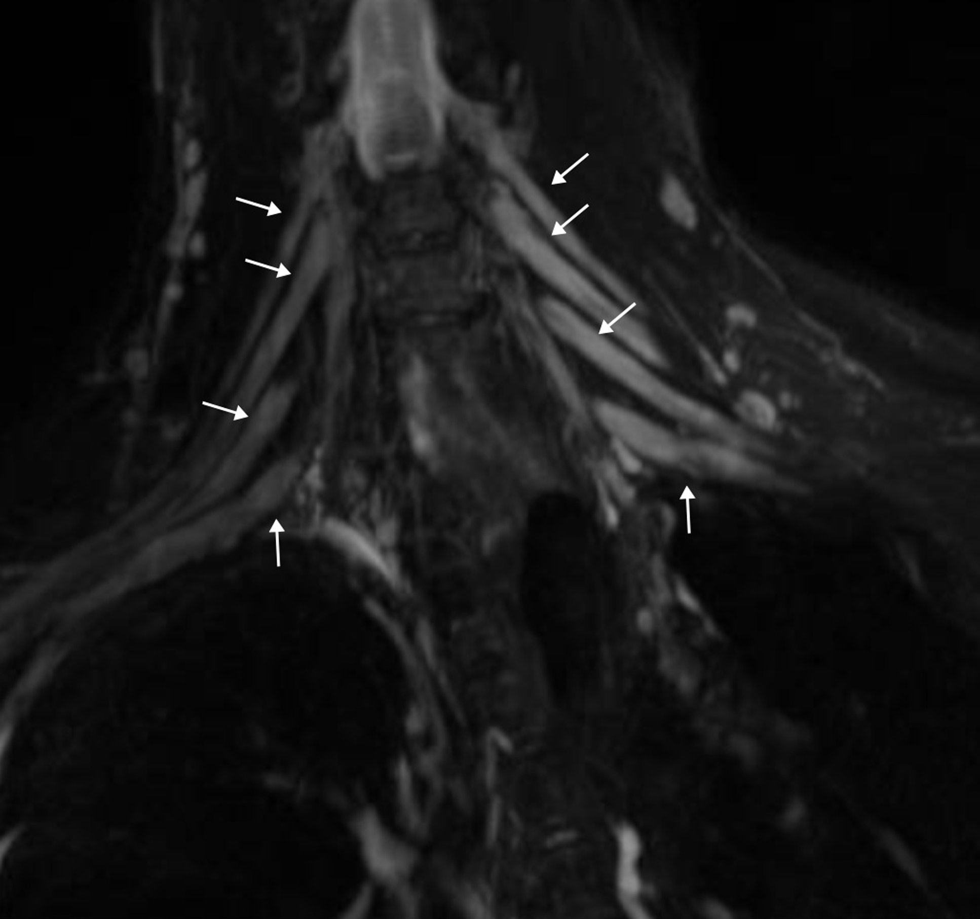
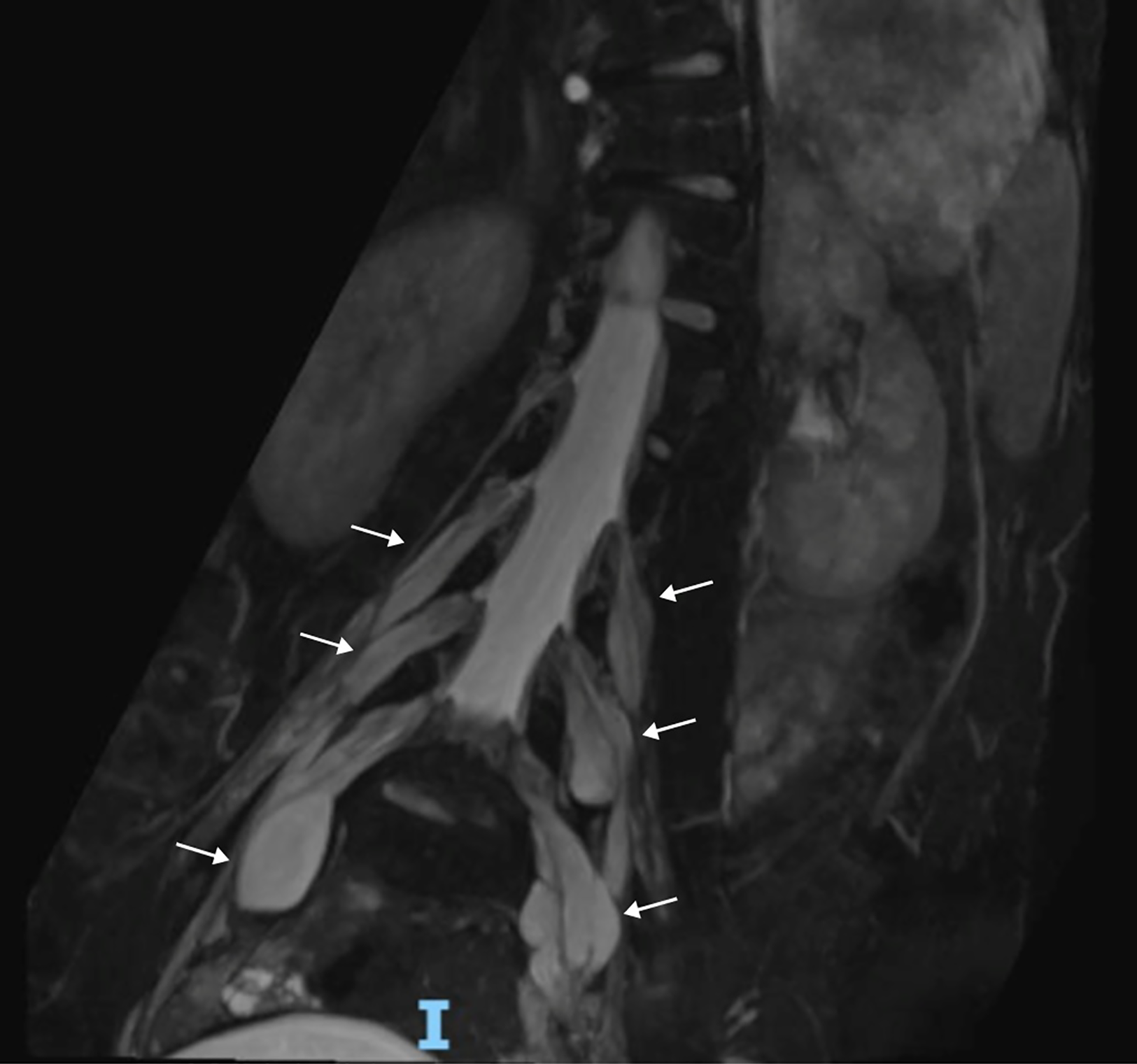
A 17-year-old girl with Charcot–Marie–Tooth disease type 1D (CMT1D) presented with progressive distal weakness since early childhood. At 7 years, targeted gene sequencing identified a pathogenic variant in EGR2 (Early Growth Response 2) gene. From infancy, the clinical picture was characterized by bilateral ptosis, ophthalmoparesis, anisocoria, facial weakness and dysphagia. Gait difficulty emerged at 3 years and worsened steadily, leading to loss of autonomous ambulation. Last neurological examination at 16 years of age showed facial weakness, distal muscle atrophy and finger and wrist flexion contractures; she was able to walk only with support with a waddling and steppage gait and presented a positive Romberg sign. Systemic features included severe left-convex thoracolumbar rotoscoliosis, respiratory impairment and orthostatic peripheral cyanosis. To assess the burden of demyelination along the peripheral nervous system, whole-spine MRI was performed, comprising sagittal T1-and T2-weighted turbo spin-echo and coronal short tau inversion recovery (STIR) sequences, the latter reformatted as maximum intensity projection (MIP). The cervical nerve roots of the brachial plexus showed bilateral, symmetric thickening (Figure 1). At the lumbosacral level, marked bilateral enlargement of the nerve roots was observed from L3 to L5, with a tortuous, multilobulated, “onion-bulb”-like morphology (Figure 2). Spinal cord signal, conus position (L1-L2) and adjacent metameres were normal. Nerve-root hypertrophy reflects chronic demyelination, with repeated demyelination–remyelination cycles producing Schwann-cell proliferation and concentric “onion-bulb” formations, documented histologically in EGR2-related neuropathies. Reference Cavalcanti, Santos, Couto, Rocha, Freitas and Nascimento1,Reference Campagnolo, Taioli and Cacciavillani2 The pattern of root enlargement is diagnostically informative. Symmetric, diffuse thickening along the longitudinal course of the roots suggests a hereditary demyelinating neuropathy, although overlap with typical Chronic Inflammatory Demyelinating Polyneuropathy (CIDP) has been reported, while asymmetric, fusiform enlargement is specific for the multifocal acquired demyelinating sensory and motor variant and other focal CIDP phenotypes. Reference Yoshii, Shibuya and Yokota3,Reference Tozza, Cassano, Erra, Muto, Habetswallner and Manganelli4 The distinction is clinically important, since hereditary forms misclassified as CIDP may lead to unnecessary immunomodulatory therapy, Reference Campagnolo, Taioli and Cacciavillani2,Reference Echaniz-Laguna, Cauquil and Chanson5 even though the clinical history and disease course should help clinicians in the differential diagnosis process. More than 100 genes have been associated with CMT. Reference Cavalcanti, Santos, Couto, Rocha, Freitas and Nascimento1 EGR2 encodes a zinc finger transcription factor essential for peripheral nerve myelination; Reference Echaniz-Laguna, Cauquil and Chanson5 pathogenic variants impair Schwann cell differentiation and produce a clinical spectrum ranging from early-onset severe variants such as congenital hypomyelinating neuropathy type 1 (MIM #605253) and Dejerine–Sottas neuropathy (MIM #145900) to late-onset moderate variants such as CMT1D (MIM #607678). Reference Echaniz-Laguna, Cauquil and Chanson5 Our patient’s phenotype resembles the EGR2-related profile described in a recent Brazilian cohort and in earlier reports. Reference Cavalcanti, Santos, Couto, Rocha, Freitas and Nascimento1,Reference Echaniz-Laguna, Cauquil and Chanson5,Reference Pareyson, Taroni and Botti6 Recurring features include early-childhood distal weakness, scoliosis, extraocular involvement, respiratory compromise and dysphagia. Reference Cavalcanti, Santos, Couto, Rocha, Freitas and Nascimento1,Reference Echaniz-Laguna, Cauquil and Chanson5,Reference Pareyson, Taroni and Botti6 Recognition of this clinical signature should prompt targeted molecular testing and multidisciplinary surveillance of pulmonary and orthopedic comorbidities. Nerve-root involvement in EGR2-related CMT1D remains rarely described. The symmetric, diffuse “onion-bulb” pattern shown here is a recognizable imaging pattern that, paired with the multisystem clinical pheontype, can direct the diagnostic workup toward targeted genetic testing while avoiding empirical immunotherapy. Whole-spine MRI thus provides a noninvasive structural readout of demyelinating burden, informative at diagnosis and suited for longitudinal follow-up.
Brachial plexus nerve-root hypertrophy. Coronal STIR MRI sequence with MIP reformat shows thickening of the cervical nerve roots of the brachial plexus (arrows), reflecting hypertrophic changes associated with chronic demyelination. STIR = short tau inversion recovery; MIP = maximum intensity projection.

Lumbosacral nerve-root hypertrophy. Coronal STIR MRI sequence with MIP reformat highlights marked bilateral enlargement of the lumbosacral nerve roots from L3 to L5 (arrows), with tortuous multilobulated “onion-bulb”-like morphology reflecting hypertrophic changes associated with chronic demyelination. STIR = short tau inversion recovery; MIP = maximum intensity projection.

Acknowledgements
The authors thank the patient and her family for their participation and for granting consent to publish the clinical and neuroimaging material.
Author contributions
F.F.C.: study concept and design; acquisition and interpretation of clinical and neuroimaging data; drafting and revising the manuscript for intellectual content.
D.D.: study concept and design; acquisition and interpretation of clinical and neuroimaging data; drafting and revising the manuscript for intellectual content; study supervision; taking full responsibility for the data and their integrity.
M.C., J.S. and F.N.: acquisition of clinical data; critical revision of the manuscript for intellectual content.
M.C.R.E.: acquisition and interpretation of neuroradiologic data; critical revision of the manuscript for intellectual content.
A.D.: study concept and design; acquisition and interpretation of clinical and neuroimaging data; drafting and revising the manuscript for intellectual content.
Funding statement
This work was supported by the Italian Ministry of Health with current research funds.
Competing interests
The authors report no disclosures relevant to the manuscript.
Ethical standards
The study was conducted in accordance with the Declaration of Helsinki and was approved for publication by our institution, Bambino Gesù Children’s Hospital, Rome, on April 26, 2026, with number RAP2026-0001. Written informed consent to disclose the clinical and neuroimaging material was obtained from the patient’s legal guardians. No identifying patient information is included in the manuscript or figures.