Noonan syndrome is an autosomal dominant disorder that was first described by Jacqueline Noonan. Reference Noonan1 Its estimated prevalence is 1:1000 to 1:2500 live births. Reference Mendez and Opitz2 It is characterised by short stature, craniofacial dysmorphism, CHD, skeletal abnormalities, developmental delay, haematologic disorder, and other abnormalities. Reference Roberts, Allanson, Tartaglia and Gelb3 Noonan syndrome arises from gene mutations that are related to the RAS mitogen-activated protein kinase signal transduction pathway. Reference Baban, Olivini and Lepri4,Reference Calcagni, Adorisio and Martinelli5 The gene mutations responsible for Noonan syndrome have been identified, and include the following: RAS family of GTPase proteins (KRAS, NRAS, RIT1, RRAS); RAS signal function modulators (PTPN11, SOS1, SOS2, CBL, RASA2, SHOC2); and downstream signal transducers (RAF1, BRAF). Reference Tekendo-Ngongang, Agenbag, Bope, Esterhuizen and Wonkam6 PTPN11 gene mutations are the most frequent mutations and have been detected in 50% of Noonan syndrome patients. Reference Yu, Luk and Leung7 The second and third most common causative genes are reported to be SOS1 and RIT1, respectively. Reference Roberts, Allanson, Tartaglia and Gelb3 RIT1 is a relatively newly identified gene that causes Noonan syndrome. Reference Aoki, Niihori and Banjo8
Heart disease is one of the most important problems in the long-term follow-up of Noonan syndrome. The incidence of cardiovascular disease in Noonan syndrome is reported to be 82–90%. Reference Prendiville, Gauvreau and Tworog-Dube9 The most common cardiovascular diseases in Noonan syndrome are pulmonary valve stenosis, hypertrophic cardiomyopathy, and atrial septal defect. Reference Roberts, Allanson, Tartaglia and Gelb3 Some patients with severe pulmonary valve stenosis need treatment to avoid negative impacts on their quality of life. Hypertrophic cardiomyopathy, which is detected in 20% of patients with Noonan syndrome, also affects patient survival. Reference Gelb, Roberts and Tartaglia10
Recently, genetic analysis to diagnose Noonan syndrome has made it possible to perform genotype–phenotype correlation analyses. For example, in the cardiovascular field, patients with the PTPN11 mutation often have pulmonary valve stenosis, and hypertrophic cardiomyopathy is more prevalent in RAF1 mutation-positive patients. Reference Tartaglia, Kalidas and Shaw11,Reference Pandit, Sarkozy and Pennacchio12 In terms of the cardiovascular system, there are few reports on prognosis and follow-up for patients with Noonan syndrome. In addition, the spectrum of cardiac morphology is not well understood in patients with newly identified gene mutations such as RIT1. Reference Aoki, Niihori and Banjo8
The aim of this study was to examine clinical manifestations in patients with Noonan syndrome, with a focus on cardiovascular disease, including an analysis of newly identified gene mutations. We also investigated the prognosis and follow-up of cardiovascular issues in Noonan syndrome.
Materials and methods
Patients
We retrospectively reviewed the medical records at Kanagawa Children’s Medical Center. Patients who were clinically and genetically diagnosed with Noonan syndrome by medical geneticists in accordance with van der Burgt’s criteria were included in the study. Reference van der Burgt13 Inclusion criteria required that a patient had undergone at least one echocardiogram. Patients suspected of having a clinically related but alternate diagnosis, such as cardio-facio-cutaneous syndrome or Costello syndrome, were excluded. The study was approved by the Ethics Committee of Kanagawa Children’s Medical Center (approval no. 2101-7) and the Ethics Committee of the Tohoku University School of Medicine (approval no. 2015-1-222, 2021-1-271).
Study design
Clinical characteristics, including age, sex, body weight, height, type of cardiovascular anomaly, echocardiogram data, information about cardiac surgery, age at first visit, and responsible mutations, were retrospectively collected from the medical records. Patients were generally followed by medical geneticists. When cardiac complications were followed at our hospital, echocardiogram results were evaluated by paediatric cardiologists. Echocardiography data, such as left ventricular end-diastolic diameter, posterior wall thickness at end-diastole, posterior wall thickness at end-systole, interventricular septal thickness at end-diastole, interventricular septal thickness at end-systole, fractional shorting, ejection fraction, pulmonary artery velocity, left ventricular outflow tract stenosis, and atrial septal defect dimensions, were extracted from the medical records.
Mutation analysis
Genomic DNA was extracted from peripheral blood samples of the patients and their family members by standard procedures. Mutation screening was performed by direct sequencing of exons and their flanking regions in the responsible genes, including PTPN11, KRAS, BRAF, MAP2K1/2, and HRAS. Reference Narumi, Aoki and Niihori14 More recently, we used either a targeted next-generation sequencing panel of 41 genes responsible for Noonan syndrome and related diseases, the TruSight One Sequencing Panel (Illumina, Inc, San Diego, CA, United States of America), or whole–exome sequencing. Reference Umeki, Niihori and Abe15,Reference Nishimura, Murakami, Hayashi, Sato and Kurosawa16 Variants identified by targeted sequencing were confirmed by Sanger sequencing.
Statistical analysis
Data are presented as the mean ± standard error of the mean for independent experiments. Statistical analysis was performed between two groups using an unpaired two-tailed Student’s t-test or an unpaired t-test with Welch’s correction. Genotype–phenotype correlations were performed using a Fisher’s exact test. p < 0.05 was considered to be statistically significant.
Results
There were 44 patients with genetically confirmed Noonan syndrome. One patient did not have cardiac data because the patient did not visit our hospital for follow-up. We analysed the data from 43 patients, and the patient information for 43 patients is presented in Table S1. The median age of the cohort was 11 years (interquartile range: 6.8–20.2 years), and 58% were male. Cardiovascular disease was present in 67.4% of patients in our cohort. The prevalence of CHD was noted in 60% of males and 72% of females. Two patients died. One of these patients was a PTPN11-positive patient who died from leukaemia, and the other was a RAF1-positive patient with hypertrophic obstructive cardiomyopathy who died suddenly.
The description of identified mutations, the prevalence of CHD, and details are listed in Table 1. The most common responsible mutation was found in PTPN11 (25 patients). The second and third most common causative genes were SOS1 (6 patients) and RIT1 (5 patients), respectively. The remaining mutations were found in RAF1, KRAS, SHOC2, and BRAF. Pulmonary valve stenosis was prevalent in PTPN11 (8 patients), SOS1 (4 patients), and RIT1 (4 patients). Hypertrophic cardiomyopathy was found in two of three patients with mutations in RAF1 whereas 5 of 25 patients with PTPN11 gene mutations showed hypertrophic cardiomyopathy. Eight patients with a PTPN11 mutation had an atrial septum defect. The remaining cardiovascular abnormalities were large ventricular septal defects, mitral valve regurgitation, and left coronary stenosis. Ten patients needed catheter intervention or surgery for cardiovascular abnormalities.
Cardiovascular disease in Noonan syndrome patients (n = 43) by gene mutation

ASD, atrial septum defect; HCM, hypertrophic cardiomyopathy; PS, pulmonary valve stenosis; VSD, ventricular septum defect.
Clinical features focused on cardiovascular disease were examined in Noonan syndrome patients with PTPN11, which was the most frequent responsible gene mutation. We compared PTPN11 mutation-positive patients and PTPN11 mutation-negative patients (Table S2). There were no significant differences in the cardiovascular events and type of CHD in patients with or without PTPN11 gene mutations.
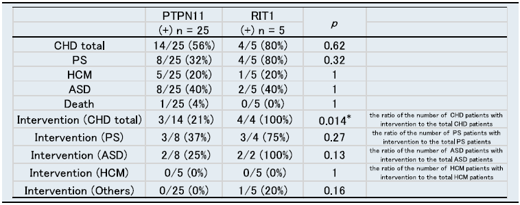
Next, we focused on the newly identified RIT1 gene mutation. We analysed the phenotype difference in the cardiovascular system between the PTPN11 gene (the most prevalent mutation) and the RIT1 gene (Table 2). The prevalence of cardiovascular phenotypes was not significantly different between the two groups. The proportion of RIT1 mutation-positive patients who underwent intervention due to cardiovascular disease was significantly higher than that of patients with PTPN11 gene mutations. A total of four patients with RIT1 gene mutation underwent cardiac intervention. Two of these patients underwent intervention for pulmonary valve stenosis. One underwent surgery for pulmonary valve stenosis and arterial septal defect, and the other underwent surgery for atrial and ventricular septum defect closure. However, the prevalence of RIT1 gene-positive patients who underwent intervention (percutaneous pulmonary valvuloplasty or surgery) for pulmonary stenosis was not significantly different from that of patients with PTPN11 gene mutations.
Prevalence of cardiovascular abnormalities compared with PTPN11 gene mutations and RIT1 gene mutations.

ASD, atrial septum defect; HCM, hypertrophic cardiomyopathy.
PS, pulmonary valve stenosis. *p < 0.05.
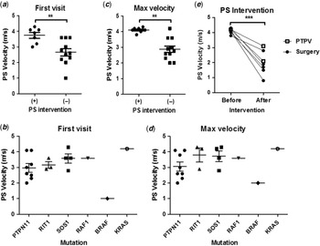
There were 20 patients who had pulmonary valve stenosis in our cohort (Table 1). Two of 20 patients did not have echocardiogram records that included transpulmonary flow velocity. Thus, we analysed the data of 18 patients. The pulmonary stenotic site in 18 patients was valvular (9 patients), supravalvular (5 patients), valvular+supravalvular (2 patients), peripheral pulmonary stenosis (1 patient), and subvalvular+supravalvular (1 patient). First, we analysed the pulmonary flow velocity in Noonan patients at first visit. Patients who underwent any intervention for pulmonary valve stenosis exhibited a significantly higher pulmonary flow velocity than those who did not undergo surgery or percutaneous pulmonary valvuloplasty (Table S3, Fig 1A). All patients who underwent surgery for pulmonary valve stenosis showed a pulmonary flow velocity of more than 3.0 m/s. There was no difference in the pulmonary flow velocity at first visit between PTPN11-positive patients and PTPN11-negative patients (Table S4). Figure 1B shows the pulmonary flow velocity among the patients with PTPN11, RIT1, SOS1, and other gene mutations at first visit. We lacked transpulmonary flow velocity data on one of the four patients with RIT1 mutation because this patient underwent surgery for pulmonary valve stenosis at another hospital. Next, we analysed the maximum pulmonary flow velocity during follow-up. Patients in the intervention group showed a greater pulmonary flow velocity than those in the non-intervention group (Table S3, Fig 1C). There was no difference in the maximum pulmonary flow velocity between PTPN11-positive patients and PTPN11-negative patients (Table S4). Figure 1D shows the maximum pulmonary flow velocity during cardiac follow-up among the patients with PTPN11, RIT1, SOS1, and other gene mutations. After intervention, the pulmonary flow velocity was significantly decreased (Fig 1E). Two of seven patients underwent percutaneous pulmonary valvuloplasty. There were no patients who needed re-intervention for residual pulmonary stenosis after surgery or percutaneous pulmonary valvuloplasty. None of the seven patients exhibited significant pulmonary insufficiency after intervention. Among these, three of these patients had no pulmonary valve regurgitation. Three of them had trivial pulmonary valve regurgitation (two surgical patients and one percutaneous pulmonary valvuloplasty patient). Finally, one of them had mild pulmonary valve regurgitation (a surgical patient).
Pulmonary valve stenosis in patients with Noonan syndrome (a) Pulmonary flow velocity at the first visit to our institution by PS intervention status. (b) Pulmonary flow velocity at the first visit to our institution among patients with PTPN11, RIT1, SOS1, and other gene mutations. (c) Maximum pulmonary flow velocity during cardiac follow-up by PS intervention status. (d) Maximum pulmonary flow velocity during cardiac follow-up among patients with PTPN11, RIT1, SOS1, and other gene mutations. (e) Decrease in pulmonary flow velocity by surgical intervention.
PS, pulmonary valve stenosis; PTPV, percutaneous pulmonary valvuloplasty; NS, not significant; **p < 0.01, ***p < 0.001.

We also analysed patients with atrial septal defect. There were 10 patients who had echocardiogram records available that included the maximum diameter of atrial septal defect during follow-up. Four of 10 patients underwent intervention for atrial septal defect. Three of four patients underwent surgery for both atrial septal defect and pulmonary valve stenosis at the same time. The one remaining patient underwent surgery for atrial septal defect and ventricular septum defects. All four patients who underwent surgery for atrial septum defect closure had large atrial septum defect with significant left-to-right shunt. Patients who underwent surgery showed a significantly larger atrial septal defect than patients who did not undergo surgery (surgery;11.1 ± 1.7 mm (n = 4) versus no surgery; 4.3 ± 1.0 mm (n = 6), p < 0.05). There was no difference in atrial septal defect diameter between PTPN11-positive patients and PTPN11-negative patients.
We also analysed cases of Noonan syndrome with hypertrophic cardiomyopathy. There were 12 patients with hypertrophic cardiomyopathy in our Noonan syndrome cohort (Table 1). Nine of 12 patients had data available that included the left ventricular wall diameter and left ventricular dimensions. All patients with recorded data had preserved left ventricular contraction. One patient who had RAF1 gene mutations exhibited progressive left ventricular wall thickening with left ventricular outflow tract stenosis. The patient underwent resection of the left ventricular outflow tract at 4 years old, and left ventricular outflow tract stenosis and left ventricular wall thickness subsequently improved. We have followed the patient for 3 years after intervention. The improvement of the left ventricular outflow tract and hypertrophy did not last. Two years after the surgery, a follow-up echocardiogram showed progressive left ventricular outflow tract stenosis.
Discussion
This Noonan syndrome cohort of Japanese patients provides new insight into the clinical manifestations of RIT1 mutations and prognosis for patients with related cardiovascular problems. Patients with RIT1 gene mutations underwent a high number of interventions to treat cardiovascular problems, especially for pulmonary valve stenosis. Our cardiac follow-up data showed that patients who have more than moderate pulmonary valve stenosis at the first visit have a higher likelihood of intervention.
A previous report suggested that 50% of Noonan syndrome cases are caused by a missense or gain-of-function mutation in PTPN11. Reference Roberts, Allanson, Tartaglia and Gelb3 Mutations in SOS1 and RAF1 genes were detected in 10% of patients with Noonan syndrome, respectively. Reference Roberts, Allanson, Tartaglia and Gelb3 The percentage of gene mutations in our cohort is similar to the prevalence described by others. Reference Roberts, Allanson, Tartaglia and Gelb3
For cardiac anomalies, the prevalence of pulmonary valve stenosis was 40% in PTPN11 mutation-positive patients in the current cohort. Previous research from Asian countries such as Hong Kong, South India, and Japan reported the prevalence of pulmonary valve stenosis with PTPN11 gene mutations to be 55.9, 35.3, and 52%, respectively. Reference Yu, Luk and Leung7,Reference Athota, Bhat and Nampoothiri17,Reference Shoji, Ida and Niihori18 Prendiville et al. reported that, at Boston Children’s Hospital, 71.9% of patients with PTPN11 gene mutations had pulmonary valve stenosis. Reference Prendiville, Gauvreau and Tworog-Dube9 A report from European countries showed that 77.0% of PTPN11 mutation-positive patients had pulmonary valve stenosis. Reference Shaw, Kalidas, Crosby, Jeffery and Patton19 The prevalence of pulmonary valve stenosis in our study was similar to that of other Asian countries. Patients with SOS1 mutations in Noonan syndrome were reported to have a high incidence of pulmonary valve stenosis. Reference Zenker, Horn and Wieczorek20 In our study, five of six patients with SOS1 gene mutations also exhibited pulmonary stenosis, and this result is consistent with previous reports.
A previous study demonstrated that patients with the RAF1 gene mutation showed a relatively good survival rate, with 94% survival after 20 years. Reference Calcagni, Limongelli and D’Ambrosio21 However, patients with Noonan syndrome with hypertrophic cardiomyopathy have a higher risk profile at presentation compared with other children with hypertrophic cardiomyopathy. Reference Wilkinson, Lowe and Salbert22 Colan et al. reported that patients with onset of the hypertrophic cardiomyopathy phenotype in infancy had a poor prognosis, with a 5-year survival of 65%. Reference Colan, Lipshultz and Lowe23 Two of the three RAF1 mutation-positive patients at our institution exhibited early-onset hypertrophic cardiomyopathy, and they did not have a good prognosis. One of these patients died and the other patient underwent left ventricular muscle resection because of left ventricular outflow tract stenosis.
We evaluated the cardiovascular system in RIT1-positive patients, and the analysis showed that, compared with PTPN11 gene mutations, patients who have RIT1 gene mutations have a higher incidence of intervention for cardiovascular disease. Yaoita et al. clearly showed a higher prevalence of cardiovascular disease, especially hypertrophic cardiomyopathy (7/13 patients), pulmonary valve stenosis (9/13 patients) and septal defect (8/13 patients) in patients with RIT1 mutations. Reference Yaoita, Niihori and Mizuno24 Kouz et al. also reported high rates of hypertrophic cardiomyopathy (14/33 patients), pulmonary valve stenosis (26/33 patients), and septal defect (13/33 patients), and found that 61% of patients with RIT1-positive mutations underwent either catheter intervention or surgery for cardiovascular disease. Reference Calcagni, Baban and Lepri25,Reference Kouz, Lissewski and Spranger26 This previous report supports our finding in Noonan syndrome with RIT1-positive mutations. The high percentage of RIT1-positive patients who undergo cardiac interventions might reflect the high incidence of CHD and high prevalence of pulmonary valve stenosis and septal defects.
A previous report provided information on the natural history of Noonan syndrome. Reference Colquitt and Noonan27 Mild pulmonary valve stenosis in Noonan syndrome patients was not progressive, and severe pulmonary valve stenosis sometimes needed therapeutic intervention. Our study included 11 patients with a pulmonary flow velocity of more than 3.0 m/s at the first visit, and 7 of these 11 patients underwent surgery or percutaneous pulmonary valvuloplasty for pulmonary valve stenosis. Our data suggest that patients with more-than-moderate pulmonary valve stenosis at the first visit will require intervention for pulmonary valve stenosis and careful follow-up in case of cardiac surgery. Pulmonary valve stenosis in Noonan syndrome is often observed with dysplastic valve leaflets, and Noonan syndrome patients undergoing percutaneous balloon pulmonary valvuloplasty have a high re-intervention rate (65%) for residual pulmonary valve stenosis. Reference Prendiville, Gauvreau and Tworog-Dube9,Reference Holzmann, Tibby and Rosenthal28 However, surgical valvotomy appears to be a highly successful treatment. Reference Linglart and Gelb29 In this study, two of seven patients underwent percutaneous pulmonary valvuloplasty rather than surgery for pulmonary valve stenosis. Although none of the seven patients required further intervention for their pulmonary valve, longer-term follow-up is required to check for re-stenosis. In our patients who underwent surgery for atrial septal defect, atrial septal defect and other cardiac anomalies (pulmonary valve stenosis and ventricular septum defect) coexisted, and all of these patients underwent surgery. For atrial septal defect alone, transcatheter closure of the atrial septal defect may be the treatment of choice. Reference Mangovski, Farkić and Jovović30
This study has some limitations. This is the experience of a single institution. RAF1, RIT1, and SOS1 had a small sample size due to the small overall sample size. Because the current study was a retrospective cohort, we could not systematically collect data from patients of the same age, and we could not retrieve enough cardiac data from patients with normal heart structure. Thus, it is difficult to show the cardiac prognosis in detail. Prospective cohorts may be difficult to perform because the age of the diagnosis is different and the diagnosis of Noonan syndrome, including genetic testing, is sometimes a sensitive issue.
In conclusion, we have described the burden of cardiovascular problems in a Japanese cohort with genetically diagnosed Noonan syndrome. We presented new information on the cardiac features of these patients with a genotype–phenotype analysis. We found that echocardiogram data at the first visit predicted intervention or surgery for cardiovascular disease. These findings suggest that genetic information can help provide a clinical prognosis in Noonan syndrome patients with heart disease, and that genotype-based follow-up should be performed for these patients in the future.
Supplementary material
To view supplementary material for this article, please visit https://doi.org/10.1017/S104795112200124X
Author contributions
YI and KK conceived of the project and designed the research. YI wrote the manuscript. HK, TI, SK, SO, KK, YA, and SY provided manuscript review. HU and KK provided assistance with the concept, design, and manuscript review.
Funding statement
This study was supported by Research on Rare and Intractable Diseases from the Ministry of Health, Labour and Welfare, Japan and AMED under grants to Y.A. (JP18ek0109241 and, JP21ek0109470), and AMED under a grant to K.K. (Initiative on Rare and Undiagnosed Diseases [IRUD] 21ek0109549).
Conflicts of interests
None.
Consent for publication
All authors approved this publication.