Surgical site infections (SSI) following implantation of prostheses are a growing problem. The total number of implant infections that occur after total hip and knee arthroplasty in the United States is projected to increase from 22,000 in 2009 to 65,000 in 2020 and with healthcare costs exceeding $1 billion per year by 2020.Reference Kurtz, Lau, Watson, Schmier and Parvizi 1
Microorganisms responsible for prosthesis infections are commonly transported in surgical theater air,Reference Edmiston, Sinski, Seabrook, Simons and Goheen 2 – Reference Wan, Chung and Tang 4 whereby they may enter incision sites during surgical procedures and cause SSI.Reference Hamilton and Jamieson 5 – Reference Ritter 7 The primary source of these airborne microorganisms is the people in the operating room, such that the number of people, door openings, and room traffic all increase the quantity of airborne colony-forming units (CFU).Reference Andersson, Bergh, Karlsson, Eriksson and Nilsson 8 – Reference Smith, Raphael, Maltenfort, Honsawek, Dolan and Younkins 10
Current methods for reducing airborne CFU in the operating room during prosthesis implantation include the use of surgical gowning with exhaust hoods, laminar flow air systems, ultraviolet light, and limiting room traffic.Reference Evans 11 , Reference Knobben, van Horn, van der Mei and Busscher 12 These methods largely attempt to alter the environment of the entire operating room in a manner that is remote from the surgical site. A more efficient tactic may be to control the airborne environment immediately surrounding the incision site, where the presence of CFU has a greater likelihood of depositing onto the prosthesis.Reference Knobben, van Horn, van der Mei and Busscher 12 – Reference Duhaime, Bonner, McGowan, Schut, Sutton and Plotkin 14 However, to our knowledge, no literature has indicated whether the density of airborne CFU measured at incision sites is related to increased incidence of SSI. Thus, the purpose of this study was to evaluate whether airborne CFU density at the incision site during operations is related to the incidence of incisional or implant infections after placing the prostheses.
METHODS
Design and Patients
This prospective, randomized clinical trial was conducted from January 19, 2012, through April 2, 2015, at the Michael E. DeBakey Veterans Affairs Medical Center using a parallel groups design. Inclusion criteria were patients at least 18 years old undergoing primary or revision total hip arthroplasty, posterior lumbar fusion or cervical laminectomy with instrumentation, or lower extremity vascular bypass with prosthetic or bovine graft. Exclusion criteria included history of prosthesis infection or presence of an active infection. Patients were evaluated for eligibility by a research associate who was not involved in their care. The institutional review board of Baylor College of Medicine approved this study, and all patients provided signed informed consent.
Patients were randomly assigned in a 1:1 ratio to either the intervention or control group using a block randomization scheme with mixed block sizes (randomly ranging from 2 to 8) to attain balanced group sizes for strata defined both by procedure type and by primary or revision implantation. Randomization lists were created using NQuery Advisor, version 7.0 (Statistical Solutions), before recruitment began by a biostatistician not involved in the care or assessment of patients. Group assignment was made after consent by the research associate. A sample size of 150 per group provided a power of 80% to detect a statistically significant (α≤.05) 3% difference in SSI rate on the basis of a historical infection rate of 6% in the facility where operations were performed. The SSI difference between groups was the planned comparison that had the lowest power, so a sample size designed to obtain adequate power for this test would have adequate power for all other planned comparisons, including the primary hypothesis of an association between CFU density and SSI.
Interventions
All implants, perioperative care, and surgical procedures were determined by the surgeons’ and facility’s routine practices and were unaffected by this investigation. Total hip arthroplasty procedures used the posterolateral approach, spinal procedures used the posterior approach, and vascular procedures used the anterior approach. All procedures were performed in operating rooms designed with conventional air handling systems compliant with the Centers for Disease Control and Prevention guidelines.Reference Sehulster and Chinn 15 Skin antiseptic varied by procedure type but was the same for both study groups: total hip arthroplasty used chlorhexidine gluconate and isopropyl alcohol solution (ChloraPrep; Becton, Dickinson); spinal procedures used iodine povacrylex and isopropyl alcohol solution (DuraPrep; 3M); vascular bypass used betadine (Purdue Products) in year 1 and ChloraPrep in years 2 and 3. All 294 patients received cefazolin or vancomycin for antibiotic prophylaxis, either as the only antibiotic (146 [49.7%]), both together (19 [6.5%]), or combined with another antibiotic (129 [43.9%]), intraoperatively and for 1 to 4 days postoperatively with weight-based dosing per the surgeons’ routine practices. Per routine care in the study facility, silver-impregnated dressings were used after all hip procedures whereas plain gauze dressings were used after all spine and vascular cases. Surgical staff wore hoods in all hip procedures but not in spinal and vascular cases. The operating surgeon and surgical team could not be masked to the intervention but were involved in neither the assignment to groups nor the evaluation of SSI.
Intervention group
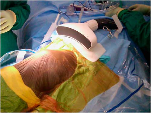
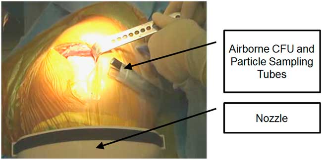
In the intervention group, all operations were performed with protection against airborne CFU and particulate at the incision site using the Air Barrier System device (ABS; Nimbic Systems). The ABS, which is cleared for sale by the US Food and Drug Administration, creates a positive pressure, nonturbulent clean air envelope that shields open surgical wounds from airborne CFU and particulate during an operation. It passes ambient air through a filter that removes 99.997% of particulate sized 0.3 µm or larger in diameter and issues it through a hose to a nozzle attached on top of the incision drape within a few inches of the incision (Figure 1). The clean air passes over the surgical field and effectively shields an area of 16 cm wide ×53 cm long.
The Air Barrier System passes ambient air through a filter that removes 99.997% of particulate sized 0.3 µm or larger in diameter and issues it through a hose to a nozzle attached on top of the incision drape within a few inches of the incision.

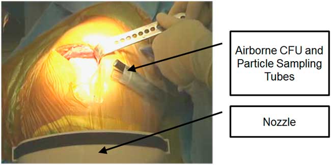
Airborne particulate and CFU were collected simultaneously through two 244-cm sterile tubes affixed at the incision site (Figure 2). A particle counter (Lasair 310; Particle Measuring Systems) drew air through 1 tube at 28.3 L/min and enumerated the particulate in the airstream. Air was drawn at 28.3 L/min through the second tube into an Andersen-style bioaerosol collection device (EMS E6; Environmental Monitoring Systems) and onto agar plates (tryptic soy agar, 5% sheep’s blood) that were exchanged every 10 minutes throughout the procedures. Control plates were exposed momentarily to ambient air as a negative control for handling and processing contamination. The plates were incubated at 35ºC for 36 hours by an independent laboratory (Hygeia Labs), and viable organisms were identified and reported as CFU per cubic meter.
Airborne particulate and colony-forming units (CFU) were collected simultaneously through two 244-cm sterile tubes affixed at the incision site.
Control group
The control group operations were conducted according to usual practice without the ABS. Airborne particulate and CFU densities were captured at 10-minute intervals exactly as described in the intervention group.
Data Collection
Patient characteristics recorded from the medical record included gender, ethnicity, age, and body mass index (calculated as weight in kilograms divided by height in meters squared). Comorbid indicators for increased SSI risk included American Society of Anesthesiologists score, medical history (cardiopulmonary disease, neurological condition, cancer, and diabetes mellitus), currently smoking, malnutrition, and history of SSI in previous year.
Operation- and procedure-related data included surgeon, operating room, antibiotic prophylaxis, number of people in the operating room at the beginning of each 10-minute interval, operative time, revision or primary procedure, and procedure type. During the operations, data were collected by a research technician who was responsible for counting the number of people and exposing and replacing the agar plates every 10 minutes.
Particulate densities were reported as particles per cubic meter at several size thresholds (0.3 µm, 0.5 µm, 1 µm, 5 µm, and ≥10 µm). Because operative time ranged widely (64 to 523 minutes), only particulate and CFU densities during the first 100 minutes of each case were analyzed. Counts obtained during the first 100 minutes did not differ significantly from counts over the full duration of each case (P≥.88).
While residing in the hospital, postoperative patients were followed up daily by a research nurse who did not get involved in patients’ care and who daily reviewed the medical records for documentation of the presence of symptoms and signs of SSI that had been established by a physician. The research nurse followed up with the patients by telephone at 1 month after the surgical procedure and then every month for 12 months so as to document the presence or absence of SSI. Diagnosis of SSI was made by a physician who was masked to the patients’ group assignment and not involved in the patients’ care by evaluating medical records using the Centers for Disease Control and Prevention criteria,Reference Mangram, Horan, Pearson, Silver and Jarvis 16 which categorize SSI into superficial incisional, deep incisional, and organ/space/implant levels. Superficial and deep incisional infections were defined and reported as “incisional” infections, whereas organ/space/implant level infections were defined and reported as “implant” infections.
Statistical Analyses
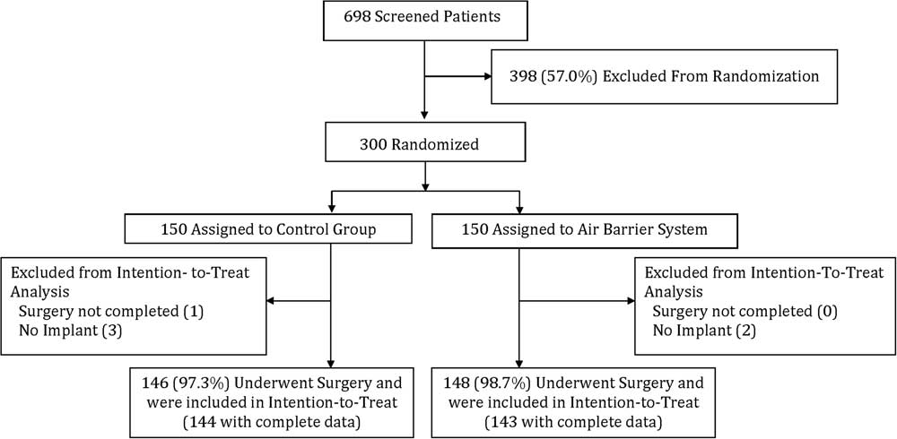
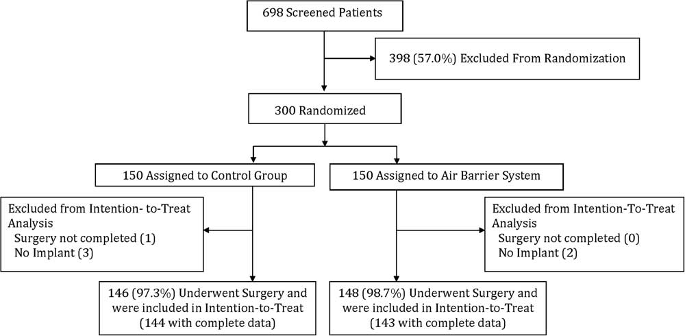
All analyses were intention to treat, which included 294 (98%) of the 300 randomized participants. Six randomized patients (2 intervention, 4 control) were excluded from analysis because they did not receive an implant and thus were not at risk for implant infection (Figure 3). One of the intervention patients was missing CFU and particulate data and was excluded from the analyses requiring those data. An additional 6 participants (4 intervention, 2 control) were missing particulate data only and were excluded from the analyses requiring the particulate data. These participants were included in all other analyses, including incidence of infection.
Flow chart of randomized controlled trial examining association of airborne microorganisms in the operating room with implant infections.
The relation of SSI to CFU density was tested using penalized maximum likelihood logistic regression.Reference Firth 17 , Reference Heinze and Schemper 18 The associations of group and covariates with particulate and CFU density were evaluated using negative binomial regression and zero-inflated negative binomial regression, respectively, with robust (Huber-White) standard errors.Reference Long 19 Group differences for incidence of infection were tested using Fisher exact tests. All data were analyzed using Stata, version 13 (StataCorp).
RESULTS
Two participants in the intervention group and 2 participants in the control group died before the 12-month follow up for reasons unrelated to their operations. These 4 participants were included in all analyses; none had been diagnosed with SSI at the time of death. All other participants (290 [98.6%] of 294) completed 1-year follow-up.
Patient and Operation Characteristics
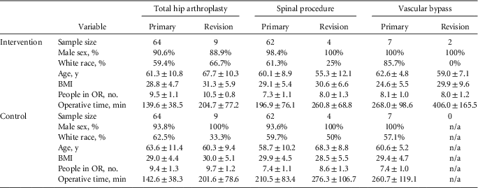
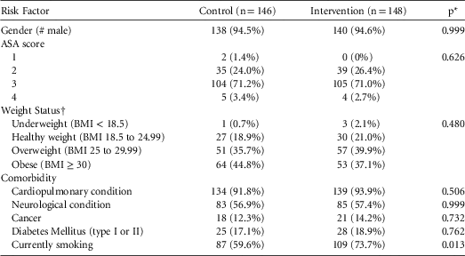
The intervention and control groups did not differ significantly with respect to patient or operation characteristics (P>.05, Table 1). The operations were performed by 9 different surgeons in 9 different operating rooms, distributed equally across the study groups. Most of the patients were men, consistent with the Veterans Affairs population, and older than 55 years. The intervention group had significantly more smokers (73.7%) than the control group (59.6%, P=.013). There were no other significant group differences for other SSI risk factors and comorbidities (P>.30, Appendix Table 1).
Descriptive Statistics by Group, Procedure Type, and Primary or Revision Operation

NOTE. Data are mean±SD unless otherwise indicated. BMI, body mass index (calculated as weight in kilograms divided by height in meters squared); n/a, not applicable; OR, operating room.
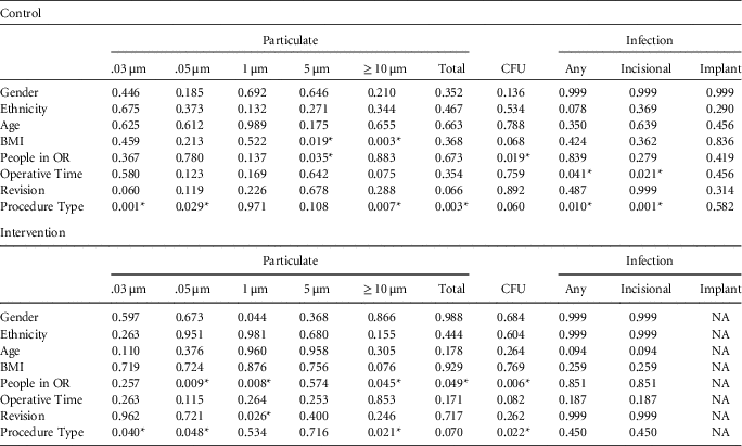
None of the patient characteristics were significantly associated with the particulate density, CFU density, or infection (P>.10) (Appendix Table 2). The number of people in the operating room was positively related to CFU density in both the intervention (P=.006) and control (P=.019) groups.
CFU and Particulate Densities
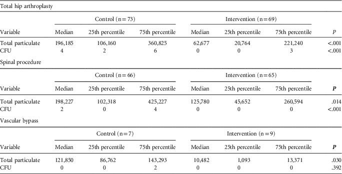
Across all procedure types, the intervention group using ABS had significantly (P<.001) lower particulate and CFU densities compared with the control group. In the intervention group, 68.1% of the 10-min surgical intervals collected during the study had 0 CFU/m3, compared with 46.1% in the control group (P<.001). Within each procedure type, the intervention group had lower median particulate density compared with the control group (Table 2).
Densities of Total Particulate and Colony-Forming Units (CFU) per Cubic Meter by Study Group and Procedure Type

NOTE. The subsample sizes (n) are the number of patients with particulate data in each subgroup; 2 patients in the control group (both hip arthroplasty cases) and 4 patients in the intervention group (1 spinal and 3 hip arthroplasty cases) were missing particulate data. One patient in the intervention group (hip arthroplasty) was missing both particulate and CFU data. P values are for comparisons of median values between control and intervention groups at each respective particulate size or CFU density.
CFU and Particulate Densities and Infection
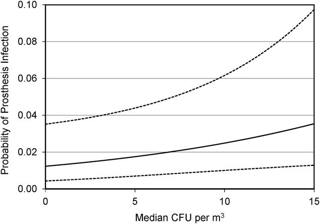
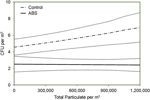
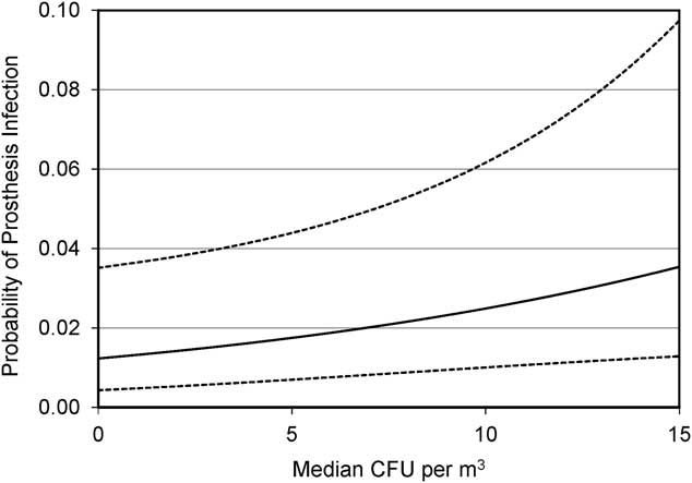
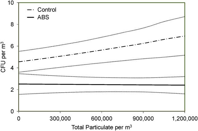
CFU density at incision sites was significantly related to incidence of implant infection (P=.021), but not of incisional infection (P=.687). Every 10 CFU/m3 increase in median CFU density approximately doubled the probability of implant infection (Figure 4). CFU density was positively related to total particulate density (P<.001) in the control group, indicating that airborne particle counts may be used as a proxy for ambient CFU density. No association between particle density and CFU could be demonstrated in the intervention group because 68.1% of the 10-minute intervals had 0 CFU and provided no significant variation in CFU levels (Figure 5).
Graph of density of colony-forming units (CFU) at incision sites and probability of implant infection (P=.021). Dashed lines represent 95% CIs.
Graph of particulate density and colony-forming units (CFU). Dotted lines represent 95% CIs. ABS, Air Barrier System.
No patients in the intervention group (0% [95% CI, 0%–2.5%]) had implant infections, whereas 4 patients in the control group (2.7% [0.7%–7.0%]) had implant infections at 8, 11, 27, and 58 days after operation, respectively. Pathogens included methicillin-resistant Staphylococcus aureus in 1 case, methicillin-sensitive S. aureus in 2 cases, and multiple organisms (Proteus mirabilis, Providencia stuartii, and Enterococcus faecalis) in 1 case. Species identification for the airborne CFU samples was too complex and was beyond the scope and purpose of the study.
Of the 148 participants in the intervention group, 4 (2.7% [95% CI, 0.7%–6.9%]) had incisional infections at 3, 8, 32, and 32 days after operation (methicillin-resistant S. aureus in 1 case, methicillin-sensitive S. aureus in 1 case, Pseudomonas in 1 case, and no cultures in 1 case). Three participants in the control group (2.0% [0.4%–6.0%]) had incisional infections at 4, 14, and 16 days after operation (methicillin-resistant S. aureus in 1 case, no growth in 1 case, and no cultures in 1 case).
The incidence of incisional infection differed by procedure type (P=.025): total hip arthroplasty (0.7%, 1/146), spinal procedure (3.0%, 4/132), and vascular bypass (12.5%, 2/16). In contrast, procedure type was not significantly associated with incidence of implant infection (P=.700): total hip arthroplasty 2.0% (3/146), spinal procedure 0.8% (1/132), and vascular bypass 0% (0/16).
DISCUSSION
To our knowledge, this is the first randomized, controlled study of the relationship between the levels of intraoperative airborne CFU adjacent to incision sites during prosthesis implantation and incidence of SSI. Analyses included CFU counts collected during 2,822 10-minute intervals, and a total of 11,039 CFU were cultured from the air during 470 hours of operative time. Our findings demonstrate that these microorganisms are significant sources of implant infections, and that reducing them within the surgical field also reduced the incidence of implant infection.
While the Centers for Disease Control and Prevention acknowledges the importance of minimizing the presence of airborne CFU in operating rooms, it provides no guidelines for acceptable limits during surgery.Reference Sehulster and Chinn 15 Our study confirms previous reports indicating that the levels of airborne CFU in the operating room are dependent on the number of people present.Reference Stocks, Self, Thompson, Adame and O’Connor 3 , Reference Andersson, Bergh, Karlsson, Eriksson and Nilsson 8 , Reference Stocks, O’Connor, Self, Marcek and Thompson 20 However, efforts to limit room traffic may not be effective in practice, as described in studies wherein operating room doors were opened 50–60 times during orthopedic joint replacement.Reference Stocks, Self, Thompson, Adame and O’Connor 3 , Reference Panahi, Stroh, Casper, Parvizi and Austin 21
A prior study demonstrated that the ABS reduced CFU levels adjacent to incision sites,Reference Stocks, O’Connor, Self, Marcek and Thompson 20 and we employed the ABS to create a significant difference in CFU levels between study groups so that our hypothesis could be tested. In our study, there were 4 implant infections of 146 procedures in the control group and no implant infections of 148 procedures in the intervention group. At an estimated additional expense of $40,000 per infection in the control group, the 4 infections cost $160,000 to treat whereas the cost of using the ABS in those procedures would be approximately $35,000. In this scenario, prevention of a single infection would have been cost-saving. The positioning of the ABS was generally acceptable to the surgeons and did not present any technical challenges to performing the procedures.
Our study indicates that airborne CFU entering incisions during operations is a likely source of contamination leading to implant infections. Incisional infections, however, may result from a broader set of intraoperative and postsurgical factors, as suggested by studies reporting that skin preparation methods were associated with incisional but not organ/space SSI,Reference Darouiche, Wall and Itani 22 , Reference Morrison, Chen, Taneja, Kucukdurmaz, Rothman and Parvizi 23 which would explain why we found no difference in rates of incisional infection. These findings indicate that appropriate control of the operating room atmosphere may complement other techniques, such as skin antisepsis, to provide a more comprehensive infection prevention strategy while implanting prostheses.
Our study design provides advantages over previous research regarding airborne contamination in the operating room. Contradictory findings in prior reports may be attributed to many factors, including nonrandomized comparisons of important risk factors and often lack of appropriate controls.Reference Andersson, Petzold, Bergh, Karlsson, Eriksson and Nilsson 24 – Reference Miner, Losina, Katz, Fossel and Platt 26 We used a stratified block random assignment that resulted in balance between the groups on relevant measured and unmeasured patient and surgical factors such that other factors could not have caused any observed differences in infection rates. The diagnosis of infection in our study was made by a physician who was masked to group assignment to minimize bias. This study, however, had some limitations. The presence of the ABS in the operating room may have affected the behavior of operating surgeons. However, the operating surgeons did not subsequently assess the presence, or lack thereof, of the occurrence of SSI. Also, the observed SSI incidence in the control group was lower than the historical average, which limited the power to detect statistically significant differences in SSI between groups. A larger randomized controlled, multicenter clinical trial would have a greater power in assessing the clinical efficacy of the ABS in reducing prosthesis-related infections.
In conclusion, our results indicate that CFU contamination of air at the incision site is a risk factor for implant but not incisional infections. CFU contamination is related to the particulate density in the air at the incision site, and both CFU and particulate density are a function of the number of people in the operating room. Limiting airborne CFU contamination at the incision site can be expected to lower implant infection risk.
ACKNOWLEDGMENTS
Financial support. National Institute of General Medical Sciences of the National Institutes of Health (award R44GM095005). Nimbic Systems provided assistance with particle count and CFU data acquisition.
Potential conflicts of interest. R.O.D. reports that he received grants from the National Institutes of Health during the conduct of the study; and that he was the director of the annual educational meetings of Multidisciplinary Alliance Against Device-Related Infection, which received education funds from Nimbic Systems, the manufacturer of the ABS. D.P.O. reports that he received consultant fees paid for statistical analyses from the National Institute of General Medical Sciences of the National Institutes of Health (award R44GM095005). All other authors report no conflicts of interest relevant to this article.
Disclaimer: The content is solely the responsibility of the authors and does not necessarily represent the official views of the National Institutes of Health.
Prevalence of risk factors and comorbidities by study group.

P-values for associations of study factors with mean particulate density, mean CFU density, and infection rates by study group.