INTRODUCTION
Cognitive impairment is common following stroke, as most first-ever ischemic stroke patients demonstrate cognitive impairment on neuropsychological tests within the first few weeks following stroke (Jaillard, Naegele, Trabucco-Miguel, LeBas, & Hommel, Reference Jaillard, Naegele, Trabucco-Miguel, LeBas and Hommel2009; Nys, van Zandvoort, de Kort, Jansen, de Haan, & Kappelle, Reference Nys, van Zandvoort, de Kort, Jansen, de Haan and Kappelle2007; Rasquin, Verhey, Lousberg, Winkens, & Lodder, Reference Rasquin, Verhey, Lousberg, Winkens and Lodder2002). The use of fast clinical rating scales in estimating cognition has been questioned, both early and later on after the stroke-onset (Bour, Rasquin, Boreas, Limburg, & Verhey, Reference Bour, Rasquin, Boreas, Limburg and Verhey2010; Jokinen et al., Reference Jokinen, Melkas, Ylikoski, Pohjasvaara, Kaste, Erkinjuntti and Hietanen2015; Kauranen et al., Reference Kauranen, Laari, Turunen, Mustanoja, Baumann and Poutiainen2014; Nys, van Zandvoort, de Kort, Jansen, et al., Reference Nys, van Zandvoort, de Kort, van der Worp, Jansen, Algra and Kappelle2005).
On more detailed neuropsychological examinations, some cognitive recovery is found within 6 months after the stroke. For example, one study found that half of the patients were impaired at baseline and improved in all cognitive domains evaluated (executive function, verbal memory, visual memory, perception/construction, language, and reasoning (Nys, van Zandvoort, de Kort, van der Worp, Jansen, Algra, & Kapelle, Reference Nys, van Zandvoort, de Kort, van der Worp, Jansen, Algra and Kappelle2005). Rasquin and colleagues (2002) found that most patients remained stable (on cognitive flexibility, memory, simple speed, and overall cognitive functioning), and up to half improved. Comparing separate patient groups (assessed <1, 1–3, and >3 months after stroke) Hurford, Charidimou, Fox, Cipolotti, and Werring (Reference Hurford, Charidimou, Fox, Cipolotti and Werring2013) saw decreasing impairment rates in executive function, speed/attention, and perceptual skills but there was no change in verbal or visual memory. In our previous study, we saw overall cognitive improvement between baseline and 6 months in all patients, but we did not analyze the data on a domain-specific basis (Kauranen et al., Reference Kauranen, Laari, Turunen, Melkas, Mustanoja, Baumann and Poutiainen2015).
Between the subacute and chronic strokes, Hochstenbach, den Otter, and Mulder (Reference Hochstenbach, den Otter and Mulder2003) found improvement in all of the cognitive domains (orientation, memory, attention, visuospatial, visuoconstructive, language, and arithmetic abilities) between 2 months and 2 years, but noted, that a small subset of patients accounted for the domain-specific improvements demonstrated in the sample, as the majority of patients remained cognitively stable or deteriorated. Sachdev, Brodaty, Valenzuela, Lorenz, and Koschera (Reference Sachdev, Brodaty, Valenzuela, Lorenz and Koschera2004) found, in addition, that in an elderly sample performance either remained stable (executive function, speed, attention, visual memory, language, reasoning) or deteriorated (verbal memory and visuoconstructive) between index assessment (at 3–6 months) and 1 year after stroke.
After 6 months of the stroke-onset, there is limited information as to whether the domain-specific cognitive functioning changes. Between 6 months and 2 years, overall cognitive performance did not improve significantly in our earlier study (Kauranen et al., Reference Kauranen, Laari, Turunen, Melkas, Mustanoja, Baumann and Poutiainen2015). Rasquin and colleagues (2004) found that in individual neuropsychological tasks, mean performance improved in almost all cognitive tests, but again, most patients’ performance remained stable between 6-month and 1-year examinations.
In studies on post-stroke cognitive status at longer follow-ups, two studies found that all or almost all of the evaluated cognitive domains were impaired at 1 year (Sachdev et al., Reference Sachdev, Brodaty, Valenzuela, Lorenz and Koschera2004) and 11 years (Schaapsmeerders et al., Reference Schaapsmeerders, Maaijwee, van Dijk, Rutten-Jacobs, Arntz, Schoonderwaldt and de Leeuw2013) post-stroke, relative to a control group. One study found only executive functions and processing speed impaired 5 years after stroke, when comparing to the norms (Barker-Collo et al., Reference Barker-Collo, Starkey, Lawes, Feigin, Senior and Parag2012).
Three of four stroke patients have reached their pension age at the time of their first stroke, and only one fourth of the stroke patients are working age (Daniel, Wolfe, Busch, & McKevitt, Reference Daniel, Wolfe, Busch and McKevitt2009). Most of the studies discussed above concern elderly patients. The exception is Schaapsmeerders and colleagues (2013) who examined working-age patients. The working-age subpopulation arguably needs special attention, as half of these stroke patients do not return to work, which inflates the indirect costs of stroke (Daniel et al., Reference Daniel, Wolfe, Busch and McKevitt2009). Returning to work is an obvious goal of rehabilitation whenever possible, and, therefore, this younger subpopulation may have differing rehabilitation needs. As we have found cognitive impairments to be an essential determinant of successful return-to-work status after stroke (Kauranen et al., Reference Kauranen, Turunen, Laari, Mustanoja, Baumann and Poutiainen2013), the evaluation of post-stroke cognitive change in working-age patients seems important.
We found little information on whether any cognitive change is to be expected beyond 6 months after the first-ever stroke. In our current study, we aimed to, first, evaluate the frequency of the overall cognitive impairment following stroke, as well as domain-specific impairment early after the stroke, through 6 months and up to 2 years. Second, we aimed to compare domain-specific cognitive change at follow-up, between healthy controls and cognitively intact and impaired stroke patients, in a working-age, first-ever stroke cohort.
METHOD
Participants
Patients were part of a consecutive working-age stroke inpatient cohort from two Finnish Central Hospitals with acute care units previously outlined (Kauranen et al., Reference Kauranen, Turunen, Laari, Mustanoja, Baumann and Poutiainen2013, Reference Kauranen, Laari, Turunen, Mustanoja, Baumann and Poutiainen2014, Reference Kauranen, Laari, Turunen, Melkas, Mustanoja, Baumann and Poutiainen2015; Pihlaja, Uimonen, Mustanoja, Tatlisumak, & Poutiainen, Reference Pihlaja, Uimonen, Mustanoja, Tatlisumak and Poutiainen2014; Turunen et al., Reference Turunen, Kauranen, Laari, Mustanoja, Tatlisumak and Poutiainen2013, Reference Turunen, Laari, Kauranen, Mustanoja, Tatlisumak and Poutiainen2016). Inclusion criteria were 18- to 65-year-old native Finnish speakers with a first-ever diagnosed supratentorial cerebral infarction. Exclusion criteria were a history of diseases known to affect cognition and severely altered state of consciousness. All patients had brain imaging done in the acute phase [brain computed tomography (CT) and/or a magnetic resonance imaging (MRI)].
This substudy consisted of the 153 patients who participated in baseline (within the first weeks), 6-month, and 2-year examinations. Initially, 223 patients were eligible for follow-up, and of these, 8 patients had died, and 13 patients had non-lethal recurrent brain damage (e.g., stroke, tumor) before the 2-year examination; these patients were excluded. An additional 49 patients were lost to follow-up for reasons such as refusal, loss of contact, or moving to another hospital district.
Fifty demographically comparable healthy subjects served as controls. Controls met the inclusion and exclusion criteria set for patients, with the exception of no stroke history. Controls were examined twice within a 3-month interval.
The Ethics Committee of the Department of Medicine, Helsinki University Central Hospital, approved the study protocol and the consent procedure. All subjects gave written informed consent for participation. Patients received standard stroke care according to institutional guidelines. All research procedures were completed in accordance with the Helsinki Declaration.
Clinical Examination, Medical Records, and Registry Data
An experienced stroke neurologist examined the patients at baseline, 6 months, and 2 years and used three neurological scales: the National Institutes of Health Stroke Scale (NIHSS) (Brott et al., Reference Brott, Adams, Olinger, Marler, Barsan, Biller and Hertzberg1989; Goldstein, Bertels, & Davis, Reference Goldstein, Bertels and Davis1989), modified Rankin Scale (mRS) (van Swieten, Koudstaal, Visser, Schouten, & van Gijn, Reference van Swieten, Koudstaal, Visser, Schouten and van Gijn1988), and Barthel Index (Mahoney & Barthel, Reference Mahoney and Barthel1965). The neurologist categorized the infarcts’ pathophysiological etiologies according to the Trial of Org 10172 in acute stroke treatment criteria (TOAST) (Adams et al., Reference Adams, Bendixen, Kappelle, Biller, Love, Gordon and Marsh1993). The infarct size (largest diameter in millimeters), side, location (cortical vs. subcortical), silent infarctions, and co-occurring white matter changes were visually evaluated from the CT or MRI axial images that were obtained for clinical decision making at baseline.
Cardiovascular risk factor data was collected from medical records and included diabetes mellitus, carotid artery stenosis over 50%, atrial fibrillation, serum cholesterol levels, and smoking habits. Patients evaluated their alcohol consumption with two questions (AUDIT-QF; Aalto, Tuunanen, Sillanaukee, & Seppä, Reference Aalto, Tuunanen, Sillanaukee and Seppä2006) at baseline, and substantial alcohol consumption was categorized when patients reported drinking either at least 5 portions per occasion or at least 3–4 portions at least 4 times a week.
Mood was assessed with a modified version of the Profile of Mood States (McNair & Lorr, Reference McNair and Lorr1964). A compact version (10 questions) was used at baseline and the full Finnish version (38 questions) at 6 months and 2 years. For comparison, a score for the compact version at 6 months and 2 years was also calculated. The length of stay in an acute care unit was recorded. Data on rehabilitation were primarily collected from medical records; in fifth of the cases, this information was obtained during an interview.
Working status before stroke and at 2 years was recorded based on registry data from the Finnish Social Insurance Institution and the Finnish Centre for Pensions, in addition to an interview. Patients were categorized into two groups: (1) being in the able workforce (employed full-time or part-time, unemployed, a student, or on parental leave); (2) being retired or on sick-leave.
Neuropsychological Examination
An experienced neuropsychologist evaluated the patients according to a written research protocol. Assessments were completed at baseline, 6 months and 2 years, unless otherwise specified. The baseline examinations were completed after a clinical neurologist had determined the patient was ready to be discharged from the acute care unit, either to home or to active rehabilitation unit. This was done in an effort to minimize potential biasing effects of fluctuating acute conditions (e.g., fatigue) on neuropsychological testing. All patients were evaluated individually in a quiet office setting, and all examinations carried out in a single session/day.
We used three measures of executive functions: Trail Making Test (time to complete form B minus time to complete form A; TM B-A; Poutiainen, Kalska, Laasonen, Närhi, & Räsänen, Reference Poutiainen, Kalska, Laasonen, Närhi and Räsänen2010; Reitan, Reference Reitan1958), Digit Span backwards of the Wechsler Adult Intelligence Scale – Third Edition (WAIS-III; Wechsler, Reference Wechsler1997, Reference Wechsler2005), and phonemic fluency (Lezak, Howieson, Bigler, & Tranel, Reference Lezak, Howieson, Bigler and Tranel2012).
We assessed verbal memory with four measures. For list learning, we used a sum score of five learning trials of ten unrelated words (Christensen, Reference Christensen1979) and a 30-min delayed recall of the same list. We assessed immediate and delayed recall of stories with the Logical Memory test I and II of the Wechsler Memory Scale – Revised (WMS-R) (Wechsler, Reference Wechsler1987, Reference Wechsler1996). We used only one story at the 2-year examination.
We evaluated visual memory with two measures of the Benton Revised Visual Retention Test (BRVRT) (Benton, Reference Benton1974). For immediate recall, we gave points for each correctly drawn geometric figure of the odd-numbered tablets. We asked patients to later draw the first and third cards’ figures for delayed recall.
For assessing psychomotor speed, we used three measures from two tests: Trail Making Test form A time (TM A) and a tapping-device on each hand.
We evaluated visuospatial functions with three measures from two tests. We used Visuospatial Searching task (Vilkki, Reference Vilkki1989), in which patients searched for parallel lines of a particular orientation among distracting lines, from four landscape orientation tablets, with different spatial orientation of the stimuli on each tablet. Patients used alternating hands when possible. We drew a score for lines found on each side. We measured the time it took patients to draw four visuospatial figures (triangle, flag, cube, 3D-cross; Lezak et al., Reference Lezak, Howieson, Bigler and Tranel2012).
For assessing basic language functions, we used three measures: an abbreviated version of the Token test (De Renzi & Faglioni, Reference De Renzi and Faglioni1978), a modified visual naming task of the Boston Diagnostic Aphasia Examination (BDAE) (Goodglass & Kaplan, Reference Goodglass and Kaplan1983; Laine, Niemi, Koivuselkä-Sallinen, & Tuomainen, Reference Laine, Niemi, Koivuselkä-Sallinen and Tuomainen1997), and repetition of a long sentence (Christensen, Reference Christensen1979). We only repeated these language assessments at 2 years for a minority of patients who were impaired at previous examination.
At 6 months and at 2 years, we assessed reasoning ability with two measures: Similarities and Block Design subtests of the WAIS-III (Wechsler, Reference Wechsler1997, Reference Wechsler2005). Reasoning was not assessed at baseline.
Transformation of Neuropsychological Data
For this substudy, we chose measures that were sufficiently normally distributed to create change scores over time. We calculated patients’ Z-scores for each test based on controls’ test score statistics. For normalizing the patients’ baseline examination scores, we used the controls’ first examination. For normalizing patients’ 6-month and 2-year examination scores, we used the controls’ second examination to control for learning effects. When controls’ test scores were not strictly normally distributed, we checked that the means and standard deviations (SDs) were in accordance with available Finnish normative data (e.g., Kivisaari & Poutiainen, Reference Kivisaari and Poutiainen2008; Wechsler, Reference Wechsler1996).
We averaged the Z-scores within each cognitive domain and categorized patients’ domain-specific performance as impaired when their domain Z-score was below the −1.65 SD level of the control group, corresponding to the 5th percentile level. As an exception, we categorized patients’ language functions as impaired when performance on at least two of the three language measures fell below the controls’ 5th percentile level, as these scores were not sufficiently normally distributed. We categorized patients as cognitively impaired when they had at least one domain-specific impairment.
To evaluate change between neuropsychological examinations in each cognitive domain among patients, we calculated difference scores by subtracting the baseline Z-score from the 6-month Z-score within every cognitive domain; and similarly by subtracting the 6-month Z-score from the 2-year Z-score. To compare patients’ change with controls’, we normalized controls’ scores with their own scores and calculated the difference scores by subtracting the first examination Z-score from the second examination Z-score within every cognitive domain.
Three patients had a missing time score but maximum points in drawing visuospatial figures. We imputed these missing time scores with the average of other patients’ scores with maximum points. For patients with other missing test scores, we calculated the domain-specific Z-score with available test scores within the domain (with two to three test scores for four patients and with a single score for seven patients). One patient had no scores in one domain, and we imputed the domain specific Z-score with the average of all patients. We imputed scores of nine mood questions with the average of other patients’ scores.
Statistical Analysis
We used IBM SPSS Statistic 23 for statistical analysis. We used Chi-square (χ2) and Mann-Whitney U tests to evaluate differences among demographical, radiological, and neurological variables. We compared the extent of change within the cognitive domains between controls, cognitively intact, and cognitively impaired stroke patients with (multiple) analysis of variance ((M)ANOVA) and further with (multiple) analysis of covariance where age and education served as covariates. We used square root transformations to obtain covariate normality. For estimates of effect sizes, we used partial eta squared (ηp 2). In pairwise post hoc analysis, we used Bonferroni correction.
RESULTS
A total of 153 patients participated at all examination time points. Baseline examination was completed, on average, within 8.1 days (SD=4.3) after stroke-onset, 6-month examination at 6.1 months (SD=0.5), and 2-year examination at 24.3 months (SD=0.6). Patient demographics did not differ from the 50 controls’: male gender (n=98 [64.1%] vs. n=31 [62.0%]; χ²=0.069, df=1; p=.794), age (M=54.4 years [SD=9.9] vs. M=54.3 [SD=9.0]; U=3604.0; p=.539), and education (M=12.3 years [SD=2.7] vs. M=12.4 [SD=2.9]; U=3796.0; p=.935).
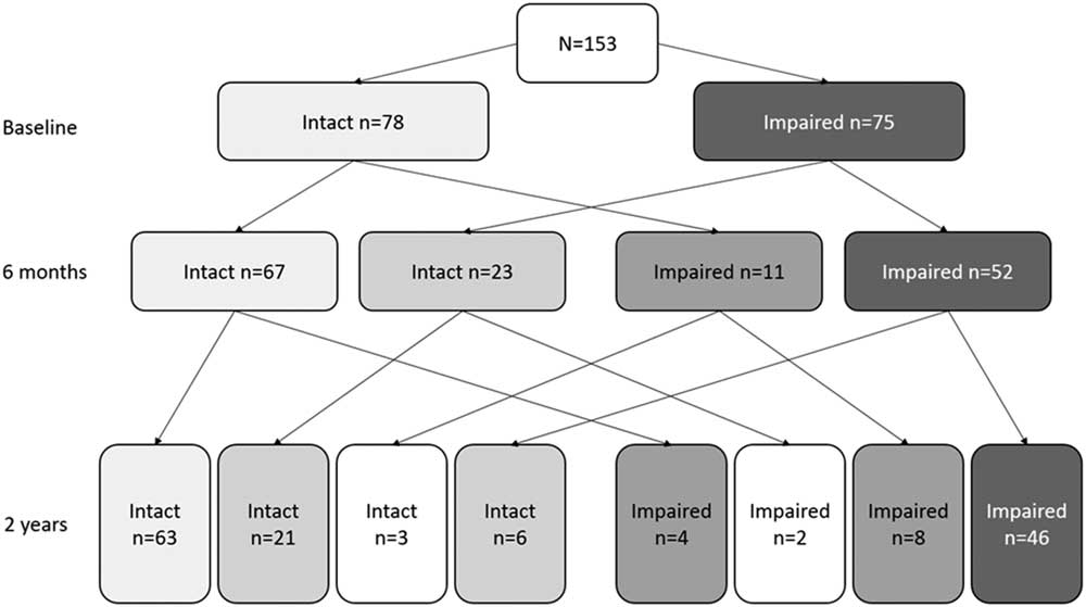
We categorized 75 patients (49.0%) as cognitively impaired at baseline, 63 patients (41.2%) were impaired at 6 months, and 60 patients (39.2%) were impaired at 2 years; see Figure 1. Between 6 months and 2 years, 138 patients (90.2%) did not alter in their cognitive status (i.e., remained either cognitively intact or impaired) while performance deteriorated (converted from intact to impaired) in 6 patients (3.9%) and improved (converted from impaired to intact) in 9 patients (5.9%).
Number of patients who were cognitively intact or impaired at different examinations.

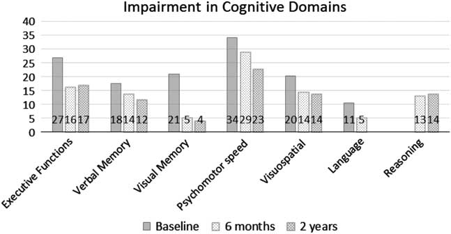
For the percentages of patients who were impaired in each cognitive domain at different examination time points, see Figure 2. Psychomotor speed and executive functions were the most commonly impaired cognitive domains and the largest decrease in impairment rate was seen in visual memory.
Percentage of patients impaired (i.e., below z of −1.65 of control data) in each cognitive domain. Language function measurements were not repeated at 2 years for the majority of patients and reasoning was not assessed at baseline.

For patients receiving neuropsychological rehabilitation (n=39; 25.5%), the mean number of sessions was 19 (median 15; range 1–94; missing data n=2). Ten patients received only short guidance (1–9 sessions), 20 patients received 12–20 sessions, and 7 received a longer neuropsychological rehabilitation (25–94 sessions). Rehabilitation started before 6 months post-stroke for 36 patients and later for 3 patients. Rehabilitation ended before 6 months post-stroke for 7 patients and lasted beyond 6 months post-stroke for 32 patients.
Patients who died or had a recurrent brain damage (n=21), or dropped out (n=49) were less educated (M=11.3 years [SD=2.6] vs. M=12.3 [2.7], U=4231.0; p=.010) and were more likely to substantially consume alcohol (36 patients [52.2%] vs. 45 patients [29.4%], χ2=10.632; p=.001) than the included patients (n=153). There were no other differences between excluded/dropped-out and included patients in demographics, risk factors, infarct characteristics, clinical characteristics, mood at baseline, or working status before stroke. The number of patients categorized as cognitively impaired at baseline did not differ significantly between excluded/dropped-out and included patients (44 patients [62.9%] vs. 75 patients [49.0%], χ2=3.695; p=.055), but excluded/dropped-out patients were more likely to be impaired in psychomotor speed (34 patients [48.6%] vs. 52 patients [34.0%], χ2=4.312; p=.038) and executive functions (28 patients [40.0%] vs. 41 patients [26.8%], χ2=3.918; p=.048) than included patients.
Comparing Cognitively Intact and Impaired Patients: Baseline
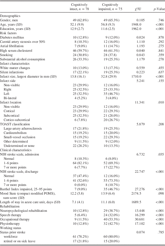
We compared demographics, risk factors, infarct characteristics, clinical characteristics, and amount of received rehabilitation between patients who were cognitively intact (n=78) and impaired (at least one domain impaired; n=75) at baseline. Cognitively intact patients were younger (p=.001), more educated (p<.001), had smaller infarcts (p<.001), and neurologically less severe strokes (e.g., lower NIH Stroke Scale scores at discharge, p<.001) compared to the cognitively impaired patients (Table 1). As expected, cognitively intact patients received less rehabilitation than cognitively impaired patients (all p<.01). We found no differences in gender, risk factors, lesion side, stroke etiology, or mood at baseline, or in working status before stroke.
Demographic and Clinical Data for Cognitively Intact and Impaired Patients at Baseline

Note. Numbers are frequencies (%), unless otherwise noted. For brain imaging (MRI scans 42.8%), high serum cholesterol, and smoking, total n=152; for Mood State, total n=144. For working status, the category workforce includes employed and unemployed patients and students. Some patients had more than one type of rehabilitation.
Cognitive Change Between Baseline and 6 Months
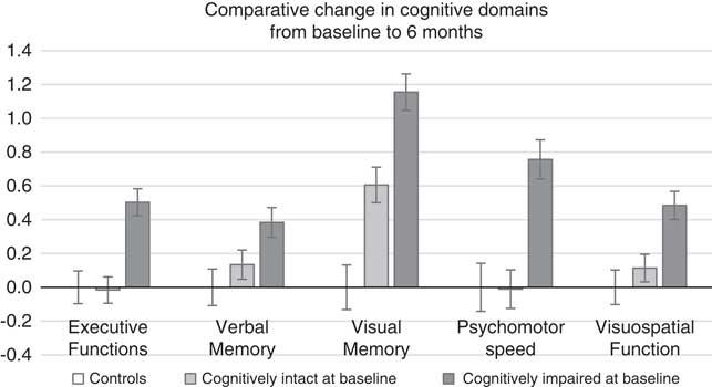
We compared domain-specific difference scores (6-month mean Z-score minus baseline mean Z-score) among controls (n=50), patients, who were cognitively intact (n=78) and patients who were cognitively impaired (n=75) at baseline. For these cognitive change scores, numbers close to zero indicate a similar change in performance compared to controls (i.e., no more of a change than between controls’ repeated measurements). Multivariate analysis of variance (MANOVA) indicated that cognitive change differed significantly between normal controls, cognitively intact patients, and cognitively impaired patients (F 10,392=8.464; p<.001; ηp 2=0.178; Wilks-Lambda=0.676).
Follow-up analysis of variance (ANOVA) revealed that the difference was significant for all cognitive domains (verbal memory p=.016; ηp 2=0.040; other domains all p<.001; ηp 2=0.076–0.189). For difference score means, see Figure 3. In post hoc analyses (pairwise Bonferroni), cognitively impaired patients showed more of an improvement than controls in all cognitive domains (verbal memory p=.019, others p ≤ .001); and also more of an improvement than the cognitively intact patients in all cognitive domains (p<.01) except for verbal memory (p=.130). Cognitively intact patients showed more of an improvement than the controls in visual memory performance (p=.001) but not in any other cognitive domain (p>.1).
Change within cognitive domains, mean change ± SEM. Change between baseline and 6 months, patients categorized as cognitively intact or impaired at the baseline examination. Reasoning was not assessed at baseline.

Controlling for age and education did not markedly change the results. The exception was for in verbal memory where cognitively impaired patients showed more of an improvement than either the controls (p<.01) or patients categorized as cognitively intact (p<.01). Removing the 27 patients with the lowest mood score (6-month mood score above the 90th percentile level of the controls’; cognitively intact patients at 6 months n=10, cognitively impaired n=17) did not markedly change the results.
Comparing Cognitively Intact and Impaired Patients: 6 Months and 2 Years
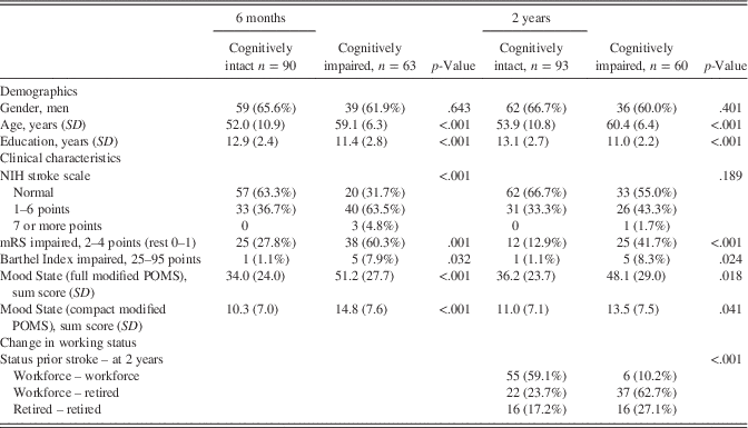
Patients who were cognitively intact at 6 months (n=90) were younger (p<.001) and more educated (p<.001) than patients still cognitively impaired at 6 months (n=63); see Table 2. Cognitively intact patients had a higher mood state compared to cognitively impaired patients (p<.001). Patients cognitively intact at 6 months were also neurologically less impaired at 6 months (e.g., National Institutes of Health Stroke Score [NIHSS] p<.001) than the cognitively impaired patients. Twenty patients with a normal NIHSS score and 25 patients with an excellent score of mRS (≤1) were categorized as cognitively impaired at 6 months. Patients who were cognitively intact or impaired at 6 months did not differ in receiving neuropsychological rehabilitation beyond 6 months from stroke (n=16; 17.8% vs. n=16; 25.4%, respectively; χ2=1.301; p=.254).
Demographic and Clinical Data for Cognitively Intact and Impaired Patients at 6 Months and at 2 Years

Note. Numbers are frequencies (%), unless otherwise noted. For Mood State, total n=146 at 6 months and n=145 at 2 years. For working status, n=152. The category workforce includes employed and unemployed patients, students, and a patient on parental leave; the category retired includes patients being on a sick-leave.
Differences in demographic variables between patients who were cognitively intact at 2 years (n=93) and patients still cognitively impaired at 2 years (n=60) were similar to the differences observed at 6 months; see Table 2. In terms of the neurological characteristics, cognitively intact and impaired patients no longer differed in the NIHSS scores (p=.189) at 2 years. Yet, 33 patients with a normal NIHSS score and 35 patients with an excellent score of mRS (≤1) were cognitively impaired at 2 years. Cognitively intact patients were less likely to be retired or on sick-leave at 2 years post-stroke after being in the able workforce before stroke, compared to patients still cognitively impaired at 2 years post-stroke. In total, 61 patients (50.8%) had returned to the able workforce at least part time, out of the 120 patients in the able workforce before stroke.
Cognitive Change Between 6 Months and 2 Years
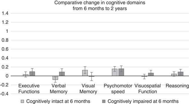
We compared domain-specific difference scores (2-year mean Z-score minus 6-month mean Z-score) between patients who were categorized as cognitively intact (n=90) or impaired (n=63) at 6-months. For these cognitive change scores, numbers close to zero indicate no change in cognitive domains between 6 months and 2 years. MANOVA indicated that cognitive change did not differ significantly between cognitively intact and impaired patients at 6-months (F 6,146=1.025; p=.411; ηp 2=0.040) and in further examination with ANOVA we found no significant domain-specific differences (p>.05 for all). For change means see Figure 4. Controlling for age and education did not change the results. As controls had only two examinations, we were not able to include the control groups’ difference score between the second and third examination. Both 6-month and 2-year patient scores were normalized with controls’ second examination scores.
Change within cognitive domains, mean change ± SEM. Change between 6 months and 2 years, patients categorized as cognitively intact or impaired at the 6-month examination.

DISCUSSION
Cognitive impairment was common in our working-age first-ever stroke cohort, as half of our patients were cognitively impaired at baseline, and 40% still demonstrated impairment 2 years post-stroke. Impairment in psychomotor speed was the most common finding at all of the time points, with more than 20% of the patients being impaired at 2 years, followed by impairment in executive functions with over 15% of the patients impaired at 2 years. As expected, patients who were cognitively impaired at baseline showed more improvement than either the controls or cognitively intact patients in all cognitive domains within 6 months. Little cognitive recovery took place after these first 6 months, as 90 % of the patients remained either cognitively intact or cognitively impaired between 6 months and 2 years.
At baseline (mean of 8 days from the stroke-onset), 49% of our first-ever stroke patients were cognitively impaired, which is in line with previous stroke studies (Jaillard et al., Reference Jaillard, Naegele, Trabucco-Miguel, LeBas and Hommel2009; Nys et al., Reference Nys, van Zandvoort, de Kort, Jansen, de Haan and Kappelle2007; Rasquin et al., Reference Rasquin, Verhey, Lousberg, Winkens and Lodder2002). Domain-specific impairments ranged from 11% in basic language processing to 18% in verbal memory to 34% in psychomotor speed. This is also in line with previous work (Nys, van Zandvoort, de Kort, van der Worp, et al., Reference Nys, van Zandvoort, de Kort, van der Worp, Jansen, Algra and Kappelle2005).
Consistent with past work (Nys, van Zandvoort, de Kort, van der Worp, et al., Reference Nys, van Zandvoort, de Kort, van der Worp, Jansen, Algra and Kappelle2005), patients who were categorized as cognitively impaired at baseline had more improvement in each of the cognitive domains between baseline and 6 months, relative to controls between repeated testing. We further showed that patients categorized as cognitively impaired at baseline had more improvement than those categorized as cognitively intact in all cognitive domains. We found the largest decreases in domain-specific impairment rates between baseline and 6 months in executive functions (from 27% to 16%) and in visual memory (from 21% to 5%).
Recovery in visual perception/construction and visual memory was found to be the most common in another stroke population as well (Nys, van Zandvoort, de Kort, van der Worp, et al., Reference Nys, van Zandvoort, de Kort, van der Worp, Jansen, Algra and Kappelle2005). However, the large decrease in the portion of our patients with visual memory impairment may be partly due to other factors than actual changes in visual memory. We measured visual memory (immediate and delayed recall) with only one test, the BRVRT (Benton, Reference Benton1974). Changes in this test were not balanced with other measures, and incidental changes may have occurred. This may also explain why patients, who were categorized as cognitively intact at baseline, showed more of an improvement relative to the controls, in visual memory performance.
Change in cognitive performance after 6 months appeared to be modest. The large majority of our patients (90%) remained either cognitively intact (55%) or impaired (35%) between the 6-month and 2-year examinations, and only 6 patients (4%) deteriorated while 9 patients (6%) improved. We observed no significant differences in domain-specific change between 6 months and 2 years between patients who were categorized as cognitively intact or impaired at 6 months. In our previous study, overall cognitive improvement was not significant between 6 months and 2 years (Kauranen et al., Reference Kauranen, Laari, Turunen, Melkas, Mustanoja, Baumann and Poutiainen2015), and we have now confirmed and specified this, as no domain-specific change emerged in the present study.
This is in line with previous studies that have found that although overall improvements are seen, most patients’ performance remains stable on individual neuropsychological measures between 6 months and 1 year (Rasquin et al., Reference Rasquin, Lodder, Ponds, Winkens, Jolles and Verhey2004), and in cognitive domains between 2-month and 2-year examinations (Hochstenbach et al., Reference Hochstenbach, den Otter and Mulder2003). In scrutinizing the domain-specific impairment rates further in our cohort, only the impairment rate of psychomotor speed decreased more than 5 percentage points between 6 months and 2 years (29% and 23% of patients impaired, respectively). This suggests that psychomotor speed had the longest trajectory of recovery among the cognitive domains.
At 2 years, 39% of patients were still cognitively impaired. The most common, long lasting domain-specific impairments were those in psychomotor speed (23%) and executive functions (17%). This corresponds well to a 5-year stroke follow-up study where patients were also impaired in executive functioning and processing speed (Barker-Collo et al., Reference Barker-Collo, Starkey, Lawes, Feigin, Senior and Parag2012). A subgroup of our patients were still impaired at 2 years, in visuospatial function (14%), reasoning (14%), verbal memory (12%), and visual memory (5%) as well. In a previous study that compared young stroke patients to a control group 11 years after stroke, patients were found to be impaired in all but one of the cognitive domains evaluated (Schaapsmeerders et al., Reference Schaapsmeerders, Maaijwee, van Dijk, Rutten-Jacobs, Arntz, Schoonderwaldt and de Leeuw2013), and the domain-specific impairment rates were slightly higher relative to our study. The criteria to denote impairment was less stringent than ours, but also some deterioration may take place between 2 to over 10 years post-stroke. Further information is needed on these long-term consequences after stroke and we are thus planning to follow-up with our patients and controls after 10 years from the initial examination.
Cognitively intact and impaired patients’ mood did not differ at baseline. However, patients who were cognitively impaired at the later follow-ups had lower mood state compared to cognitively intact patients. Nys and colleagues (Reference Nys, van Zandvoort, van der Worp, de Haan, de Kort, Jansen and Kappelle2006) found that early cognitive impairment predicts later depressive symptoms. At least part of the cognitively impaired patients’ low mood found in our sample might be related to the impairments due to stroke. Post-stroke depression has correlated with cognitive impairment at 3 months and 1 year post-stroke (Kauhanen et al., Reference Kauhanen, Korpelainen, Hiltunen, Brusin, Mononen, Määttä and Myllylä1999), and in our patient cohort, post-stroke fatigue at 3 months has been associated with impaired processing speed and memory performance later (Pihlaja et al., Reference Pihlaja, Uimonen, Mustanoja, Tatlisumak and Poutiainen2014). The association between altered mood and cognitive deficits highlights the need to assess and treat mood symptoms in clinical practice in patients with cognitive impairments.
Within the current sample, 25% of patients received neuropsychological rehabilitation. For most, rehabilitation lasted beyond 6 months post-stroke. Cessation of rehabilitation did not seem to explain why patients’ cognition did not further improve after 6 months. We stress the need to start neuropsychological rehabilitation early to guide cognitive recovery, most of which seemed to take place within the first 6 months. Furthermore, sufficient length of rehabilitation is encouraged to help patients process the changes in mood during recovery and to later adjust to their possibly permanently impaired cognitive status.
Even if recovering from physical disability, stroke survivors might be unable to return to previous occupations or independent life due to cognitive impairments (Gottesman & Hillis, Reference Gottesman and Hillis2010). Our previous findings showed that 32% of patients with a normal NIHSS score at discharge from hospital were still cognitively impaired 6 months after stroke (Kauranen et al., Reference Kauranen, Laari, Turunen, Mustanoja, Baumann and Poutiainen2014). We now further showed that 14–16% of all patients at 6 months, and 22–23% of all patients at 2 years, had recovered clinically well (i.e., have a normal NIHSS or an excellent mRS score) but were still cognitively impaired according to neuropsychological test performance. In an older sample, 71% of patients had excellent clinical recovery (mRS ≤1) at 3 months after stroke, but had cognitive impairment on detailed neuropsychological examination (Jokinen et al., Reference Jokinen, Melkas, Ylikoski, Pohjasvaara, Kaste, Erkinjuntti and Hietanen2015).
The precision of short clinical evaluation methods, such as the Mini-Mental State Examination (MMSE) or NIHSS, are questioned in estimating cognitive functioning after stroke (Bour et al., Reference Bour, Rasquin, Boreas, Limburg and Verhey2010; Hurford et al., Reference Hurford, Charidimou, Fox, Cipolotti and Werring2013; Jokinen et al., Reference Jokinen, Melkas, Ylikoski, Pohjasvaara, Kaste, Erkinjuntti and Hietanen2015; Kauranen et al., Reference Kauranen, Laari, Turunen, Mustanoja, Baumann and Poutiainen2014; Nys, van Zandvoort, de Kort, Jansen, et al., Reference Nys, van Zandvoort, de Kort, van der Worp, Jansen, Algra and Kappelle2005), and this highlights the importance of detailed neuropsychological evaluation instead. In our previous study, despite having relatively minor neurological and functional impairments (as assessed with, e.g., the NIHSS), only 41% of patients working before stroke had returned to work 6 months after stroke (Kauranen et al., Reference Kauranen, Turunen, Laari, Mustanoja, Baumann and Poutiainen2013). The number of domain-specific cognitive impairments was the only significant predictor of returning to work in a model including neurological impairments and risk factors (Kauranen et al., Reference Kauranen, Turunen, Laari, Mustanoja, Baumann and Poutiainen2013). Now, using the registry data, we were able to show that cognitively impaired patients at 2 years were more likely to have left the workforce for retirement or sick-leave than those who were cognitively intact post-stroke. Overall, 51% of our patients in this substudy had returned at least part-time to the able workforce by 2 years post-stroke.
Several strengths and limitations require consideration. As a strength, we had several patients completing three neuropsychological assessments in a 2-year follow-up period. As a limitation, patients who dropped-out or were excluded were less educated, consumed more alcohol, and were more likely to be impaired in both psychomotor speed and executive functions at baseline, than the patients who remained in the study. All other demographic and clinical variables were comparable between these groups, and our results should be rather well generalizable to a working-age population.
We analyzed a substantial amount of background information without using statistical corrections, albeit with less sensitive non-parametric methods, and these results should be interpreted with caution. The time difference between assessments of controls was shorter than that of patients, and may have caused slightly different learning effects. Also, some of the recovery seen between baseline and 6 months may have been due to effects of fluctuating acute conditions, like fatigue. We did, however, try to minimize these effects by evaluating patients when they were ready to be discharged home or to active rehabilitation. Previously, Nys and colleagues (Nys, van Zandvoort, de Kort, van der Worp, et al., 2005) have shown that neuropsychological data from the first weeks of stroke-onset predicts cognitive outcomes adequately.
As a strength, we were able to show that a large portion of clinically, reasonably well recovered patients have long lasting cognitive impairments at 2 years post-stroke. The portion of patients with cognitive impairment might even have been slightly higher if we had been able to follow all the patients. We used a rather stringent cutoff to denote domain-specific impairment (Z-score below −1.65 standard deviations relative to the control group), similar to one previous study (Nys, van Zandvoort, de Kort, van der Worp, et al., Reference Nys, van Zandvoort, de Kort, van der Worp, Jansen, Algra and Kappelle2005). This minimized the chances of overinflating the numbers of cognitively impaired patients. However, part of the patients categorized as cognitively intact likely had some minor cognitive dysfunction. This may partially explain why patients categorized as cognitively intact had some cognitive improvement between baseline and 6 months when compared to the controls, and why some of the patients categorized as cognitively intact were referred to neuropsychological rehabilitation following medical staff evaluations.
In conclusion, 2 years after a stroke almost 40% of our patients had long lasting cognitive impairment although half of them had neurologically recovered well according to the neurological scales. The majority of recovery took place within the first 6 months after stoke, and cognitive status remained stable after this time for 90% of patients. These findings stress the need for an early, detailed neuropsychological examination and for a speedy start, and sufficient length, of rehabilitation.
ACKNOWLEDGMENTS
This work was supported by Kela, The Social Insurance Institution of Finland (grant number 37/26/2007); and Adult Education Allowance by the Education Fund, which is financed by the Unemployment Insurance Fund and the state (K.T., grant numbers 0075508-04, 0075508-05). Dr. Lindsay Miller and INS ILC Research Editing and Consulting Program are gratefully acknowledged for English language editing. The authors have no conflicts of interest to disclose.