

Introduction
Trichinella spiralis (T. spiralis) is a zoonotic parasitic worm that may parasitise more than 150 animal species globally (Pozio Reference Pozio2007). T. spiralis infection poses a threat to public health, and it has a major negative impact on the livestock industry, especially swine production, potentially causing large financial losses (Rostami et al. Reference Rostami, Gamble, Dupouy-Camet, Khazan and Bruschi2017). The diagnosis of animal infection with Trichinella falls into two categories: direct demonstration of Trichinella larvae in muscle samples (Gajadhar et al. Reference Gajadhar, Noeckler, Boireau, Rossi, Scandrett and Gamble2019; Różycki et al. Reference Różycki, Korpysa-Dzirba, Bełcik, Bilska-Zając, Kochanowski, Karamon, Sroka and Cencek2022) and indirect methods such as serodiagnostic tests for detection of specific antibodies to Trichinella spp. in blood or serum (Gajadhar et al. Reference Gajadhar, Pozio, Gamble, Nöckler, Maddox-Hyttel, Forbes, Vallee, Rossi, Marinculić and Boireau2009), and can be confirmed by PCR (Lin et al. Reference Lin, Cao, Zhang, Zhou, Deng, Li and Zhou2013) (summarised in Barlow et al. Reference Barlow, Roy, Hawkins, Ankarah and Rosenthal2021).
Trichinella larvae can be quantitatively detected in muscle samples using the magnetic stirring digestion method (MSDM), sometimes referred to as the magnetic stir-bar artificial digestion method. This approach is still the gold standard worldwide (Nöckler et al. Reference Nöckler, Reckinger, Szabo, Maddox-Hyttel, Pozio, Der Giessen, Vallee and Boireau2009). In field conditions, this technique, which is based on controlled artificial digestion, sedimentation, and microscopic analysis, provides high specificity (~97%) and a reasonable sensitivity (~86%), with a detection limit as low as one larva per sample and accurate quantification at three larvae per gram (Różycki et al. Reference Różycki, Korpysa-Dzirba, Bełcik, Bilska-Zając, Kochanowski, Karamon, Sroka and Cencek2022). Trichinoscopy or compression method was the original method employed for Trichinella detection and surveillance. Compression method allows encysted larvae to be directly seen without needing a lot of sample processing (Forbes et al. Reference Forbes, Parker and Scandrett2003). However, it relies on the operator’s familiarity with the organism to be effective (Dupouy-Camet and Murrell Reference Dupouy-Camet and Murrell2007). Most studies, evaluating these different methods, are concerned with epidemiological studies as Trichinella surveillance programs (Barlow et al. Reference Barlow, Roy, Hawkins, Ankarah and Rosenthal2021) or food safety-related purposes (Basso et al. Reference Basso, Marreros, Hofmann, Salvisberg, Lundström-Stadelmann and Frey2022).
Trichinella is an excellent model for a wide range of experimental studies. This is due to several unique features of its life cycle that can be completed in a single host; distinct intestinal and muscular phases of infection; and it can be easily maintained in laboratory animals. Moreover, the great impact of trichinellosis on humans and livestock encourages researchers to evaluate the efficacy of new drugs or vaccines against Trichinella infection (Abdel-Hafeez et al. Reference Abdel-Hafeez, Kamal, Abdelgelil and Abdel-Fatah2015).
However, for evaluation of the efficacy of a new drug or vaccine for Trichinella, we need an accurate method for quantification of Trichinella larvae in muscles of experimental animals. Therefore, the current work is mainly designed in order to evaluate the procedures used for Trichinella larval counting in experimental studies. Comparing MSDM to individual muscle digestion and compression techniques, using standardised, methodical procedures under controlled settings, would provide insight into their respective effectiveness, sensitivity, and applicability.
Materials and methods
Animals and parasite
Fifteen male Swiss albino mice (outbred, laboratory-bred, parasite-free) were used in this study. They were 6–8 weeks old and weighed 18–20 g each. The animals were purchased from the Theodore Bilharz Research Institute (Giza, Egypt) and kept according to the institutional and national guidelines. The parasite, T. spiralis, was maintained in the laboratory of the Medical Parasitology Department at Tanta Faculty of Medicine through serial passages in rats and mice. Its original host was domestic pigs from the Cairo slaughterhouse, Egypt. The European Union Reference Laboratory for Parasites at the Superior Institute of Health in Rome, Italy, genotyped this strain as T. spiralis (code: ISS6158).
Mice remained on a standard commercial pelleted diet with free accessible water in the animal house. All mice were orally infected with T. spiralis 200 larvae/mouse according to Dunn and Wright (Reference Dunn and Wright1985). Mice were sacrificed 35 days post-infection for larval count after being anaesthetised using isoflurane, by this time, T. spiralis larvae are fully developed and completely encysted inside the muscle tissues (El-Wakil et al. Reference El-Wakil, Abdelmaksoud, AbouShousha and Ghallab2021; Abou Rayia et al. Reference Abou Rayia, Othman, Harras, Helal, Dawood and Soliman2022). They were divided into three main groups (five mice each): group 1: mice were subjected to the MSDM for T. spiralis total larval count in muscles; group 2: mice were subjected to individual separate muscle digestion of masseter (M), diaphragm (D), gastrocnemius (G), and tongue (T) muscles; and group 3: mice were subjected to slide compression method for the previously mentioned muscles.
Experimental procedures
Magnetic stirring digestion method
The MSDM was carried out according to Blair (Reference Blair and Campbell1983) and Forbes et al. (Reference Forbes, Parker and Scandrett2003). In brief, after dissection and evisceration of each sacrificed mouse, the whole muscles were weighed, then chopped and digested in 200 millilitres of distilled water with 1% concentrated HCl and 1% pepsin (1/10 000) (Figure 1A). The mixture was incubated at 37°C for one hour while being continuously stirred with an electric stirrer (Figure 1C). Each mouse was digested separately. A 50-mesh/inch sieve was used to filter out the coarse particles from the digested product. After being gathered on a 200-mesh/inch sieve and twice cleaned with tap water, the larvae were suspended in 150 millilitres of tap water in a conical flask (Figure 1D). A McMaster counting chamber was used to count the larvae in the sediment using light microscope set to ×10 magnification for the total larval count and can be expressed as larvae/gram muscles (Figure 1E).
Steps of digestion of Trichinella spiralis larvae in muscles. (A–C) Digestion of muscles in an artificial gastric fluid at 37°C for 2 hours with stirring. (D) Filtration through a 50-mesh sieve. (E) Larvae washed 2–3 times, sedimented in a conical flask, and supernatant discarded. D, diaphragm; G, gastrocnemius; M, masseter; MSDM, magnetic stirring digestion method; T, tongue; IMSDM, individual muscle sample digestion method.

Individual muscle digestion
The selected muscles from each mouse, namely gastrocnemius, masseter, diaphragm, and tongue, were dissected and weighed separately. Then, they were subjected to digestion by the same artificial digestion solution as previously detailed in a proportion of 15 ml/g of sample (Figure 1). The larval count was expressed as larvae per gram of muscle (Vignau et al. Reference Vignau, Guardis, Risso and Eiras1997; Mayer-Scholl et al. Reference Mayer-Scholl, Pozio, Gayda, Thaben, Bahn and Nöckler2017).
Compression method
The selected muscles from each mouse—gastrocnemius, masseter, diaphragm, and tongue—were dissected and weighed separately. These muscles were compressed between two tightly bound slides until the tissue sample turned translucent. The presence of encysted Trichinella larvae was microscopically assessed. The larval count was expressed as larvae per gram of muscle (Forbes et al. Reference Forbes, Parker and Scandrett2003).
Statistical analysis
All data were tabulated and statistically analysed by the statistical package for the social sciences software program, IBM SPSS Statistics for Windows, version 27 (IBM Corp., Armonk, N.Y., USA). The numerical data were first tested for normality by the kurtosis coefficient, histogram, Q-Q plot, and boxplots. Normally distributed numerical data were expressed as mean ± standard deviation. The mean larval count in each individual muscle subgroup was compared to the mean larval count obtained by the multi-sample digestion method by the Independent Samples T test. To control for type I error inflation due to multiple testing, we applied the Bonferroni correction. The significance threshold (α) was adjusted by dividing the conventional level of 0.05 by the number of comparisons (8), resulting in a Bonferroni-adjusted α of 0.006. A mixed ANOVA model was conducted, with the different muscle sites treated as the within-subject factor and the method used (individual muscle digestion or compression) as the between-subject factor. The multivariate tests revealed a significant interaction between these two factors. Consequently, post hoc analyses were performed to examine individual muscle differences within each method. Furthermore, correlation matrix was generated by Pearson’s correlation to investigate the linear relationships between various digestion and compression methods for larval counting in different muscles and the total larval count by the standard digestion method. Agreement between the studied digestion and compression methods for larval count in relation to total larval count by digestion method was accomplished and results were displayed as Concordance Correlation Coefficient (CCC), its 95% confidence interval (CI), precision, and accuracy. The CCC measures the degree of agreement and correlation between two sets of continuous variables. By using the CCC, we can determine how well the new methods align with the established “gold standard” one. CCC cutoffs of <0.90, 0.90–0.95, 0.95–0.99, and >0.99 indicate poor, moderate, substantial, and almost perfect agreement, respectively. Precision measures how well the data points fall on a straight line (correlation between values), while accuracy measures closeness to the line of perfect agreement (45-degree line through the origin). P values of <0.05 were considered statistically significant.
Results
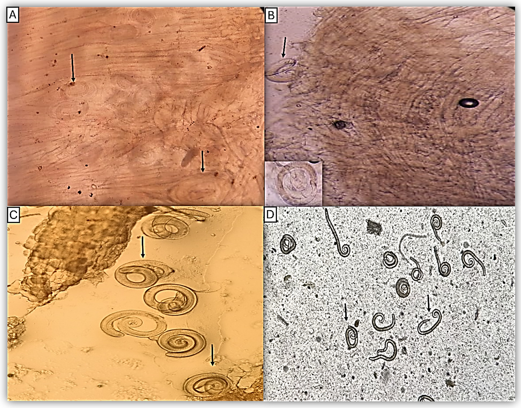
The digestion method revealed first-stage larvae, digested free from muscle cells. These larvae are around 1 mm long. The stichosome, a collection of discoid cells that line the oesophagus and take up the anterior half of the worm’s body, is the most distinctive characteristic of Trichinella larvae. Live larvae can be distinguished by their motility, and if cold, they are tightly coiled. Dead larvae assume a C-shape. The compression method revealed Trichinella encysted larvae (Figure 2).
Microscopic visualisation of Trichinella spiralis larvae within muscle tissue (black arrows). (A, B) Compression method (×10; inset in B, ×40). (C, D) Digestion method (×10).
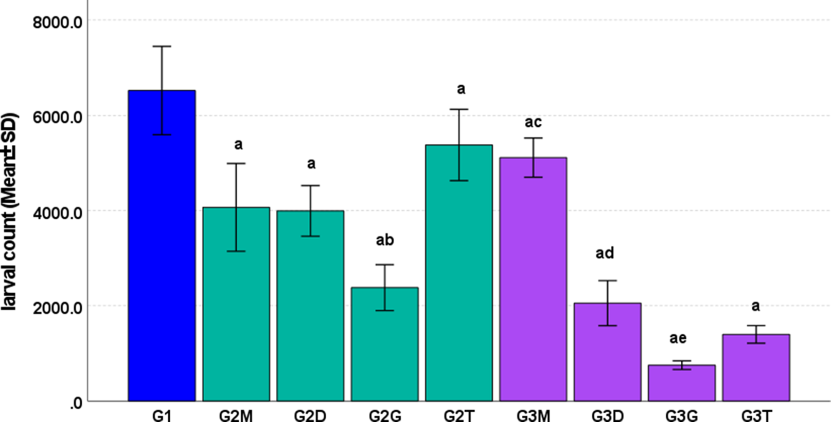
The total larvae count by the MSDM in the whole carcass ranged from 35,266 to 39,732 larvae. Comparison of digestion and compression methods for Trichinella larval count/gram muscles in individual muscles versus the larval count in the whole muscles by the MSDM was presented in Table 1. MSDM (group 1) yielded the highest mean larval count/gram muscles. Individual muscle digestion in group 2 produced significantly lower larval counts in different muscles than MSDM, except for the tongue (2T), which showed no statistically significant difference from MSDM (P = 0.064). Compression methods in group 3 consistently yielded much lower counts than MSDM with statistically significant differences (P <0.006), with the exception of the masseter (3M), which showed no statistically significant difference from MSDM (P = 0.015). However, it is important to mention that the tongue showed uneven distribution of Trichinella larvae among its parts.
Comparison of the digestion and compression methods for larval count in individual muscles and the total larval count by digestion method

* Significant at p ≤ Bonferroni-adjusted α of 0.006.
D, Diaphragm; G, Gastrocnemius; M, Masseter; MSDM, Magnetic Stirring Digestion Method; T, Tongue.
Repeated measures ANOVA for individual muscle digestion showed significant differences between masseter digestion method (2M) and gastrocnemius digestion method (2G), as well as gastrocnemius muscle (2G) vs the tongue (2T); otherwise, no significant differences were detected between other pairwise comparisons of different muscles. Alternatively, repeated measures ANOVA for compression method subgroups revealed significant differences between most muscles except the diaphragm (3D) vs the tongue (3T) (P = 0.306) (Table 1 and Figure 3).
Comparison of Trichinella larval counts per gram of individual muscles using digestion and compression methods versus total larval counts in whole muscles by MSDM. ‘a’ indicates significant differences between G1 and all subgroups, ‘b’ indicates significant differences between G2G and each of G2M and G2T, ‘c’ indicates significant differences between G3M and each of G3D, G3G, and G3T, ‘d’ indicates significant differences between G3D and G3G. ‘e’ indicates a significant difference between G3G and G3T.

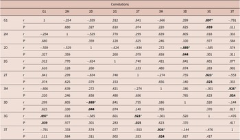
Pearson correlation was conducted to investigate the linear relationships between various digestion and compression methods for T. spiralis larval counting in different muscles and the total larval count by the standard digestion method (G1). Notably, the gastrocnemius compression method (3G) showed a strong and statistically significant positive correlation with G1 (r = 0.897, P = 0.039), indicating that this method closely aligns with the reference standard and may be a reliable alternative for larval quantification. Additionally, significant positive correlations were observed between the tongue digestion (2T) and gastrocnemius compression (3G) methods (r = 0.923, P = 0.025), and between the masseter and tongue compression methods (3M and 3T) (r = 0.926, P = 0.024), suggesting consistency within these alternative techniques. Conversely, a significant negative correlation existed between diaphragm digestion and compression methods (2D and 3D) (r = –0.889, P = 0.044), implying that these two approaches yielded inversely related results for the diaphragm muscle. Overall, while most methods do not show significant correlations with the total digestion method, the gastrocnemius compression method stands out as the most promising alternative based on its strong and significant correlation (Table 2).
Correlations between the digestion and compression methods for larval count in different muscles and the total larval count by digestion method

* Correlation is significant at the 0.05 level.
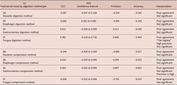
The Concordance Correlation Coefficient (CCC) was used to evaluate the agreement between various larval count methods compared to the total larval count by digestion method (G1). Results indicate that muscle digestion and compression methods for larval count in individual muscles showed poor agreement with the total larval count by digestion method, with only the tongue digestion method (2T) showing the highest agreement but lacking statistical significance. Most methods are not interchangeable with reference methods due to poor concordance (Table 3).
Agreement between the digestion and compression methods for larval count in relation to total larval count by digestion method

Discussion
The magnetic stirrer digestion method (MSDM) is considered the gold standard method for detection of Trichinella that allows effective recovery of encysted larvae from muscle samples for counting (Gamble et al. Reference Gamble, Bessonov, Cuperlovic, Gajadhar, Van Knapen, Noeckler, Schenone and Zhu2000; Mayer-Scholl et al. Reference Mayer-Scholl, Pozio, Gayda, Thaben, Bahn and Nöckler2017; Barlow et al. Reference Barlow, Roy, Hawkins, Ankarah and Rosenthal2021). Moreover, Barlow et al. (Reference Barlow, Roy, Hawkins, Ankarah and Rosenthal2021) reported that MSDM has a lower cost than most methods used for detection of Trichinella larvae in muscles (at about 6 cents/sample). Notably, some experimental studies use the digestion of the whole muscles of the experimental animals to express the total larval burden as a parasitological parameter to show the efficacy of drugs (Ashour et al. Reference Ashour, Abou Rayia, Saad and El-Bakary2016; Othman et al. Reference Othman, Abou Rayia, Ashour, Saied, Zineldeen and El-Ebiary2016) or vaccines (Ashour et al. Reference Ashour, Ibrahim, Elshamy and Zoghroban2022) for Trichinella infection. Otherwise, some researchers use selected muscles for digestion (Mostafa et al. Reference Mostafa, Hassan and Ahmed2020; Wang et al. Reference Wang, Bai, Tang, Yang, Wang, Zhu, Luo, Yan, Jia, Liu and Liu2020; Codina et al. Reference Codina, Indelman, Hinrichsen and Lamas2025). Therefore, the current study aimed to compare MSDM of the whole muscles to other methods using individual muscles either by digestion or compression methods.
Our results showed that artificial digestion method of the whole carcass had the highest mean larval count/gram muscles as compared to the individual muscle digestion or compression methods. This may be attributed to several factors. First, the weight of muscle tissue digested is greater than that examined by trichinoscopy or individual muscle digestion. Second, proper digestion of muscles can attain almost all Trichinella larvae encysted in the muscles. Previous studies have reported that when large amounts of tissues are available for digestion, the sensitivity is further increased (Gajadhar et al. Reference Gajadhar, Pozio, Gamble, Nöckler, Maddox-Hyttel, Forbes, Vallee, Rossi, Marinculić and Boireau2009, Reference Gajadhar, Noeckler, Boireau, Rossi, Scandrett and Gamble2019; Noeckler et al. Reference Noeckler, Pozio, van der Giessen, Hill and Gamble2019). Moreover, total larval count is suitable in experimental studies where low burden of larvae is achieved in the effectively treated/vaccinated experimental animals. Kapel et al. (Reference Kapel, Webster and Gamble2005) reported that a larger mass of muscle must be digested to confidently detect sparser infections.
Predilection sites of Trichinella larvae vary by host species, but in most species, diaphragm, masseter, and tongue are among the commonest sites. This muscle preference leads to the irregular distribution of larvae in the skeletal muscles (Gajadhar et al. Reference Gajadhar, Noeckler, Boireau, Rossi, Scandrett and Gamble2019). Therefore, digestion of the whole muscles for the total larval count overcomes this limitation of other techniques using individual muscles.
Our results revealed that individual digestion of the tongue showed comparable results to the total larval count, with no statistically significant difference. The tongue is a preferred site of larval settlement in mice (Codina et al. Reference Codina, Indelman, Hinrichsen and Lamas2025). Meanwhile, by compression method, the tongue showed much lower numbers of larvae as well as the uneven distribution of Trichinella larvae among its parts. Similar findings were reported by Forbes et al. (Reference Forbes, Parker and Scandrett2003). They stated that digestion assays of selected muscles consistently detected more larvae/gm tissue than trichinoscopic techniques. In contrast, Vignau et al. (Reference Vignau, Guardis, Risso and Eiras1997) mentioned that artificial digestion of selected muscles is not superior to trichinoscopy and that there are no statistically significant differences between them. They suggested the use of samples not less than 1 g of muscle, and to divide them into subsamples of 5 mm × 1 mm thickness to obtain comparable results by trichinoscopy.
Detection of Trichinella larvae by compression of muscle biopsy between two slides is the simplest way of parasitological diagnosis without a lot of sample processing. But this method is not sensitive enough to detect larvae especially when larval density is low or for not yet encapsulated larvae or larvae from non-encapsulated species. In this case, there is a risk of confusing the larvae with the muscle fibres making larval detection more difficult resulting in false-negative results (Pozio et al. Reference Pozio and Zarlenga2013). Despite such drawbacks, we agree that trichinoscopy is useful for confirmation of infection of experimental animals before starting the digestion process. The diaphragm is ideal for this purpose because it is a thin muscle that is easily compressed (Blair Reference Blair and Campbell1983; Mostafa et al. Reference Mostafa, Hassan and Ahmed2020). Similarly, Barlow et al. (Reference Barlow, Roy, Hawkins, Ankarah and Rosenthal2021) mentioned that trichinoscopy is useful for quick analysis when performed in the field in surveillance programs.
The current study showed that digestion and compression methods for larval count in individual muscles are not interchangeable with the total larval count by the conventional digestion method due to poor concordance, with only the tongue digestion and gastrocnemius compression methods that may be used as alternatives, despite their overall poor agreement with the reference method.
We must emphasise that the current study focused on small animals that are used regularly in experimental studies. For food safety or survey studies in larger animals, in which the muscle mass of an individual muscle is large, different findings may be obtained. For example, Leclair et al. (Reference Leclair, Forbes, Suppa and Gajadhar2003) assessed the tongue, among different muscles, for the detection of Trichinella larvae in large animals such as walrus. They showed that the apex (anterior tip of the tongue) had greater larvae than the root (posterior base of the tongue). The apex samples consistently had the highest numbers of larvae when compared with the other body muscles. Moreover, they reported that 100% detection rate was achieved when the entire tongue was homogenised and tested.
Interestingly, Makrutzki et al. (Reference Makrutzki, Riehn, Hamedy, Petroff, Hasenclever, Meiler and Lücker2014) used a modified MSDM using inverse microscopy and a modified larval counting basin known as ‘Trichoview’, which allows efficient visualisation of larvae in the sediment. They suggested that these modifications reduced some preparatory steps (supernatant withdrawal, fluid transfer, and washing) and reduced examination time, as the Trichoview counting basin has a surface area seven times smaller than that of the standard basin used in the MSDM.
However, MSDM may have some constraints such as dependence on the pepsin quality and availability and the skills of the personnel (Noeckler et al. Reference Noeckler, Pozio, van der Giessen, Hill and Gamble2019). Also, if Trichinella larvae are immature, there is possibility for their destruction by the digestion process (Riehn et al. Reference Riehn, Hasenclever, Petroff, Nöckler, Mayer-Scholl, Makrutzki and Lücker2013). Therefore, commercial digestion kits, Alternative Artificial Digestion (AAD) Kit such as PrioCHECK™ Trichinella, were developed. This assay uses a recombinant enzyme (serine-endopeptidase of the enzyme group subtilisin) instead of pepsin. It reduces the digestion time and limits the environmental contamination and occupational exposure, since the higher digestion temperatures (60°C) kill the larvae (Barlow et al. Reference Barlow, Roy, Hawkins, Ankarah and Rosenthal2021).
Only three studies have compared the AAD kit with the standard pepsin-HCl digestion method. Two of these studies reported similar results between the two methods (Konecsni et al. Reference Konecsni, Scheller, Scandrett, Buholzer and Gajadhar2017; Gajadhar et al. Reference Gajadhar, Konecsni, Scandrett and Buholzer2018), whereas Franssen et al. (Reference Franssen, Johne, Van der Giessen, Nöckler and Mayer-Scholl2019) reported a lesser efficacy of the AAD kit, particularly in detecting larvae of T. pseudospiralis. In addition, more debris resulted from digesting muscles when using AAD Kit which necessitated larger rinse volumes than the manufacturer’s recommendation and could increase assay time and introduce further human error (Gajadhar et al. Reference Gajadhar, Konecsni, Scandrett and Buholzer2018; Konecsni et al. Reference Konecsni, Scheller, Scandrett, Buholzer and Gajadhar2017). Moreover, killing the larvae makes this method unsuitable for experimental studies where viability is studied and limits the ability to distinguish species by PCR assays (Franssen et al. Reference Franssen, Johne, Van der Giessen, Nöckler and Mayer-Scholl2019). Recently, the automated artificial digestion with pepsin-HCl in the Trichomatic-35 instrument was evaluated and proved equivalent to the standard method using the magnetic stirrer (Basso et al. Reference Basso, Marreros, Hofmann, Salvisberg, Lundström-Stadelmann and Frey2022).
However, all these promising methods are still under evaluation and yet there are some technical issues such as the need for well-equipped laboratories as well as personnel training and standardisation of the instruments. Hence, they may not be ideal for research performed in many laboratories, especially in developing countries.
In conclusion, the present study recommends estimating the total larval count in the whole carcass using MSDM as the most accurate method for experimental studies when evaluating the efficacy of drugs or vaccines against T. spirallis. In contrast, larval counts using individual muscle digestion or compression methods may represent an approximate indicator of infection level in infected animals, but not a reliable measure of the absolute number of larvae in whole muscles.
Funding support
This study is not funded.
Competing interests
On behalf of all authors, the corresponding author states that there is no conflict of interest.
Ethical standards
Every procedure used in this work complies with the applicable national and institutional guidelines on the care and use of laboratory animals’ ethical standards. The Laboratory Animal Centre for Research Ethics Committee of Tanta University’s Faculty of Medicine gave its approval to the study protocol (Approval code: 36264PR1375/9/25).