





Impact statement
This study advances mycelium-based bio-coatings on cotton textiles as a sustainable alternative to chemical finishes, leveraging cotton’s bioreceptive properties to propagate Ganoderma lucidum mycelium and transfer multifunctional attributes, such as hydrophobicity and biodegradability. Through interdisciplinary collaboration between designers and biotechnologists, we demonstrate proof-of-concept prototypes that preserve textile flexibility while introducing low-impact aesthetic and performance enhancements, addressing persistent challenges in scalability, durability, and environmental persistence of existing coatings. By bridging creative biodesign exploration with rigorous material testing, this work proposes mycelium finishing as a pathway to textile innovation, addressing the textile industry’s urgent sustainability challenges, from waste generation to resource-intensive coatings, potentially enabling scalable, circular processes that valorise agricultural by-products and reduce reliance on fossil fuels.
Introduction
Environmental and social concerns in the textile industry have driven a robust research and development effort into alternative textile materials, with a strong trend towards the valorisation of vegan and bio-based options; among these, lab-grown textiles show promising properties for developing multifunctional and sustainable alternatives (Liu et al. Reference Liu, Zhang, Wang, Yue and Jiang2025). In this field, mycelium biofabrication is among the most studied processes, as shown in the patent review conducted by Elsacker et al. (Reference Elsacker, Vandelook and Peeters2023), which highlights innovative technological approaches to develop mycelium leather, including biofabrication processes such as solid-state surface fermentation (Raman et al. Reference Raman, Kim, Kim, Oh and Shin2022), liquid-state surface fermentation (Gandia et al. Reference Gandia, van den Brandhof, Appels and Jones2021) and stirred submerged liquid fermentation (Szilvay et al. Reference Szilvay, Laine, Barrantes, Suhonen, Boer and Penttilä2021). Similarly, biodesign (Myers Reference Myers2012) has increasingly explored the potential of grown textiles, often focusing on mycelium as an active collaborator in material creation (Collet Reference Collet2017; Crawford et al. Reference Crawford, Stefanov, Miller, Leary and Johnson2024b; Toussaint et al. Reference Toussaint, Bruggeman and van den Eijnde2024).
Non-biofabricated textiles still play an important role in this material exploration. In fact, fungal materials are generally classified into pure fungal materials and composites (Romero-Cedillo et al. Reference Romero-Cedillo, Robledo-Leal, Aguilar-Marcelino, Acosta-Urdapilleta and Téllez-Téllez2025); among the latter, textile-mycelium biocomposites have been tested for the efficient conversion of textile waste into biocomposites to envision biobricks (Kaniyamparambil et al. Reference Kaniyamparambil, Ziyan, AlBlooshi, Dali, Lizundia, Mettu, Mabrook, Salim and Tardy2026) or as alternatives to plastic packaging products (Saini et al. Reference Saini, Kaur and Brar2024). Textiles have also been used for layering in mycelium-based biocomposite sandwiches (Jiang et al. Reference Jiang, Walczyk, McIntyre, Bucinell and Tudryn2017) or as support in multi-layer lamination to enhance material strength (Crawford et al. Reference Crawford, Stefanov, Miller, Leary and Johnson2024b; Kniep et al. Reference Kniep, Graupner, Reimer and Müssig2024).
However, the direct cultivation of fungal mycelium on existing textile substrates to enhance textiles’ original features remains little explored. Mycelium has demonstrated multifunctional properties as a textile alternative, including hydrophobicity, thermal and acoustic insulation, fire resistance, biodegradability and water repellence (Manan et al. Reference Manan, Ullah, Ul-Islam, Atta and Yang2021; Udayanga and Miriyagalla Reference Udayanga, Miriyagalla, Bhatt, Gangola, Udayanga and Kumar2021; Alemu et al. Reference Alemu, Tafesse and Mondal2022; Amstislavski et al. Reference Amstislavski, Pöhler, Valtonen, Wikström, Harlin, Salo, Jetsu and Szilvay2024; Karthikeyan et al. Reference Karthikeyan, Raja, Sri, Bilal, Pooja and Sindhuja2025; Rahman et al. Reference Rahman, Tawiah, Wei, Fei, Fei, George and Tawiah2026). In the hypothesis that some of these remarkable features can be transferred to conventional textiles through a mycelium finishing, in this proof-of-concept study, we explore the potential of mycelium (Ganoderma lucidum) to form a bio-coating by propagating across cotton textile fabrics. The aim of this study is to test mycelium’s ability to transform the fabric itself, exploring the possibility that mycelium-based coatings could provide new functionalities and aesthetic qualities while preserving key textile characteristics, such as flexibility.
This research builds on a design-driven exploration that investigates bioreceptivity as a material quality for textiles (Guillitte Reference Guillitte1995; Cruz and Beckett Reference Cruz and Beckett2016; Pollini and Rognoli Reference Pollini and Rognoli2021; Keune et al. Reference Keune, Lim, Dumitrescu and Thomsen2025); this preliminary study investigated how textiles can serve as substrates for the interaction and growth of living organisms (Brunelli et al., under review, expected Reference Brunelli, Pollini, Harvey and Marseglia2026). As part of this hands-on research, early prototypes of mycelium-coated textiles were developed, providing initial proof-of-concept. This work set the stage for our central research question: Can textiles act as substrates for mycelial growth to produce a low-impact, thin biofabricated coating, as an alternative to conventional chemical finishing processes in the textile industry?
The development of sustainable textile finishing technologies remains a pressing challenge, given the environmental toll of conventional approaches and persistent inefficiencies in emerging alternatives. Polyurethane (PU)-based hydrophobic coatings, favoured for their durability and water repellency on bio-based textiles, exemplify this issue; moreover, bio-based alternative hydrophobic coatings, despite showing a good material performance, still face challenges related to large-scale production, economic feasibility, recycling technologies, and environmental performance effectiveness dependent by feedstock and formulation (Jayalath et al. Reference Jayalath, Ananthakrishnan, Jeong, Shibu, Zhang, Kumar, Yoo, Shamshina and Therasme2025). Likewise, fire-resistant coatings frequently incorporate non-environmentally friendly chemicals, which pose environmental persistence and toxicity risks. Despite advances in greener formulations, challenges persist in achieving wash durability, minimising ecological impacts, and standardising testing protocols across applications (Patel et al. Reference Patel, Chaudhary, Patel, Chaudhari and Gupta2025). The aim of this study on mycelium-derived coatings is to offer a new pathway for addressing these shortcomings. Thanks to mycelium’s ability to grow on a wide range of organic substrates, including textiles, and to its intrinsic properties, as well as its aesthetic potential, mycelium emerges as a strong candidate for replacing conventional fossil-based textile coatings, offering a lower-impact alternative.
To address this possibility, this study is rooted in an interdisciplinary collaboration between designers and scientists. It explores textiles as bioreceptive surfaces and aims to develop and test a thin mycelium-based coating that provides new functional and aesthetic properties through a biofabrication process. In particular, the first author, with a design background, actively collaborated with biochemists from VTT Technical Research Centre of Finland and chemists from the School of Chemical Engineering of Aalto University. For this reason, this research was carried out within multiple biodesign and scientific laboratory environments, where the designer assumed the role of Biodesigner and Designer in Lab (Pollini Reference Pollini2024), working closely with scientific experts, especially to run material testing of the mycelium-coated textile prototypes developed.
The hybrid and interdisciplinary nature of the study is visible in the following sections, where literature, case studies, research approaches and reflections from multiple disciplines coexist and complement each other, bridging the initial creative biodesign exploration with a more technical analysis of the mycelium-coated textiles performances.
State of the art
The combination of mycelium and textiles has been actively explored in the field of biodesign, yet it has received comparatively little attention in the scientific literature. This study positions itself as an interdisciplinary exploration within biodesign, bridging creative exploration with scientific inquiry. Therefore, the following overview offers the state of the art by drawing on contributions from both creative and scientific research. Furthermore, a key distinction must be drawn within mycelium-textile biocomposites, as this section focuses specifically on mycelium deployed as a surface coating or texturising agent rather than, for instance, as a biobinder or structural element. To our knowledge no scientific studies and only a limited number of biodesign projects have explored textiles as bioreceptive surfaces for cultivating a mycelium coating directly onto the textile itself. Nonetheless, few scientific studies present approaches partially related to this study. However, all these examples remain at an early laboratory or speculative stage, without systematic material validation.
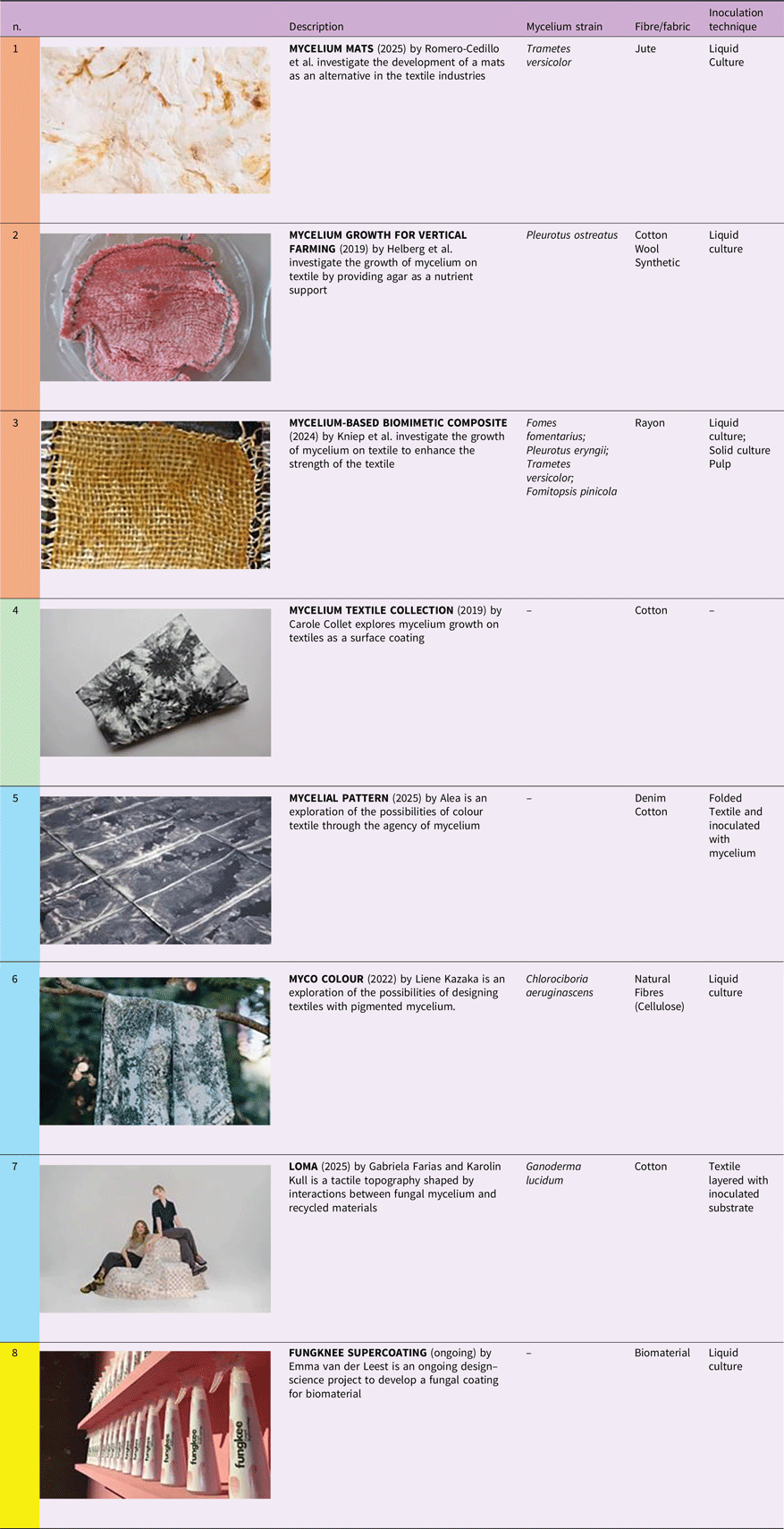
The desk research, conducted from September 2025 to February 2026, included both academic and grey literature on academic search engines such as Google Scholar, ResearchGate, and Scopus, as well as design-oriented platforms including Dezeen, Designboom, Futures Materials Bank and biodesigners’ websites and portfolios. Eight examples were identified and are presented in Table 1.
Examples shown in orange derive from scientific research; those in green represent scientific studies developed through interdisciplinary collaboration involving designers; those in blue refer to biodesign applications; and those in yellow refer to start-up initiatives

The first three examples reported in Table 1 are derived from scientific literature, although they do not directly investigate the development of mycelium-based coatings for textiles, their hypotheses and/or material outcomes are comparable to such an application. The study of Romero-Cedillo et al. (n.1) describes the development of flexible mycelium mats through the growth of Trametes versicolor on jute fabric, and reports testing of different post-treatments and evaluation of material properties including water absorption. The authors highlight that mycelium growth on jute shows a “higher water absorption capacity, so it is possible to use them in some areas that are not exposed to direct contact with water, which should be evaluated” (Romero-Cedillo et al. Reference Romero-Cedillo, Robledo-Leal, Aguilar-Marcelino, Acosta-Urdapilleta and Téllez-Téllez2025). Helberg et al. (Reference Helberg, Klöcker, Sabantina, Storck, Böttjer, Brockhagen, Kinzel, Rattenholl and Ehrmann2019) (n.2) investigated the growth of Pleurotus ostreatus on textile fabrics for vertical farming and observed that the presence of nutrient agar can facilitate the growth of a thicker mycelium layer on the textile. However, when the textiles were submerged in agar prior to inoculation, a rigid composite material was produced. Kniep et al. (Reference Kniep, Graupner, Reimer and Müssig2024) (n.3) explored the growth of mycelium on textiles to enhance their mechanical strength and develop alternatives to animal leather, highlighting the importance of the selection of mycelium strain, as not all methods are compatible with all fungal species. Furthermore, the presence of lignin in the textile substrate (rayon) increases the integration between the mycelium and the fabric. Switching to more designerly explorations, in the research of Carole Collet (n.4), Mycelium Textile (Collet Reference Collet2017), the designer, working in a laboratory environment, explores a collaborative approach with the organism, allowing mycelium to grow on natural textiles as a surface coating. Through this process, she investigates how mycelium growth can generate new surface finishes and aesthetic qualities, while integrating traditional textile techniques into the biofabrication workflow. She experiments with mycelial growth on different waste materials, including cotton to create living patterns. Beyond academic research, few biodesign-driven experiments and prototypes documented in grey literature further explore mycelium as a textile coating. Examples 5 and 6 in Table 1 focus on the aesthetic potential of living growth: Mycelial Pattern by Alea investigates mycelium’s capacity to transform or partially degrade textile dyes, suggesting new aesthetic possibilities and extended material life cycles through biological intervention, while Myco Colour explores pigment release during fungal growth, such as the blue-green coloration produced by Chlorociboria aeruginascens on textiles, highlighting the chromatic potential of mycelium-based finishings. Loma by Gabriela Farías and Karolin Kull (n.7) investigates the growth of Ganoderma lucidum on cotton fabric to create tactile surface topographies through a layered system in which inoculated substrate progressively colonises the stretched textile. Finally, Emma van der Leest’s start-up initiative, Fungkee Supercoating (n.8), is developing a fully bio-based fungal coating designed to render biomaterials water-repellent while preserving biodegradability.
Some of the scientific studies included in Table 1, as well as other studies that explore mycelium leather or mycelium-textile biocomposites adopt both pre- and post-treatment processes. Pre-treatment processes applied to the textile substrates, in addition to autoclave sterilisation (which is common to all the examples and aims to eliminate microbial contamination), are intended to increase nutrients and facilitate mycelial growth. In particular, nutrient-based treatments such as agar (Helberg et al. Reference Helberg, Klöcker, Sabantina, Storck, Böttjer, Brockhagen, Kinzel, Rattenholl and Ehrmann2019) and nutrient solutions containing urea, peptone, and soybean (Wang et al. Reference Wang, Kaur, Pensupa, Uisan, Du, Yang and Lin2018) are used. Post-treatments, such as physical, enzymatic and chemical processes, are applied to improve the mechanical and aesthetic properties of the developed material, as flexibility, durability and absorbent properties (Benetti et al. Reference Benetti, Conti and Dimitriadis2025). The most common post-treatment is the application of glycerol or similar plasticiser solutions to increase material flexibility and elasticity (Crawford et al. Reference Crawford, Ruthanna Miller, Branco, Fletcher and Stefanov2024a; Pertile et al. Reference Pertile, Fontana, Isoton, Brandalise and Camassola2025; Romero-Cedillo et al. Reference Romero-Cedillo, Robledo-Leal, Aguilar-Marcelino, Acosta-Urdapilleta and Téllez-Téllez2025), while recent studies, such as Appels et al. (Reference Appels, Camere, Montalti, Karana, Jansen, Dijksterhuis, Krijgsheld and Wösten2019), highlight the use of heat pressing for improving the flexural strength properties of the samples. To improve water resistance, the samples are often treated with wax and oil coatings (Farranhoor et al., in Benetti et al. Reference Benetti, Conti and Dimitriadis2025).
This study originated from a wider material exploration of bioreceptive textiles, guided by the RQ: can textiles be a bioreceptive surface for living organisms? This practice-based biodesign inquiry focused on how textile substrates can host and support different living agents, while examining the aesthetic outcomes of such colonisation (Brunelli et al., under review, expected Reference Brunelli, Pollini, Harvey and Marseglia2026). After observing successful prototypes in which the mycelium stood out for its performance creating valuable proofs of concept, a subsequent study aimed at strengthening the validation of this initial hypothesis through the following RQ: in which way can mycelium grow as a coating on textile surfaces, and can this growth enhance or modify the material features of the textile? Early hypotheses were that a mycelium coating could improve textile hydrophobicity and alter aesthetic qualities, such as look and feel, while maintaining flexibility.
Materials and methods
This research on mycelium as a textile coating emerged from an interdisciplinary approach that originated in the creative exploration of bioreceptive textiles and subsequently in a preliminary scientific validation of the most promising proof-of-concept developed with mycelium.
Biotinkering with mycelium as a textile coating
This study starts with an exploratory phase led by the designer through biotinkering activities (Pollini Reference Pollini2024; Marseglia et al. Reference Marseglia, Cantini, Celli, Brunelli and Lotti2025; Cianfano et al. Reference Cianfano, Celli, Marseglia, Rognoli, Nimkulrat, Ferraris and Mattioli2025), investigating the growth of different mycelial species (among other organisms), such as Pleurotus ostreatus, Ganoderma lucidum, Trametes versicolor, and Trametes sanguinea, on several textile substrates, both natural and synthetic, such as cotton, jute, lycra, denim and wool. Assuming the role of the biodesigner (Pollini Reference Pollini2024), the researcher combined creative experimentation with laboratory-based practices within a scientific environment, specifically the BioMakerStudioFootnote 1 of Aalto University, a DIY Biolab setting. These biotinkering activities were primarily oriented toward understanding the potential of textiles as a bioreceptive surface, exploring different techniques to grow mycelium directly on textile substrates as a transformative coating. A following research phase explored the aesthetic potential of surfaces generated through fungal growth, leveraging the ability of different mycelial species to release pigments and to develop soft, tactile, and visually distinctive coatings on textile surfaces. During the biotinkering phase, iterative and intuitive evaluation guided material development. Specifically, a sensory evaluation was carried out during the biotinkering phase as an exploratory, qualitative assessment to identify promising candidates for subsequent quantitative testing.
Application-oriented and validation through interdisciplinary collaboration
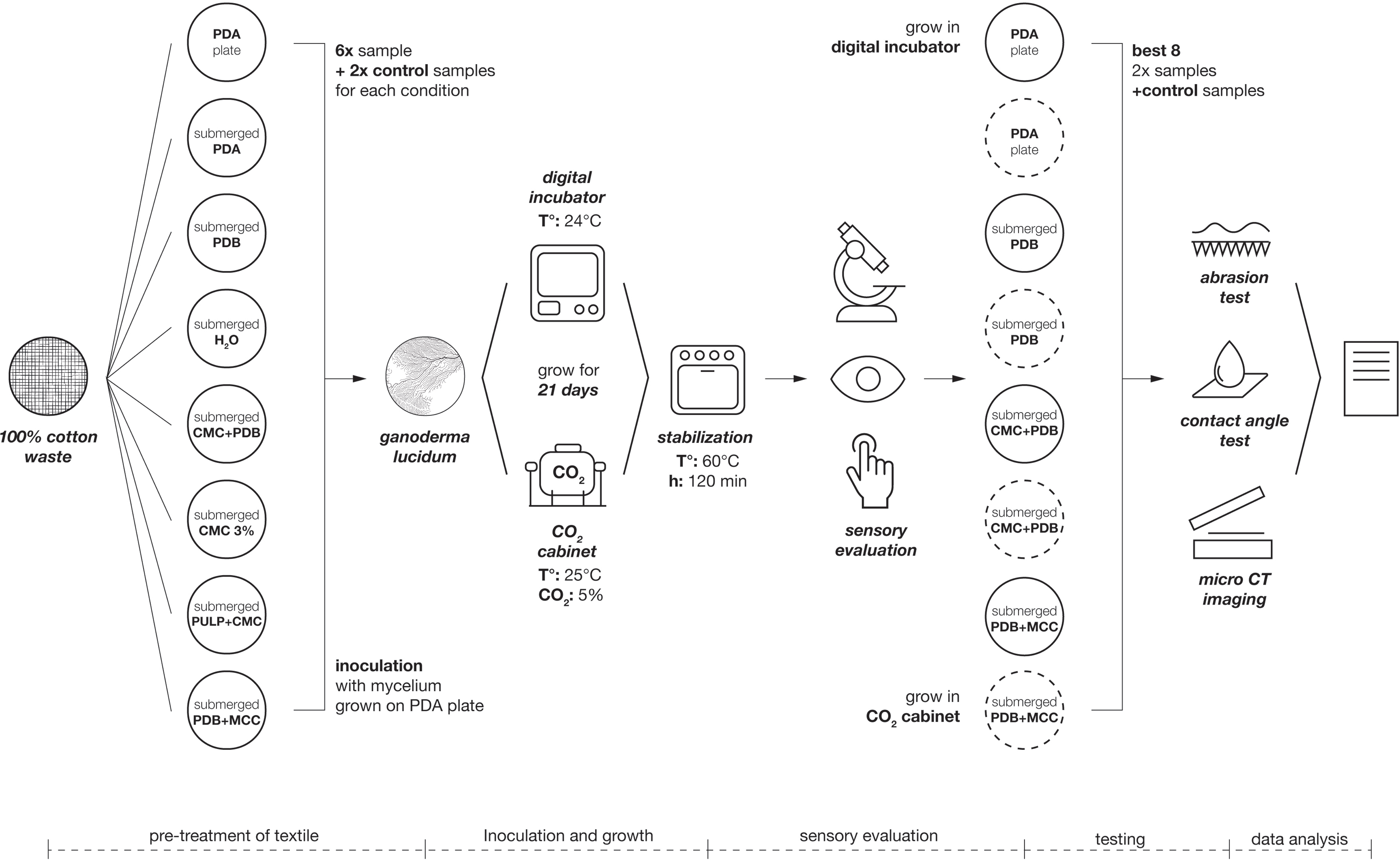
To further explore mycelium’s potential in the context of bioreceptive textiles, the designer initiated an interdisciplinary collaboration with scientists from Aalto University and VTT Technical Research Centre of Finland to develop a mycelium coating grown directly on the textile surface. This collaboration introduced the first author into a highly controlled scientific environment, where standardised methods and rigorous protocols were required to obtain quantitative data to analyse. The interdisciplinary research follows successive stages, as illustrated in Figure 1. First, an initial experimental phase was conducted, in which textile substrates were pre-treated with different nutrient solutions. The inoculated textiles were then cultivated under two distinct environmental conditions. This was followed by a preliminary selection phase based on sensory evaluation to identify the most promising samples. Subsequently, a testing phase focused on the selected samples and finally, a data analysis phase integrated the experimental findings to evaluate performance and material behaviour.
Phases of the research after activating interdisciplinary collaboration with scientists from Aalto’ Design and Chemistry Departments and VTT.

Preparation of textile substrates
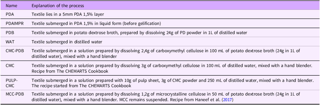
The experiments were conducted on a textile substrate composed of 100% white plainweave cotton, sourced from textile waste from the Costume Workshop at Aalto Studios, Aalto University. The material was selected for its natural fibre composition and its compatibility with fungal colonisation, as demonstrated in previous biotinkering explorations. All samples were cut into standardised dimensions (55 × 55 mm) and autoclaved at 121°C for 60 min before use. To explore the influence of different nutrients on mycelium growth and coating formation, a total of 8 different pre-treatments of the textile, presented in Table 2 were prepared. Agar-based media were selected as they are among the most commonly used substrates for fungal growth, being rich in simple sugars that are readily metabolised by mycelium. Cellulose-based media were included because cellulose is the most abundant natural polymer and represents a suitable carbon source for mycelial growth. The cellulose-based media were prepared in the CHEMARTS laboratories of Aalto UniversityFootnote 2 .
All the pre-treatment techniques tested for textile

In the submerged conditions, the textiles were fully immersed for approximately 2 seconds in a beaker containing the selected nutrients, using sterilised tweezers and working under a laminar flow hood in a lab environment. All the textile substrates thus obtained were then autoclaved at 121°C for 60 min before use to prevent contamination.
Growth of mycelium coating
After pre-treatment, all the textile substrates were inoculated with Ganoderma lucidum. For this, mycelium was first grown on potato dextrose agar (PDA 1.5%) plates and stored at 4°C for 3 weeks before inoculation. For inoculation, a small section of growing mycelium, including the underlying agar, were transferred from the PDA plates onto the centre of the textile surface. All inoculation procedures were performed in a BSL1 Lab – Biofilia of Aalto School of ARTS Footnote 3 – under a laminar flow hood, and all equipment was sterilised with ethanol to minimise the risk of contamination. For each condition, six replicate samples and two control samples without mycelium inoculation were prepared.
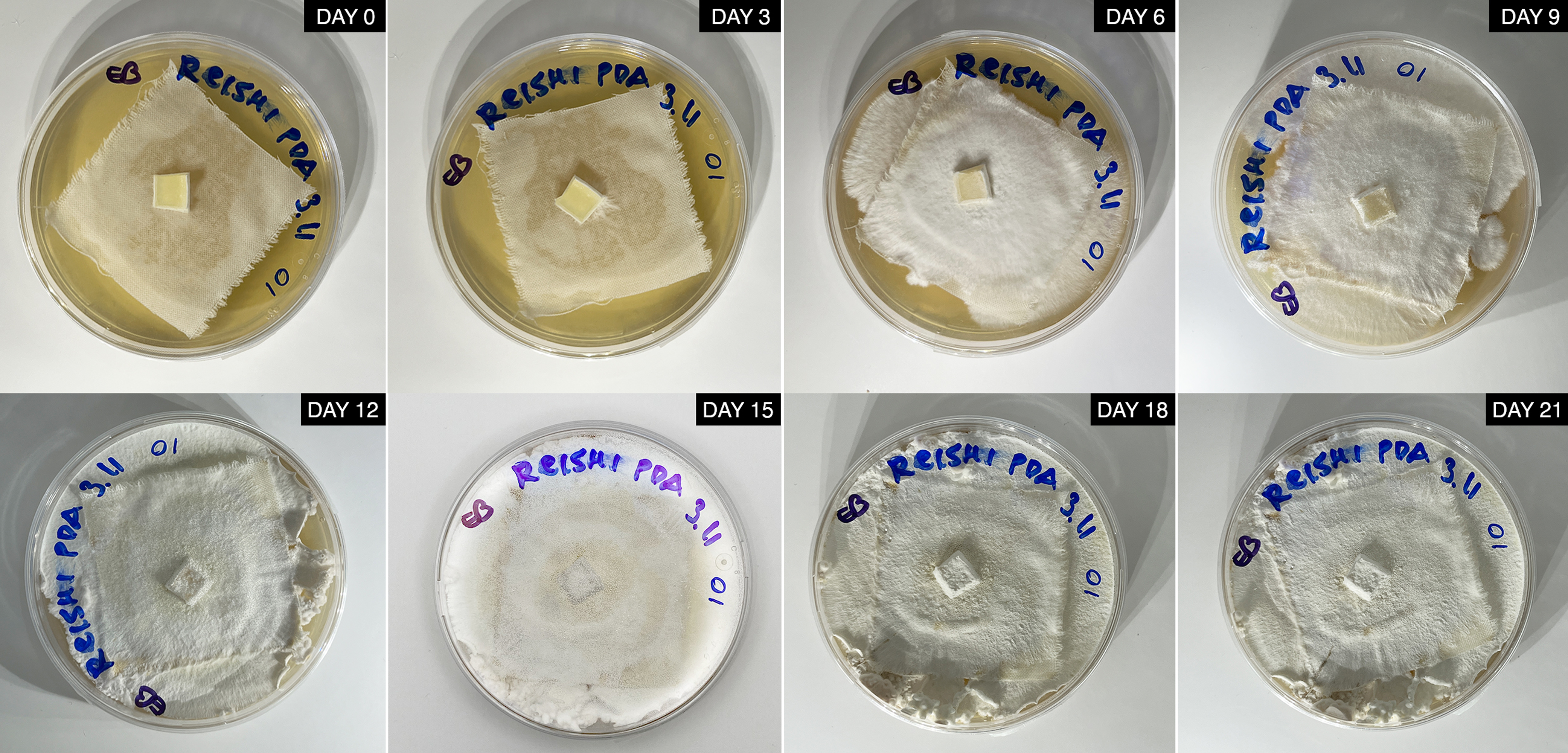
In this study, two different growing conditions were compared: a digital incubator and a CO2 incubator cabinet. Similar samples were grown half in a digital incubator and half in a CO2 incubator cabinet. According to literature (Zadrazil Reference Zadrazil1975) growing at elevated CO2 concentrations were reported to increase growth rate. For the first growth condition, the inoculated substrates were incubated at 24°C for 21 days at atmospheric CO2 concentration in a digital reptile egg incubator (VEVOR). For the second growth condition, the inoculated substrates were incubated in Model 3111 (Forma Scientific, United States) CO2 incubator cabinet at 25°C, 5 % CO2 concentration and 75–85 % relative humidity. All samples were grown for 21 days. During the growth phase, photographs were taken every 2–4 days to monitor the development of the mycelial coating on the textile substrates (Figure 2).
Sequential photographic documentation of mycelial coating growth on textile substrates cultured on PDA and incubated in a digital incubator, showing the progression of coating formation over time.

Drying
At the end of the growth period, the mycelium-coated textile samples were removed from the Petri dishes and left in an oven at 60°C for 120 min to stop further biological growth, following the advice from the scientist group and the stabilisation procedure described by Haneef et al. (Reference Haneef, Ceseracciu, Canale, Bayer, Heredia-Guerrero and Athanassiou2017). After drying no post-treatments were applied to the material samples.
Qualitative assessment for sample selection
An initial qualitatively preliminary screening was conducted through a sensory evaluation, based primarily on tactile and visual assessment. The sensorial evaluation was carried out collaboratively between two designers and two biotechnologists. The evaluation criteria served as a common basis for this analysis included: coating continuity and coverage (visual assessment); coating thickness (visual and tactile assessment); textile flexibility retention (tactile bending test); surface texture characteristics (tactile assessment). This qualitative screening did not employ formal sensory evaluation protocols – e.g., standardised scoring scales, blinded evaluation, or trained sensory panels (Sülar and Okur Reference Sülar and Okur2007; Stone et al. Reference Stone, Bleibaum and Thomas2020) – as it served as an exploratory phase within the biodesign tinkering methodology to guide subsequent rigorous material testing. Visual observations were conducted both during the growth phase and after the drying process, combining qualitative observation with the use of scientific environments and tools. Observations were performed using both the naked eye and Model 570 (American Optical, US) optical microscope to support the qualitative assessment of coating coverage and surface morphology. This analysis served as a preliminary screening to compare the different growth conditions and the effectiveness of the different pre-treatment techniques, identifying the most representative samples for subsequent quantitative testing, including abrasion and water contact angle measurements.
Scientific evaluation of the mycelium-coated textile samples
For the quantitative and scientific evaluation of the mycelium coating ability on textiles, the designer activated an additional interdisciplinary collaboration with scientists from the Department of Chemistry at Aalto University. In particular, based on the selected samples resulting from the sensory evaluation, a further qualitative and preliminary quantitative analysis has been subsequently carried out to validate the effect of mycelium as a textile coating. Specifically, scientific evaluation included Water Contact Angle (WCA) measurement, Abrasion Resistance Testing, and X-ray Microtomography (Micro-CT).
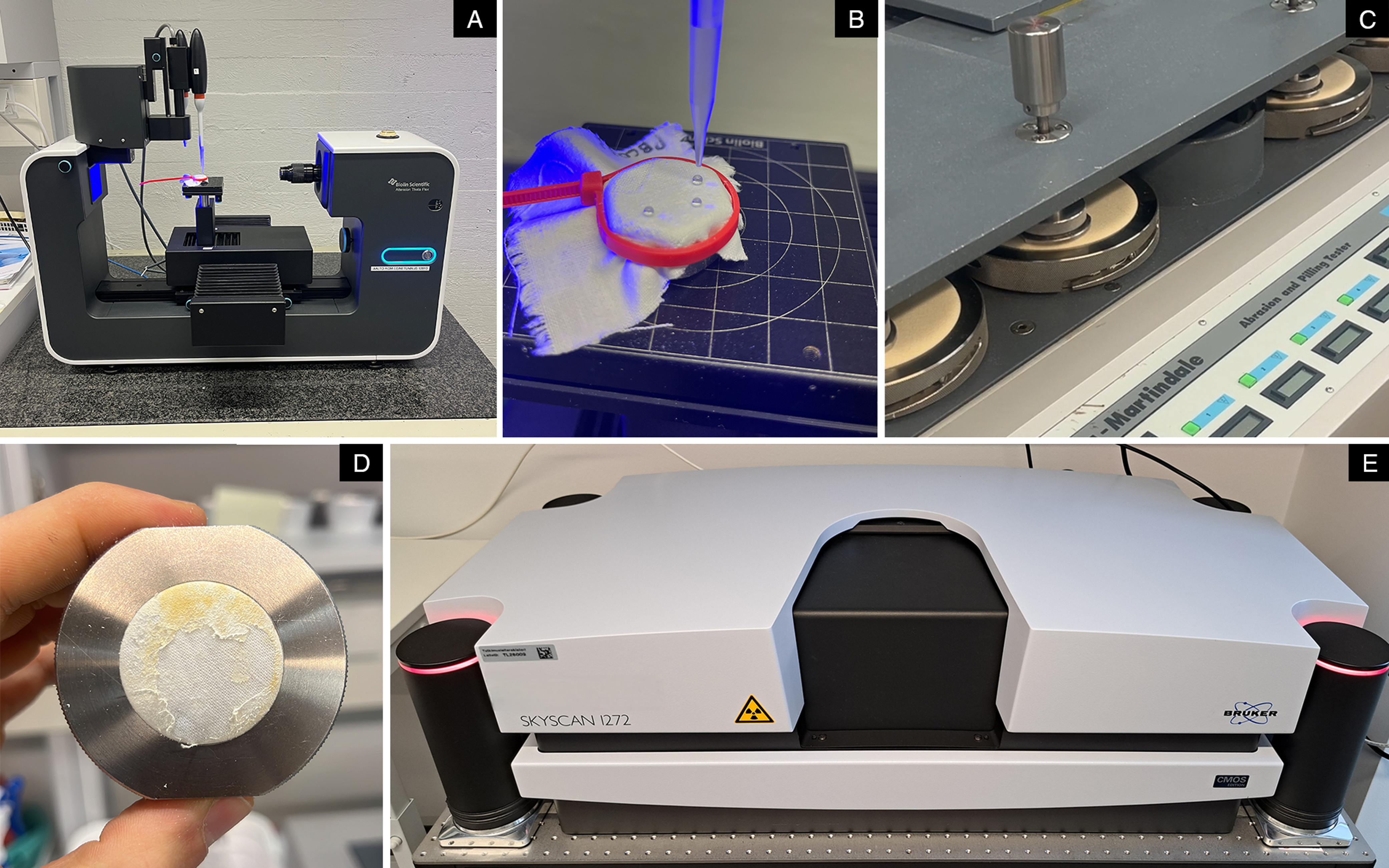
To measure the wetting properties of the mycelium-coated textile, the water contact angle (WCA) was measured using a Theta Flex (Biolin Scientific), applying the sessile drop method with distilled water (Lauren Reference Lauren2020). The WCA values are recorded from the shape of the droplet sitting on the sample surface. Three measurements were taken for each sample (Figure 3a and b).
Material samples under testing: a) WCA test with Theta Flex (Biolin Scientific); b) Material samples after test completion; c) Abrasion Test with Martindale; d) Material samples check during testing; e) X-ray microtomography scanning with Skyscan 1272.

The abrasion resistance of the mycelium-coated fabric was tested with Martindale equipment using a 9 kPa weight (Figure 3c and d). The test was based on the standard EN ISO 12947-1:1998 with some modifications, taking into account the coated nature of the sample. A circular sample with a diameter of 4 cm was rubbed against an abrasive medium and checked at pre-determined intervals (10 rubs until 50, then 50 until 500, then 100 until 1000, then 500 until 1500) until the coating was rubbed away.
Finally, X-ray microtomography qualitative analysis was carried out in collaboration with scientists at VTT to investigate the integration between the mycelium layer and the textile fibres, and evaluate the surface and the extensiveness of the mycelium growth within the samples. A Skyscan 1272 (Bruker, United States) Micro-CT X-ray microtomograph was used to capture 3D images of chosen samples utilising SkyScan 1272 (Bruker) control software (Figure 3e). The scans covered the whole 360° view in a central camera position with pixel size of 1 µm, voltage of 30 kV, current of 150 µA, rotation step of 0.2°, and frame averaging of 4 frames. Using NRecon (Bruker) software, these 360° images were reconstructed into cross-sectional images with a pixel size of 1 µm. These images were used for rendering the 3D view with a voxel size of 1 µm and capturing images from the render using CTVox (Bruker).
Results
In this study, eight experimental setups were developed and replicated to identify the optimal conditions for producing a mycelium-based coating on textiles. The main variables considered were the different nutrient-based pre-treatments applied to the textile to promote mycelial growth, and the two different growth environments adopted during cultivation. This section reports both qualitative and quantitative data obtained from tests performed on mycelium-coated samples. An initial biotinkering activity was followed by the assessment of a total of 58 samples through different analyses and tests. The assessment began with a sensory evaluation, which supported the preliminary qualitative evaluation of coating thickness, cohesion, and surface continuity. This phase was followed by laboratory testing conducted using scientific tools and controlled environments to quantitatively assess mycelium-coating properties, including abrasion resistance and wetting properties. Throughout all testing phases, collaboration between design and scientific disciplines played a central role, enabling the integration of experiential, qualitative observations with quantitative experimental data and supporting a comprehensive interpretation of the results.
Biotinkering activities
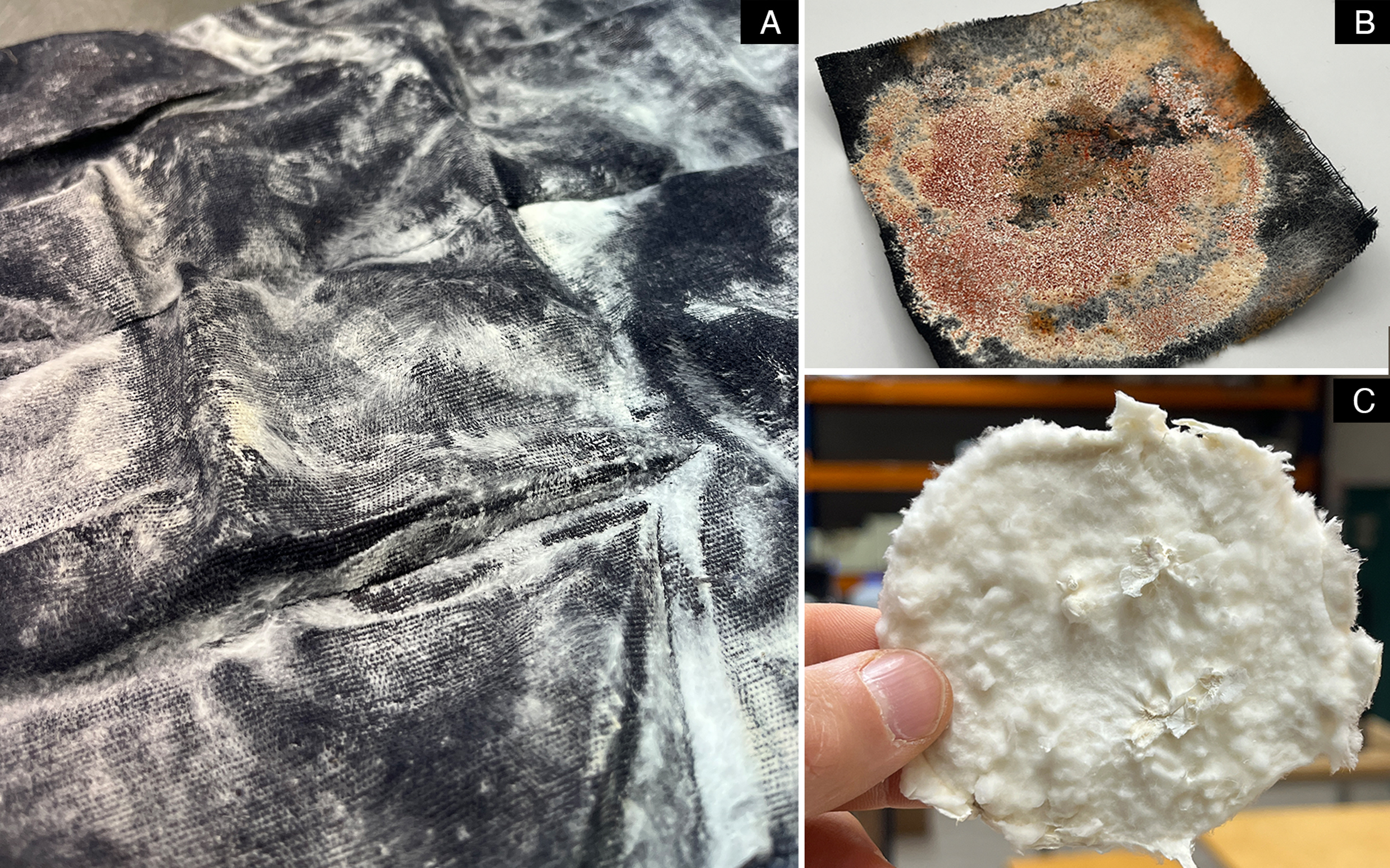
The results revealed that the use of different textiles as bioreceptive surfaces for mycelium growth produced a variety of biocolonisation patterns and coating outcomes. Different fungal strains produced new aesthetic and tactile qualities for the textiles. The use of Pleurotus ostreatus generally resulted in softer, fluffier coatings, whereas Ganoderma lucidum tended to form thinner, denser, and more compact layers. In particular, the presence of nutrients on textiles and the adoption of different cultivation techniques significantly affected the resulting coating. Moreover, when textile substrates were submerged in nutrient agar, mycelial growth tended to develop more homogeneously and continuously compared to samples grown without nutrients. These early exploratory activities not only generated preliminary material samples and situated observations, as shown in Figure 4, but also laid the groundwork for a mycelium-based coating for textile application and further experimental development.
Representative material samples from the biotinkering activities results: a) mycelium growth pattern on denim textile substrate; b) fluffy coating on cotton substrate using Pleurotus ostreatus; c) Red mycelium-based pigmentation and pattern developed with the growth of Trametes Sanguinea mycelium on a black cotton textile surface.

Sensory evaluation
An interdisciplinary sensory evaluation aimed to provide an initial intuitive assessment of the samples, and to support the selection of the most promising techniques and growing conditions for further testing. All the 58 samples were assessed by two designers and two scientists. The analysis primarily focused on the presence and continuity of the mycelium layer, as well as on the preservation of the flexibility of the original cotton textile substrate. The photographic documentation during the growth showed that the samples cultivated on PDA medium exhibited the fastest colonisation, both in the digital incubator and in the CO2 cabinet. As illustrated in Figure 2, mycelial growth was already visible after 3 days, and after 6 days, a continuous mycelium layer had already colonised the entire cotton textile surface. Similarly, samples immersed in PDA (PDAIMPR) showed rapid growth, with visible mycelium appearing within the first few days.
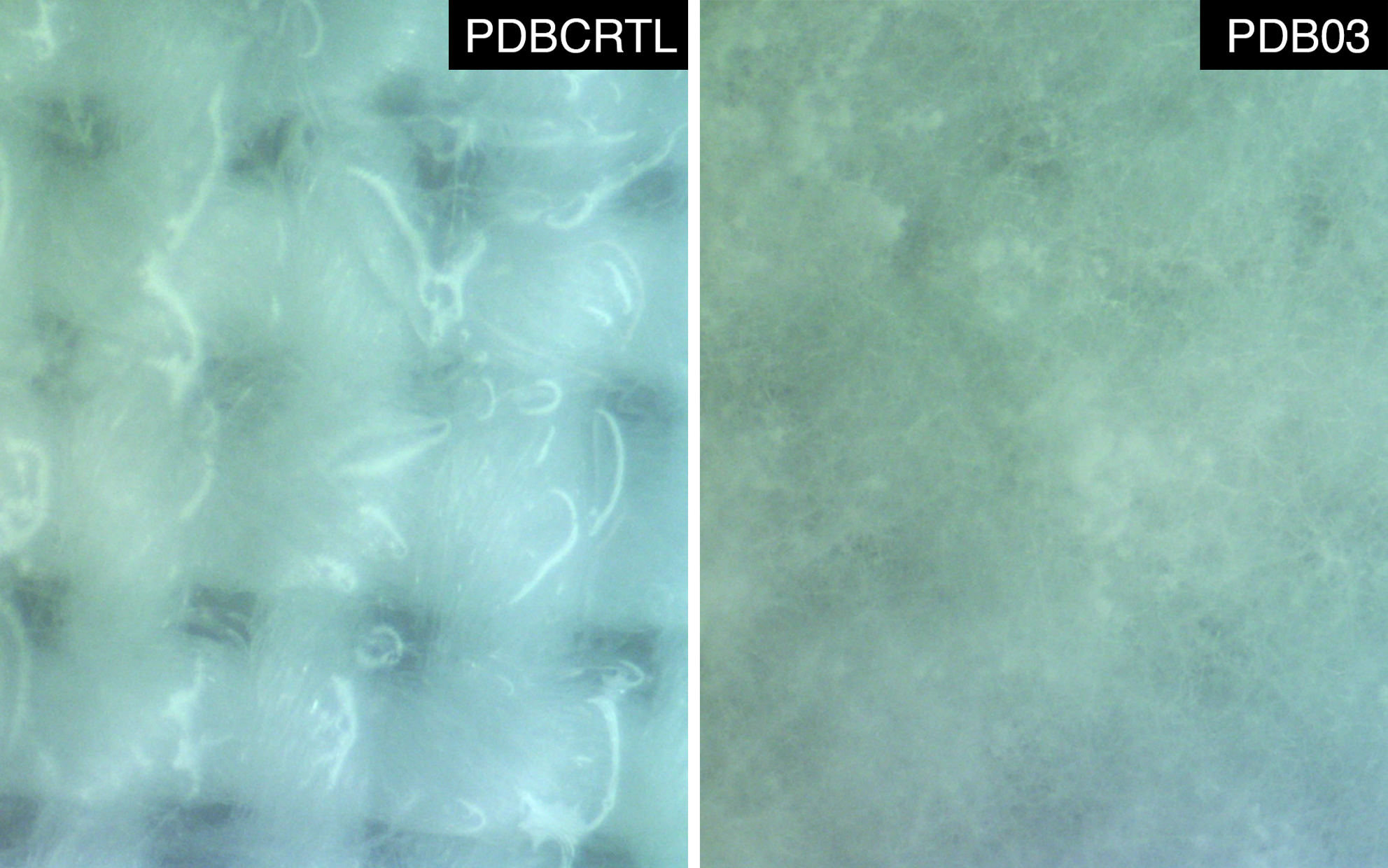
In some cases, the visual assessment of mycelial growth was challenging, as the structures generated by the nutrient pre-treatment of the textile could be mistaken for hyphal colonisation within the fabric. For this reason, an optical microscope was used, as shown in Figure 5, to monitor the development of the mycelium in detail over time and to compare inoculated samples with control samples.
Comparison between control samples and inoculated samples submerged in PDB and cultivated in a CO₂ cabinet (PDBCO2). Optical microscope image captured after 15 days of growth. The image facilitates the analysis, clearly showing how the mycelium grows over and progressively covers the textile surface, forming a continuous coating layer.

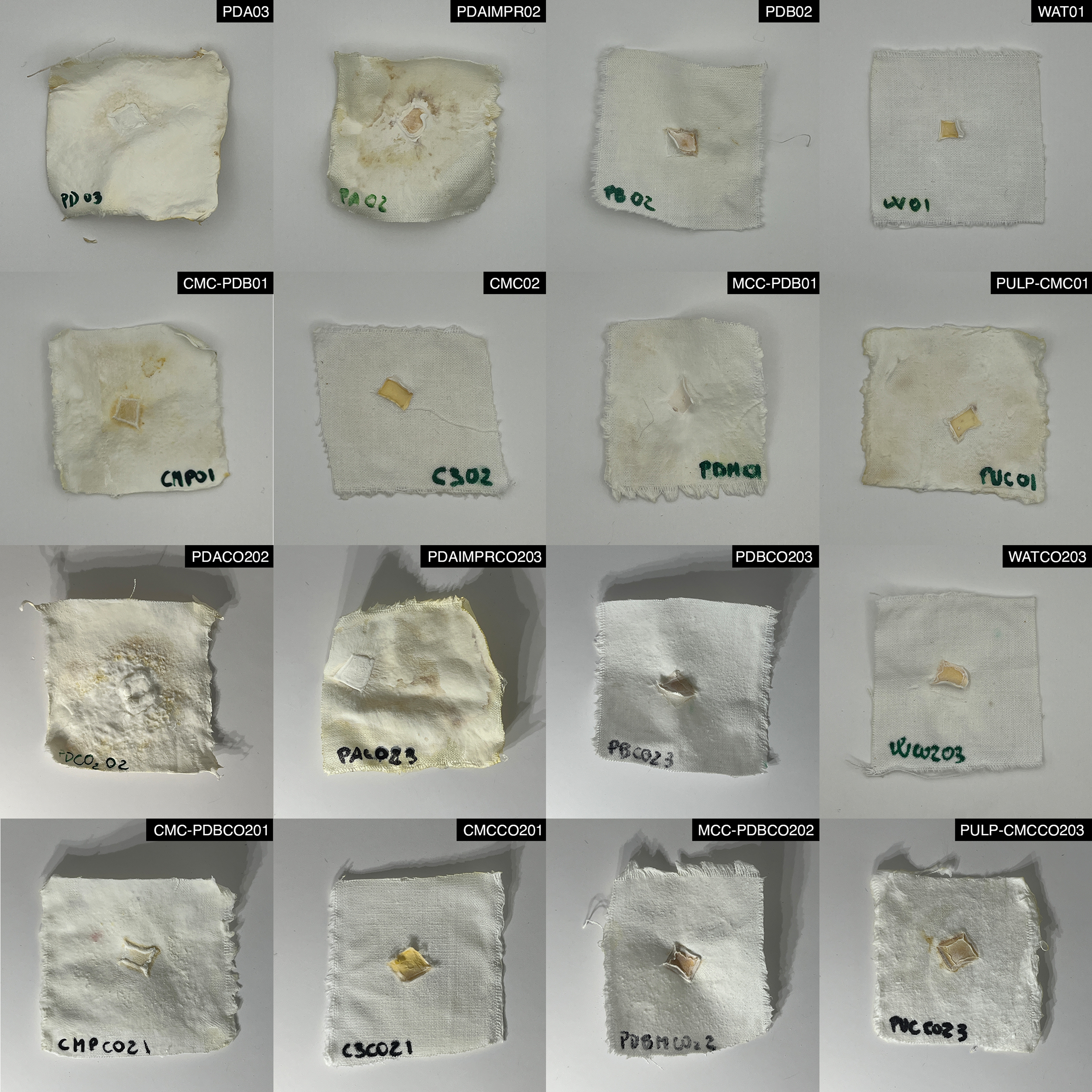
After oven drying, all samples showed reduced mass and increased stiffness. No significant differences were observed between textiles grown in the digital incubator and those cultivated in the CO2 cabinet in terms of coating formation or overall material behaviour (Figure 6).
Representative images of samples of mycelium-coated textiles after drying.

Across all samples with visible mycelial growth, a clear colour change was observed; the original white cotton progressively yellowed, indicating that mycelial colonisation directly affects the chromatic properties of the textile substrate. The mycelium coating did not produce a uniform or standardised finish, instead, it introduced variations in colour, texture, irregularities, and localised densities reflecting the biological growth of the organism. Regarding growth evaluation, PDA samples developed a relatively uniform mycelial layer, fully colonising the textile. PDAIMPR and PDB-treated textiles showed a thinner colonisation concentrated near the inoculation point at the centre of the fabric, while CMC-PDB samples also exhibited satisfactory growth. MCC-PDB samples displayed visible mycelial development but already showed signs of fragility and surface powdering. Samples treated only with distilled water (WAT) or CMC in water (CMC) showed minimal or no mycelial growth and were immediately discarded. In terms of flexibility, PDB samples remained highly flexible, comparable to untreated cotton, likely due to the thin mycelial layer. PDA samples retained good flexibility and present softness and a velvet surface. CMC-PDB and MCC-PDB samples were slightly less flexible, while PDAIMPR samples showed a noticeable reduction in flexibility. Textiles treated with pulp and CMC (PULP-CMC) became completely rigid after drying.
This initial screening thus led to the elimination of PDAIMPR, WAT, CMC, and PULP-CMC samples from subsequent testing, focusing on the most promising growth conditions. Overall, the results suggest that both the presence and the composition of nutrients on the textile influence the growth of the mycelial coating, affecting not only the thickness and growth rate of the mycelium layer but also the flexibility of the resulting bio-coated textile samples.
Contact angle test
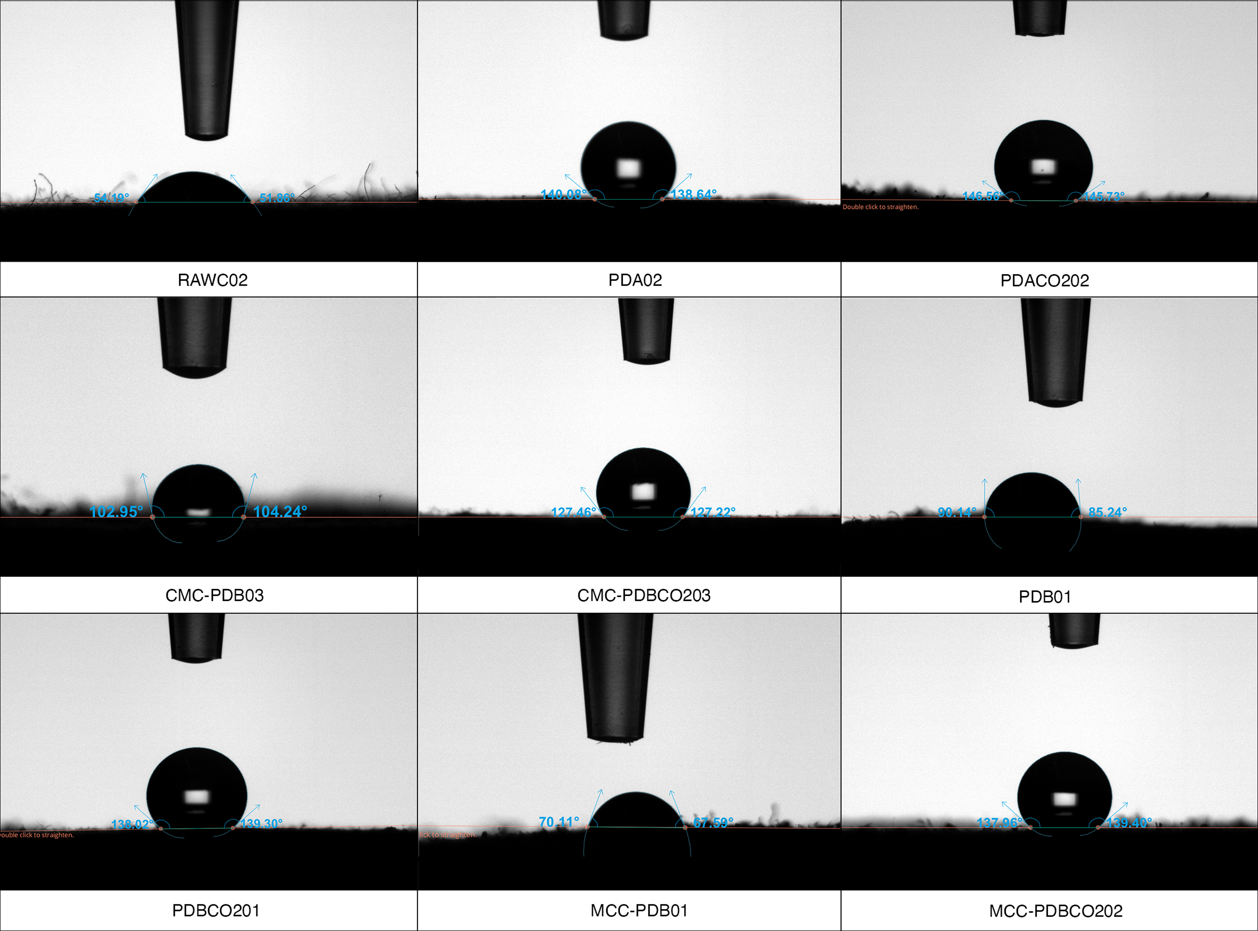
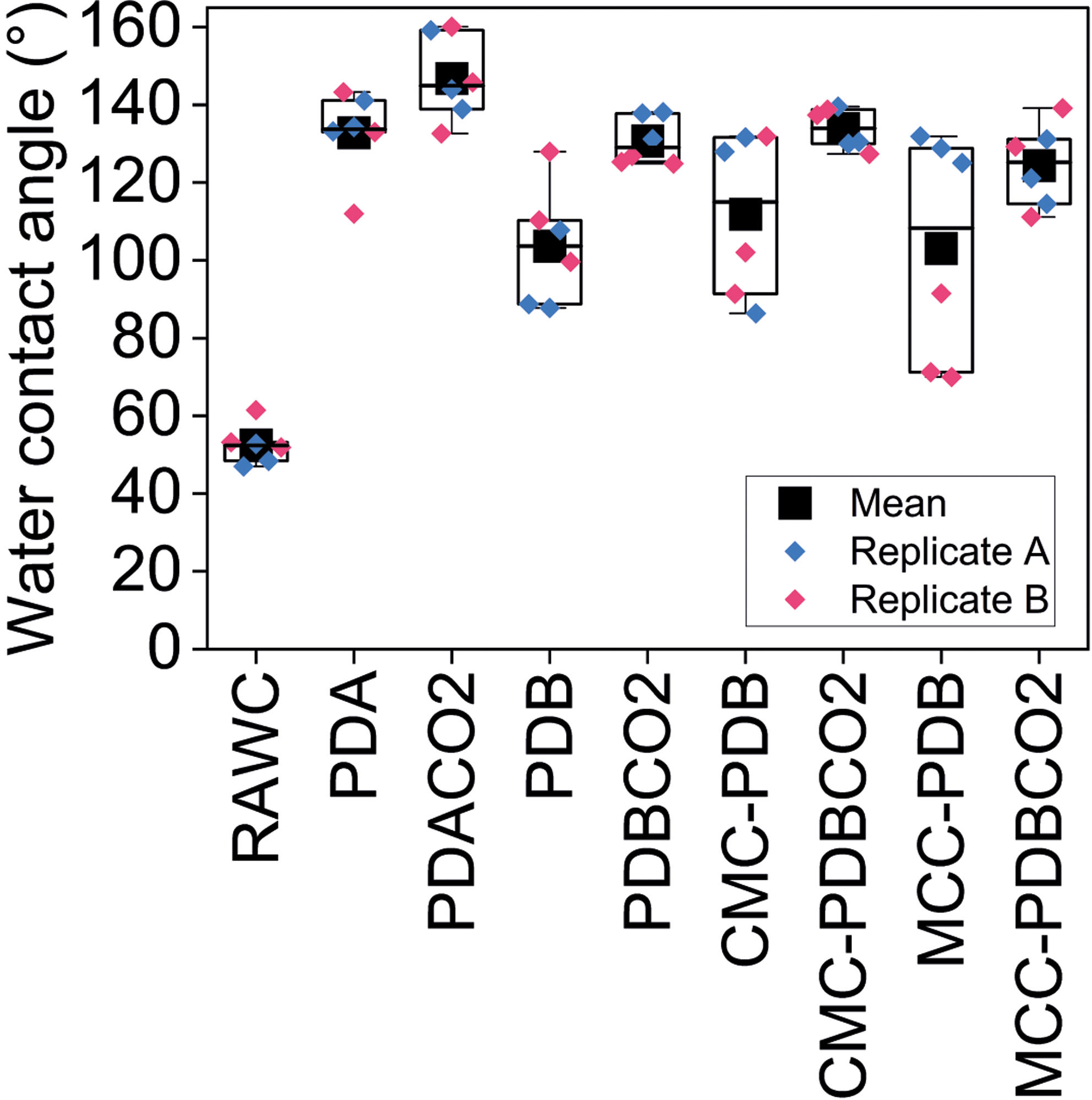
The water-repellent properties of the bio-coated textile surfaces were tested by determining WCA values. In total, 26 samples were tested, including two samples for the raw cotton (the cotton textile without any treatment or mycelial growth used as substrate in this study) and two samples for each of the best-performing techniques . Raw cotton (RAWC) showed a WCA of 53° while all tested mycelium-coated textiles showed clearly increased WCA values ranging from 103–147° (Figures 7 and 8). The results show that, when mycelium was grown at atmospheric CO2 levels, the highest surface WCA reached was 133° on textile/PDA. Cultivation on textiles with PDB, CMC-PDB and MCC-PDB the mycelium layer WCA was lower ranging from 103–112°, but still clearly hydrophobic in nature. Interestingly, when the samples were grown at elevated CO2 levels, the WCA values increased for all samples. The highest WCA at 147° was measured for mycelium coating grown on textile/PDA and 5 % CO2. Particularly for CMC-PDB and MCC-PDB, when the samples were placed under tension during measurement, cracks formed in the mycelium coating. This affected the measurement process but also confirmed the increased stiffness observed in some of the material samples.
Representative images of sessile water drops on treated and untreated textile surfaces for determining WCA values. The RAWC correspond to the cotton textile without any treatment or mycelial growth.

WCA values of the mycelium-coated samples and control samples. Two replicate samples (blue and red diamonds) were measured in three locations. RAWC corresponds to cotton textile without any treatment or mycelial growth used as substrate in this study. Mean values are shown as black squares, median values by horizontal line, 25 and 75 % percentiles as boxes, and the 1.5x interquartile range indicated by whisker lines.

To test whether the media composition had a direct influence on the textile properties, we also determined the WCA values for textiles incubated in each media without mycelium in the same conditions as the mycelium samples. The WCA values for PDA, PDB, and CMC-PDB were similar to those of untreated cotton (49–55°). For cotton incubated with MCC-PDB the WCA was slightly higher at 62°, however, being still clearly hydrophilic.
Taken together, the results show that mycelium grown on a textile surface can provide a hydrophobic layer with increased WCA values. Cultivation in the CO2 incubator was observed to be beneficial for reaching higher surface hydrophobicity. The hydrophobic property of mycelium has been ascribed to the interfacial self-assembly of hydrophobin proteins in the cell wall of aerial hyphae (Wösten et al. Reference Wösten, De Vries, Van Der Mei, Busscher and Wessels1994; Linder et al. Reference Linder, Szilvay, Nakari-Setälä and Penttilä2005). Media composition and growth conditions are likely to influence the production of hydrophobins and other hydrophobic metabolites, as well as on mycelium density and surface microstructure, and consequently surface hydrophobicity. The biological mechanisms underlying this condition-dependent hydrophobicity remain to be elucidated in future work.
Abrasion test
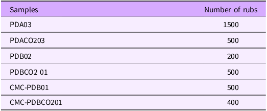
Abrasion tests were conducted to assess the structural resistance of the mycelium coating on the textile substrate under mechanical stress. In order to assess which technique performed best under abrasion, one sample for each of the best-performing techniques identified through the sensory evaluation, 6 samples in total, were tested until the coating was completely removed (Table 3). The absence of replicants provides some statistical limitations to this research but gives indicative results on the functionality of the different coating techniques, which can be used for future optimisation. The results show that the coating grown on PDA had the highest resistance to abrasion with 1500 rubs, this confirms the outcomes of the sensory evaluation, in which these samples were characterised by a thicker and more cohesive mycelial coating.
Resistance to abrasion of the samples tested

Samples with coating grown on textiles submerged in PDB had the poorest resistance to abrasion among all tested samples. This can be attributed to the thin and fragile mycelium layer developed. The other samples show a similar resistance with no substantial differences observed among them. Across all tested samples, especially in solutions where the layer was thicker (as with PDA, PDACO2, CMC-PDB and CMC-PDBCO2) the primary point of coating removal was consistently initiated from the inoculation area, where the coating was absent.
These findings suggest that the abrasion resistance observed represents a promising preliminary indicator of the mechanical performance of mycelium-based textile coatings; this is also relevant when considering the scalability and applicability of this biofabricated coating solution. However, future experiments should focus on eliminating or redesigning the inoculation area in order to improve coating continuity and mechanical durability. Moreover, due to the exploratory nature of this proof-of-concept study, single samples were tested to provide comparative indications of coating performance, with the understanding that additional replicates would be required for comprehensive statistical validation.
X-ray microtomography
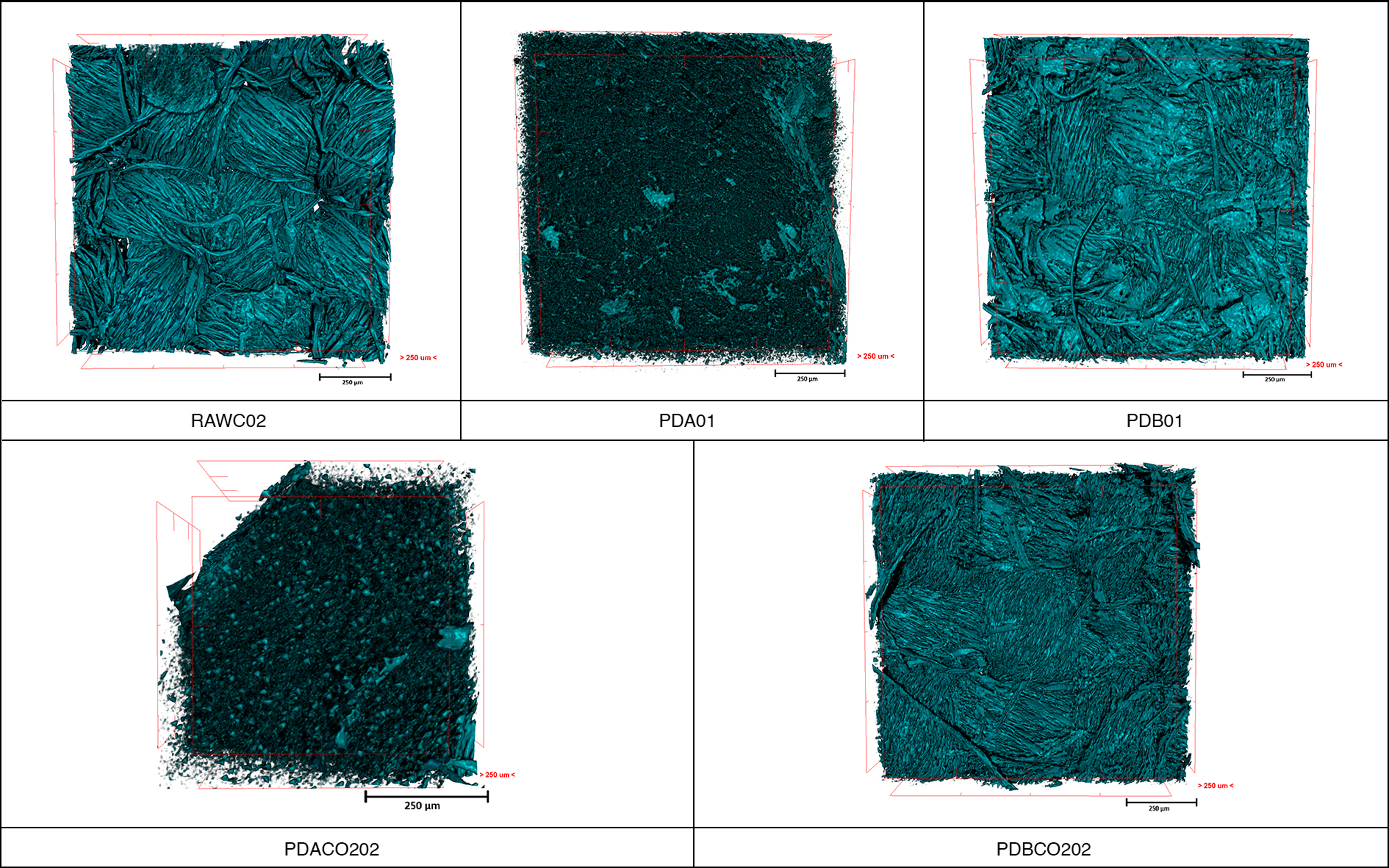
X-ray microtomography was utilised to qualitatively evaluate how well the grown mycelium integrated with the cotton textile and the coverage of mycelium on the surface. A total of 9 samples were analysed, including raw cotton and control samples for comparison. The samples tested consisted of PDA and PDB, grown under the two incubation conditions, the digital incubator and the CO2 cabinet. The 3D reconstructions show that when mycelium was grown using PDB and atmospheric CO2 levels, the mycelium coverage on the textile surface was low and the textile weave pattern remained visible (Figure 9), although to the naked eye the growth of the mycelium on some samples appeared homogeneous. Increasing the CO2 to 5% appeared to promote surface growth, as indicated by a surface layer formation that masked the underlying textile. On the other hand, when grown using PDA the mycelium formed a uniform layer fully covering the textile surface. At elevated CO2, the layer appeared thicker; however, reliable thickness determination was not feasible due to the uneven surfaces and similar densities of the textile and mycelium. Taken together, the results clearly show that PDA promotes mycelium growth on the textile surface and forms a uniform layer. While the imaging provided a preliminary understanding of how mycelium colonises the textile surface, further research is needed to quantitatively assess the thickness and density of the coating layer.
Representative images of X-ray microtomography of untreated cotton and mycelium-coated textile samples, showing the formation and thickness of the developed coating layer. The RAWC correspond to the cotton textile without any treatment or mycelial growth.

Discussion
Mycelium as a transformative agent for textiles
The results and the preliminary test demonstrate that textile can function as a bioreceptive surface for mycelium growth and that biofabrication process involving mycelium may represent a sustainable and regenerative alternative to conventional chemical and environmentally harmful coating processes. In particular, the possibility of growing a mycelium-based coating highlights how mycelium can act as a transformative agent capable of introducing new functional properties and new aesthetic qualities to the textile material, while preserving key textile characteristics such as flexibility. Water contact angle measurements show that water-repellent properties emerge through the growth of a mycelium coating layer on a textile that originally did not present such properties. Similarly, surface hydrophobisation has been observed previously for other mycelium-based materials (Antinori et al. Reference Antinori, Ceseracciu, Mancini, Heredia-Guerrero and Athanassiou2020). The highest WCA values obtained in this study (WCA ∼147°C) for samples cultivated at elevated CO₂ concentrations are comparable to those of other bio-based coating solutions, for example Greenland wax coatings (WCA ∼120°) or colloidal wax coating (WCA ∼145°) (Forsman et al. Reference Forsman, Johansson, Koivula, Tuure, Kääriäinen and Österberg2020).
Although our quantitative tests remain preliminary, the study’s results are particularly promising. Considering the water-repellent behaviour emerged directly through biological growth, rather than through the application of synthetic chemical finishes, a wide range of potential applications might take into consideration the mycelium bio-coating technique, ranging from fashion to packaging. Abrasion test and X-ray microtomography images further confirmed that the mycelium layer establishes physical adhesion to the textile structure by penetrating between the fibres, generating a good integration and mechanical resistance to abrasion of the mycelium coating. This aspect represents an important preliminary parameter for future application scenarios (e.g., in textile and furniture design), although additional testing is required to better assess the durability of the coating and have an appropriate statistical analysis.
Beyond functional performance, mycelium growth strongly influences the aesthetic and tactile qualities of the textile samples. This was particularly evident during the biotinkering activities, where the material samples presented colour variations, fluffy or compact coatings, and emerging living patterns. Also during the interdisciplinary and scientific experiments the agency of the living organism became visible in the final aesthetic of the material samples. The organism responds to the conditions and inputs provided by the designer and scientist, such as nutrient composition, substrate preparation, and growth environment, yet the living organism contributes to the expression and formation of the final aesthetics (Collet Reference Collet2017). Surface irregularities, density variations, and any differences in softness reflect a process in which biological growth actively participates in defining the final aesthetic and the tactile qualities of the textile. As also suggested by the intervention “Imperfection as requirement not defect” of Maurizio Montalti at Aalto University in February 2026 during the Mycelium Design SeminarFootnote 4 , rather than producing perfect and standardised finishes, mycelium introduces a living aesthetic in which variation, imperfections, and the influence of spontaneous and dynamic natural processes become material qualities. This perspective highlights the expressive potential of working with living organisms and supports a new aesthetic paradigm grounded in biological growth processes (Dade-Robertson et al. Reference Dade-Robertson, Kolodkin-Gal and Parker2023).
In this sense, we can state that new fields of action are opening up for designers, situated between a materiality that can be deliberately designed and another that evolves through the spontaneous variability driven by the living microorganisms involved.
Material samples as a facilitator for design and science collaboration
The interdisciplinary collaboration approach adopted in this study positioned material samples not only as outcomes of the research but also as central facilitators of dialogue between design and science. The development and testing of material samples required specialised methods, tools, and disciplinary knowledge that extended beyond traditional design practice, embracing different scientific environments and experimental protocols. This brought the first author, a designer, to work across different scientific environments with varying levels of technological complexity and scientific rigour, moving from a DIY fabrication space, to highly controlled scientific laboratories in order to understand how textiles could function as substrates for mycelial growth and how this process could support the development and a preliminary validation of a low-impact mycelium-based coating for textiles.
Initial exploratory biotinkering practices and material investigations conducted within the BioMakerStudio environment, generated preliminary samples that allowed the designer to reflect, discuss, and develop practical and experience based knowledge (Parisi et al. Reference Parisi, Rognoli and Sonneveld2017) on the possibilities of using mycelium as a coating. These early samples also expanded potential application scenarios and made the designer aware of the functional and aesthetic potential of mycelium-coatings on textiles. Through these exploratory practices, material samples also enabled an initial understanding of the organisms’ needs and behaviours, observing how different substrates, nutrient conditions, and growth parameters influenced biocolonisation patterns and material outcomes.
The experimental phase and the practice of biotinkering proved essential in supporting the evolution of mycelium coating from an initial exploratory dimension into a more substantive and theoretically grounded subject of interdisciplinary inquiry. This experimental biodesign phase enabled the generation of numerous material variables for intuitive assessment, supporting the creative and speculative analysis of diverse applications. This creative investigative stage proved pivotal in informing the subsequent development of a more rigorous and interdisciplinary analytical inquiry. The increasing complexity of the observed results and the need to also validate material performance, lead to the integration of scientific methods, laboratory environments, and standardised testing.
As the study progressed, material samples became central to structuring and facilitating interdisciplinary collaboration. Their presence during interdisciplinary brainstorming and laboratory meetings facilitated communication across disciplines, helping to overcome language barriers by providing tangible objects around which a discussion could take place. To develop the experiments, planned and designed through interdisciplinary brainstorming between designer and scientists, balancing scientific rigour with design-oriented objectives (Marseglia et al. Reference Marseglia, Cantini, Celli, Brunelli and Lotti2025), the designer worked in more structured laboratory environments, such as the Biofilia Lab, a BSL-1 laboratory, assuming the role of “designer in lab,” following scientific protocols and under the supervision of scientists (Sawa Reference Sawa2016; Stefanova Reference Stefanova, Chakrabarti, Poovaiah, Bokil and Kant2021; Pollini Reference Pollini2024).
During the mycelium growth phase, observation and comparison of the material samples, including optical microscope analysis, were conducted in collaboration with VTT Technical Research Centre of Finland, supporting shared reflection and interpretation of growth dynamics. This collaborative setting helped to identify factors that could positively or negatively influence mycelium growth on textiles, such as humidity conditions and substrate treatments. All the information and knowledge that the designer gained from these interdisciplinary sessions were also applied when monitoring the samples growing in the digital incubator placed in the BioMakerStudio, trying to identify similarities, connections, and divergences through the insights previously acquired during the collaborative discussions with the scientists.
In the testing facilities of the School of Chemical Engineering, material samples were evaluated through standardised procedures, including abrasion tests and contact angle measurements, in order to assess the performance of the mycelium-based coating. In these environments, the designer assumed a more observational and assistant role within the laboratory (Crawford Reference Crawford2023), as the use of specialised analytical instruments required advanced technical expertise and specific training.
Throughout all phases, the process was systematically documented through photographs, videos, and detailed written notes recording ingredients, techniques, quantities, measured data, and personal observations about the samples, enabling effective communication and interpretation of the results. Being physically present in the laboratory, even when taking a relatively passive role in the experimental activities, was essential for the designer to gain a deeper understanding of the samples’ material features. This unique position also supported the acquisition of the scientific language necessary, always one of the main challenges in collaboration (Langella Reference Langella2019), to discuss the results with other researchers and interdisciplinary collaborators. Most importantly, scientific protocols, direct observation and replication of the process enabled the designer to immediately recognise the potential of the mycelium coating for textile applications, fostering the ability to propose further tests, expand material design and research lines, as well as possible application scenarios (Rust, 2007 in Driver et al. Reference Driver, Peralta and Moultrie2011; Quijano et al. Reference Quijano, Jing, Ferrero-Regis, Navone, Golberg and Zhang2026).
Overall, the material scenarios developed in this study enabled the scientific disciplines involved to envision concrete application pathways through the systemic perspective of design, translating laboratory concepts and results into potential use contexts and technological development trajectories. In this sense, the project acted as a mediating device, transforming scientific knowledge into operational applications capable of anticipating future technology transfer.
This interdisciplinary process significantly expanded the designer’s role. Starting from an exploratory and intuition based on hands-on material experimentation, the collaboration with scientists enabled the translation of early design insights into scientific protocols and measurable material samples. In this study, interdisciplinary collaboration functioned as a validation for the exploratory phase of the design process, scaling up intuitive material exploration into scientific and transferable knowledge that opens up pathways for further research, as well as new speculative and application-oriented scenarios for mycelium-coated textiles.
Finally, the established collaborations enabled other scientists involved to take part in the ideation process of material design, in turn being influenced by the distinctive divergence of the design process.
Within this study, some limitations and future opportunities have emerged. The aim of the study was primarily to explore, through an interdisciplinary approach, the potential of mycelium-based coating for textiles and to provide insight to further research directions rather than to develop a fully validated solution for industrial or market applications. Although preliminary, the results obtained in terms of mycelium growth, surface adhesion, and hydrophobic performance provide indicative evidence of the feasibility and potential of mycelium-coated textiles.
This study presents limitations that should be addressed in future research; one concerns the number of samples tested, which should be increased to improve the robustness, reproducibility, and statistical reliability of the results. Moreover, the durability of the mycelium-based coating under laundering conditions was not evaluated. Since wash resistance represents a critical requirement for textile finishing applications, future studies should investigate the stability, adhesion, and performance of the mycelium coating after repeated washing cycles.
Another limitation relates to the verification of biological deactivation after the drying process. Although the drying process for mycelium applied in this study (60°C for 120 min) was intended to deactivate biological activity, following the procedure described by Haneef et al. (Reference Haneef, Ceseracciu, Canale, Bayer, Heredia-Guerrero and Athanassiou2017), the viability of the fungal material after drying was not microbiologically verified. Further investigations should therefore include viability assays and safety assessments to confirm the complete inactivation of the mycelium, particularly considering potential applications involving skin-contact textiles and related regulatory requirements.
While this work primarily assessed the technical feasibility of mycelial coating formation, future research should investigate additional performance and user-experience aspects of mycelium-coated textiles, including wearer comfort, breathability, fire resistance, and the broader sensory characterisation as tactile perception and softness. These evaluations, combined with process optimisation, will be essential to understand the scalability and applicability of mycelium-coated textiles in both textile and furniture contexts. Regarding material bioreceptivity studies, further investigations could also establish the minimal nutrient composition needed for mycelium growth and examine how fibre composition, colour, weave, geometry, and porosity might influence mycelium colonisation, deepening the understanding of textile structures as mycelium’s bioreceptive surfaces.
Conclusion
This study demonstrates that mycelium can grow on textiles to produce a biofabricated coating capable of transforming both the material performance and the aesthetic of the textile substrate without relying on synthetic chemical finishes. Overall, the study provides a foundation for future developments in sustainable material alternatives at the intersection of textile and biodesign, suggesting that mycelium-based coatings could contribute to rethinking textile finishing processes in real-world applications, opening the way for future applications in both research and practice within the design and science context.
Beyond its material outcomes, this study presents an interdisciplinary approach in which biodesign research originated from biotinkering, further evolved through scientific collaboration, and ultimately returned to design as an expanded field of action. This cyclical integration not only bridges creative exploration with empirical rigour but also models a collaborative paradigm for sustainable innovation in material biodesign practices.
Data availability statement
For Data available within the article or its supplementary materials: The authors confirm that the data supporting the findings of this study are available within the article.
Acknowledgments
We are grateful to N. Lukkarinen for her technical assistance in conducting the WCA tests and to N. Nygren-Sundell for her support in performing the abrasion tests. We also thank L. Chernyaeva for her guidance throughout the study and for granting access to the Biofilia Lab of Aalto University.
Author contributions
Conceptualisation: Edoardo Brunelli; Barbara Pollini; Szilvay Géza. Methodology: Edoardo Brunelli; Barbara Pollini; Szilvay Géza. Data curation: Edoardo Brunelli, Szilvay Géza, Peuhkurinen Lauri. Investigation: Edoardo Brunelli, Peuhkurinen Lauri, Nygren-Sundell Nicole. Data visualisation: Edoardo Brunelli, Szilvay Géza, Peuhkurinen Lauri. Writing original draft: All auhors. All authors approved the final submitted draft.
Financial support
This work was partly carried out under the Academy of Finland Center of Excellence Program (2022–2029) in Life-Inspired Hybrid Materials (LIBER), project number (346106).
Competing interests
None.
Ethical standards
The research meets all ethical guidelines, including adherence to the legal requirements of the study country.

 Open access

