Aberrant right subclavian artery is an anomaly of the aortic arch that may cause oesophageal compression resulting in dysphagia lusoria. Traditional surgical exposure is via lateral thoracotomy. Reference Austin and Wolfe1 Due to the morbidity associated with this approach, hybrid management incorporating endovascular techniques has been used instead of open surgical repair in adults at some centres. Reference Verzini, Isernia and Simonte2–Reference Saran, Dearani and Said5 Hybrid approaches have not been readily adopted by surgeons for use in the paediatric population given challenges related to both device dimensions and the patient’s growth potential. Furthermore, the aberrant subclavian artery is not resected in hybrid approaches, and there remains the potential for residual mass effect on the oesophagus resulting in persistent symptoms. However, this approach holds some promise in populations of patients who otherwise would not be candidates for open surgical repair. We present a case of dysphagia lusoria in a paediatric patient with Duchenne’s muscular dystrophy and a left arch with aberrant right subclavian artery managed with a hybrid approach.
Case report
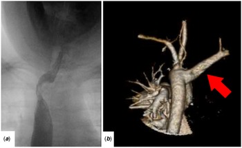
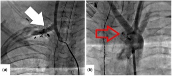
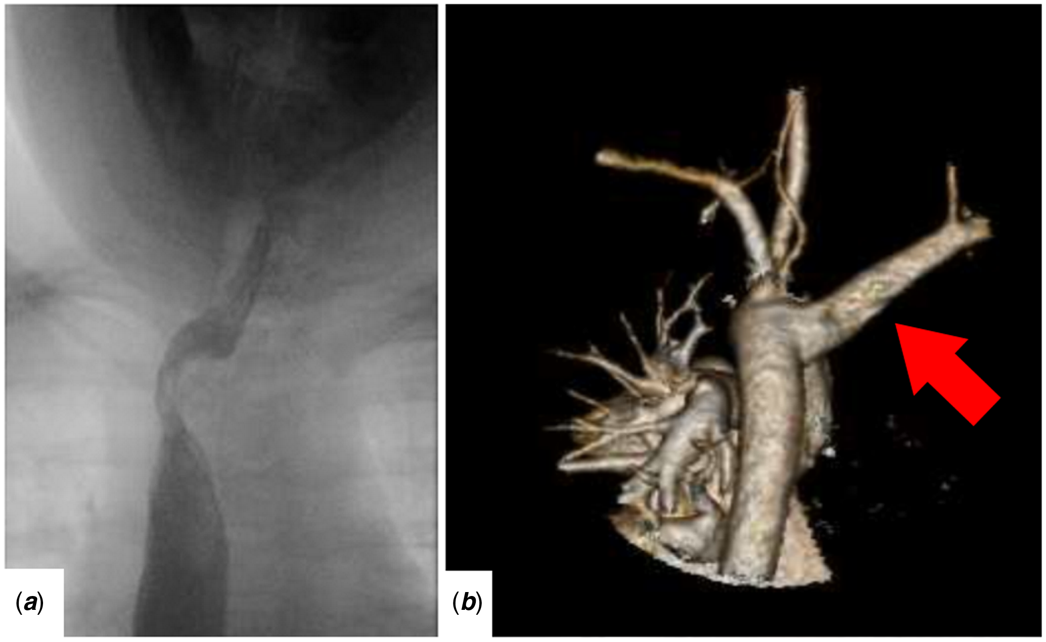
A 9-year-old wheelchair-bound boy with Duchenne’s muscular dystrophy was referred for management of dysphagia in the setting of a left arch with an aberrant right subclavian artery. An oesophagram (Fig 1a) revealed a large oblique impression in the upper thoracic oesophagus with no other evidence of motility issues. Subsequently, CT angiography delineated the retroesophageal course of the right subclavian artery; the base of the aberrant vessel was not aneurysmal (Fig 1b). The boy had progressive symptoms of dysphagia including the inability to swallow meat. As such, an intervention was indicated. However, the patient’s limited mobility, restrictive lung disease, and high-dose prednisone regimen for treatment of Duchenne’s muscular dystrophy made him a high-risk candidate for open repair of the lesion via left thoracotomy. Hybrid revascularisation was discussed with the patient and his parents, and the decision was made jointly to proceed.
Preoperative imaging studies. ( a ) Oesophagram demonstrating distortion of the oesophagus. ( b ) 3-dimensional reconstruction of the aorta and head vessels demonstrating the aberrant course of the right subclavian artery, filled red arrow indicates aberrant right subclavian artery.
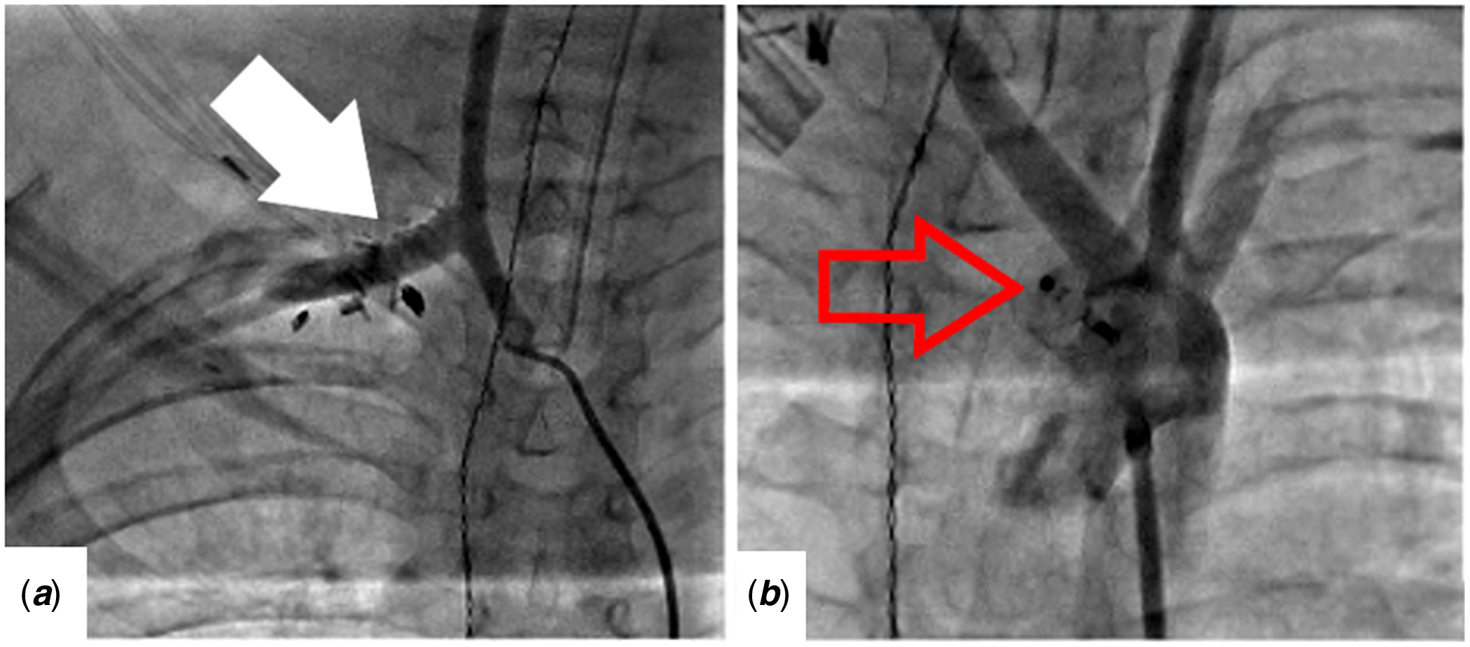
In the hybrid catheterisation laboratory, surgical right carotid-to-subclavian bypass was performed with a 6 mm dacron graft via right supraclavicular incision. The aberrant right subclavian was ligated proximal to the right vertebral artery (Fig 2a). The right common femoral artery was accessed. Due to the short landing zone and proximity of the trachea and oesophagus, a 10 mm × 7 mm Amplatzer Vascular Plug II (St. Jude Medical, Saint Paul, MN) was initially selected. The device was positioned using the oesophageal temperature probe as a landmark and deployed. Shortly thereafter, the device embolised, and endovascular retrieval was performed. A 14 mm × 10 mm device was subsequently selected and deployed making sure to avoid oesophageal or tracheal compression (Fig 2b). Of note, the conical nature of the landing zone resulted in elongation of the device with projection into the lumen of the descending thoracic aorta without any evidence of stenosis or obstruction. The patient subsequently had an uneventful recovery. At discharge, the patient was tolerating a regular diet. He was last seen in follow-up 18 months post-operatively with no symptoms of dysphagia.
Completed reconstruction. ( a ) Right carotid artery angiography demonstrating appropriate flow into the right subclavian artery through the bypass graft, filled white arrow demonstrates opacification of the carotid-subclavian bypass. ( b ) Completion aortogram following deployment of a 14 mm Amplatzer Vascular Plug II (St. Jude Medical, Saint Paul, MN), indicated by hollow red arrow, demonstrating no opacification of the retroesophageal component of the aberrant right subclavian artery.
Discussion
Dysphagia lusoria is a rare entity in the paediatric population, and management has traditionally required thoracotomy for repair. However, open surgical repair has been associated with a high rate of morbidity and mortality among adults. Reference Austin and Wolfe1 Management strategies among adults have evolved to incorporate endovascular techniques for hybrid repair. Reference Verzini, Isernia and Simonte2,Reference van Bogerijen, Patel and Eliason3 However, it should be noted that after hybrid repair, the decompressed aberrant subclavian remains posterior to the oesophagus.
Our patient’s small size introduced concerns about both proximal migration and embolisation of the occluder device on the one hand, and – on the other hand – mass effect on the oesophagus or trachea in the setting of a short landing zone. This underlined the importance of appropriate positioning and sizing. Reference Soo Hoo, Rokkas and Rossi6 An important feature to account for was that oversizing the device elongated the distal aspect of the plug resulting in elongation of the device. This conformational change necessitated projection of the device into the lumen of the descending thoracic aorta. However, oversizing reduced the risk of either migration or embolisation. This presents one of the unique challenges in adapting adult devices to use in paediatric populations as off-the-shelf devices may not be of the appropriate length, diameter, or combination of the two for paediatric anatomy.
In the adult population, aneurysms of the aberrant right subclavian artery are more common, and placement of a thoracic endograft is required in almost all cases. Reference Verzini, Isernia and Simonte2,Reference van Bogerijen, Patel and Eliason3,Reference Jalaie, Grommes and Sailer7 Thoracic endovascular aortic repair was not pursued in this patient due to the lack of growth potential for a covered stent graft in a very young patient. The 6 mm bypass graft used in this patient has the same lack of growth potential, and this represents a limitation of this technique.
Utilisation of existing hybrid techniques is challenging in children. Increasing experience with these techniques in children will potentially allow for a reduction in morbidity and mortality when operating on high-risk populations. Use of less-invasive techniques may allow surgeons to offer interventions to patients not suitable for open repair; however, this technique is not a replacement for traditional open surgical repair in patients who are surgical candidates. As the adult experience evolves and additional devices are brought to market, there will be increasing opportunities for adaptation of endovascular technologies to children.
Acknowledgements
None.
Financial support
This research received no specific grant from any funding agency, commercial or not-for-profit sectors.
Conflicts of interest
None.
Ethical standards
All procedures performed in studies involving human participants were in accordance with the ethical standards of the institutional and/or national research committee and with the 1964 Helsinki declaration and its later amendments or comparable ethical standards.
Informed consent
The institutional review board of Stanford University waived the need for informed consent on account of the study being a case study and not qualifying as research.