Introduction
Ichthyophthirius multifiliis is a holotrichous protozoan that invades the gills and skin surfaces of fish and can cause morbidity and high mortality in most species of freshwater fish worldwide. The morbidity rate due to this disease may reach up to 100%, causing great economic losses in aquaculture and ornamental fish breeding (Osman et al., Reference Osman, El-Bana, Noor El Deen and Abd El-Hady2009).
To cope with the parasitism and deleterious consequences, various parasiticides have been used in aquaculture. Formerly, ichthyophthiriasis was treated effectively with malachite green and mercurous nitrate, however, due to their genotoxic carcinogenesis, the two agents have been banned in most countries (Alderman, Reference Alderman1985; Wahli et al., Reference Wahli, Schmitt and Meier1993; Li et al., Reference Li, Wise and Robinson1996). Since the ban, other chemotherapeutants, such as formalin (Rowland et al., Reference Rowland, Mifsud, Nixon, Read and Landos2009), chlorophyllin (Wohllebe et al., Reference Wohllebe, Richter and Häder2012), chloramine-T (Rintamäki-Kinnunen et al., Reference Rintamäki-Kinnunen, Rahkonen, Mannermaa-Keränen, Suo-malainen, Mykrä and Valtonen2005), copper sulphate (Rowland et al., Reference Rowland, Mifsud, Nixon, Read and Landos2009), potassium ferrate (VI) (Ling et al., Reference Ling, Wang, Wang and Gong2011), potassium permanganate (Straus and Griffin, Reference Straus and Griffin2002), tricaine methanesulfonate (Xu et al., Reference Xu, Shoemaker and Klesius2008) and bronopol (Shinn et al., Reference Shinn, Camacho, Bron, Conway, Yoon, Guo, Taylor and Ni2012) have been evaluated for their antiparasitic efficacy. However, the treatments commonly used in commercial aquaculture as alternatives to malachite green are of low efficacy, cause environmental problems or are unlikely to receive regulatory approval (Dickerson and Dawe, Reference Dickerson, Dawe and Woo1995; Tieman and Goodwin, Reference Tieman and Goodwin2001). Vaccination against the parasites may provide an alternative to chemical treatment (Goven et al., Reference Goven, Gilbert and Gratzek1980; Klinger and Floyd, Reference Klinger and Floyd2002). But the cost is high and this cannot be ignored for fish farmer, restraining its widespread use. Therefore, an effective therapy has to be improved and new drugs with novel mechanisms of action are thus urgently required.
Recently, there have been increased research activities into the utilization of environment-friendly products to control I. multifiliis infection (Yao et al., Reference Yao, Shen, Li, Xu, Hao, Pan, Wang and Yin2010, Reference Yao, Zhou, Li, Yin, Ru, Pan, Hao, Xu and Shen2011; Yi et al., Reference Yi, Lu, Hu, Ling and Wang2012). Effective compounds of natural original expected to be more advantageous than synthetic antiparasitic agent, as they have generally a lower environmental impact and are easily biodegradable. Thus, a great number of biological control microorganisms have been screened and researched, including fungi (Crump et al., Reference Crump, Sayre and Young1983; Chen and Dickson, Reference Chen and Dickson1996), bacteria (Becker et al., Reference Becker, Zavaleta, Colbert, Schroth, Weinhold, Hancock and Vangundy1988), ray fungi and protozoon (Chen and Liu, Reference Chen and Liu2005; Sun et al., Reference Sun, Gao, Shi, Li and Liu2006). Streptomyces are distributed widely in terrestrial and marine habitats and are of commercial interest due to their unique capacity to produce novel metabolites. The genus Streptomyces was classified under the family Streptomycetaceae, which includes Gram-positive aerobic members of the order Actinomycetales and suborder Streptomycineae within the new class Actinobacteria (Anderson and Wellington, Reference Anderson and Wellington2001). They produce approximately 75% of commercially and medically useful antibiotics and 60% of antibiotics used in agriculture (Watve et al., Reference Watve, Tickoo, Jog and Bhole2001).
In our previous study, we investigated the anti-I. multifiliis efficacy of extracellular products of Streptomyces griseus SDX-4 against I. multifiliis, and an active compound, Nystatin, was isolated by using the strategy of bioactivity-guided isolation (Yao et al., Reference Yao, Li, Li, Xu, Ai and Shen2015). It lead us to consider if Streptomyces albus, as another species of this genus, is also capable of controlling the parasite. The principal objective of this study was to assess the antiparasitic properties of S. albus JSY-2 and isolate active constituents responsible for the activity using in vitro antiparasitic assay associated with bioassay-guided fractionation. Additionally, the acute toxicity of the active compounds against Erythroculter ilishaeformis was evaluated.
Materials and methods
Parasites and hosts
Strains of I. multifiliis were maintained in our laboratory. The collection and propagation of I. multifiliis cysts were directed following the protocol as described in our previous study (Yao et al., Reference Yao, Li, Li, Xu, Ai and Shen2014). Several heavy infected E. ilishaeformis were placed into filtered aquarium water for 30–60 min. Mature trophonts were allowed to dislodge from the host by body movements of the fish whilst in close proximity. Isolated trophonts were randomly divided into two batches, one was used to assay the activity of the fraction and active compound isolated from S. albus JSY-2 for killing the tomonts, and the other placed in plastic beaker with aerated groundwater filtered and incubated at 23.5 ± 0.5 °C for 18–20 h, and theronts were allowed to emerge naturally. After theronts were released from cysts in water, the total number of hatched theronts were estimated.
Source of uninfected fish
Erythroculter ilishaeformis, weighing approximately 50 g each, were obtained from comprehensive test base of our institute and maintained in a 1 m3 tank at 24 ± 1 °C. All fish were acclimatized to laboratory conditions (dissolved oxygen >6 mg L−1; nitrites <0.02 mg L−1; ammonia <0.1 mg L−1) for 7 days. Prior to experiments, the skin surface and gills of 10 randomly selected fish were examined to confirm that they were not infected with gill or skin parasites.
Bacterial strains
Strain S. albus JSY-2, isolated from a soil sample of East Sea in Zhoushan, Zhejinag province, China, was inoculated in ISP Medium No.1.
In vitro anti-parasitic efficacy against the theronts and tomonts
In vitro anti-parasitic efficacy of fractions and isotated compounds from S. albus JSY-2 against theronts and tomonts were performed as described in our previous work (Yao et al., Reference Yao, Shen, Li, Xu, Hao, Pan, Wang and Yin2010; Ling et al., Reference Ling, Wang, Lu, Wang, Lui and Gong2012). Briefly, approximately 100 viable theronts were distributed to each well of a 24-well tissue culture plate (Becton Dickinson Labware, NJ, USA) and exposed to test samples. Mortality of theronts in each well was recorded by microscopic examination (×100 magnification) at 10 min, 1, 2, 3 and 4 h after exposure. A negative control was included using aerated groundwater containing the same amount of DMSO as the maximum concentration test group. The trial was repeated three times.
As for tomont, 30 tomonts were distributed to each well of a 24-well tissue culture plate and exposed to various concentrations of test samples. After treating for 6 h, the test samples were replaced by aerated groundwater containing no test sample. A negative control was also conducted as described in theronts test. Then the plates were incubated at 23 °C throughout the trial. When all the tomonts in the control group were incubated as theront stage, the trail is stopped. At the end of the trial, the dead tomonts were recorded and the mortality was counted. The parasites with the absence of internal cell motility or abnormal cell division and the ones that cannot produce the theronts were considered dead. All treatment and control groups were conducted with three replicates.
Bioactivity-guide isolation of pure compound from S. albus JSY-2
The chromatographic separation was monitored by the strategy of bioactivity-guided isolation (in vitro tests guided), only the extracts or fractions that showed strong activity against I. multifiliis were subjected to further separation and purification.
The 348 L fermentation broth of S. albus JSY-2 was filtered and then concentrated to dryness (187.9 g) by a vacuum freeze dryer (Chemical-free, Operon Co., Ltd., Korea). The dry powder was suspended in distilled water, and then was extracted successively in a separating funnel with petroleum ether, ethyl acetate, chloroform and n-butanol. Each extract was collected and then subjected to in vitro test; chloroform extracts that showed the highest antiparasitic efficacy were then subject to column chromatography on a silica gel and sequentially eluted with ethyl acetate and methanol with increasing polarity which resulted in five fractions. These five fractions were submitted to in vitro test, and Fr. C was the most active (EC50 for theronts and tomonts were 32.5 and 38.8 mg L−1). Fr. C was then applied to macroporous resin HP20 (lvbaicao chemical Co., Ltd., Beijing province, China) as determined by preliminary experiments and eluted with 20% methanol (5.0 mL min−1 flow rate, 30 °C column temperature). A crystal was obtained from Fr. C which was isolated by the above method. The chemical structure confirmations of the pure compound were accomplished by comparing its ESI-MS, 1H NMR and 13C NMR data with the literature values, the compound was identified as a known chemical, salinomycin (SAL), with the molecular formula of C42H70O11, the molecular weight is 571.
In vivo efficacy of SAL against Ich
One hundred and twenty uninfected E. ilishaeformis, weighing approximately 30 g each, were transferred to 100 L tanks and then approximately 6000 theronts/fish were put into the tanks, the fish were held in the tank for 6 h with gentle aeration to promote infection. After exposure, the fish were divided into four groups and each group was transferred with 10 infected grass carp and corresponding concentrations of SAL (2.0, 4.0, 8.0 mg L−1), the control group is treated with no chemical. Triplicated tanks were used in each concentration. The SAL in each tank was replaced on days 3 and 5 with a fresh solution at the same concentrations.
On day 5 after exposure, three fish of each group were placed into 50 L tanks containing filtered aquarium water without extracellular products, 10 glass slides were placed in each tank to collect trophonts and the number of trophonts was counted. For each treatment, 20 trophonts were distributed to each well (N = 3) of a six-well tissue culture plate and allowed to attach. The water in each well was removed carefully, and 5 mL of filtered aquarium water was added to each well. Mortality and reproduction of tomonts in each well were determined, the test were terminated when the parasites in the control groups reached the theront stage.
Ten days after exposure, fish mortality in each group was recorded and the number of parasites on surface (gills and fins) of fish were also counted. Fish were carefully observed every hour for any signs of distress indicative of erratic behavior, the fish was picked up to count the trophonts on the gills and fins as soon as dead. Fish mortality was recorded daily and the parasites on the gills and fins of fish were counted.
Acute toxicity of SAL on E. ilishaeformis
The acute toxicity of SAL was carried out according to the method of Yao et al. (Reference Yao, Shen, Li, Xu, Hao, Pan, Wang and Yin2010) with slight modifications. The tests were conducted in 80 L glass tanks, each containing 50 L of test solution and 10 healthy E. ilishaeformis. Dilutions were prepared from the stock solution as the following concentrations: 30.0, 35.0, 40.0, 45.0, 50.0, 55.0 and 60.0 mg L−1 for SAL. The tests were conducted in triplicate, as well as controls (under the same test conditions with no chemicals). Fish mortalities in the treatment and control groups were recorded after 48 h of exposure, median lethal dose (LD50) was calculated.
Data analysis
All data in this study were performed using the SPSS 19.0 probit procedure, and the homogeneity of the replicates of the samples was checked by the Mann–Whitney U test. Tomont survival and tomont reproduction were compared with Student–Newman–Keuls test procedure for multiple comparisons (α = 0.05).
Results
In vitro antiparasitic efficacy of SAL against I. multifiliis theronts and tomonts
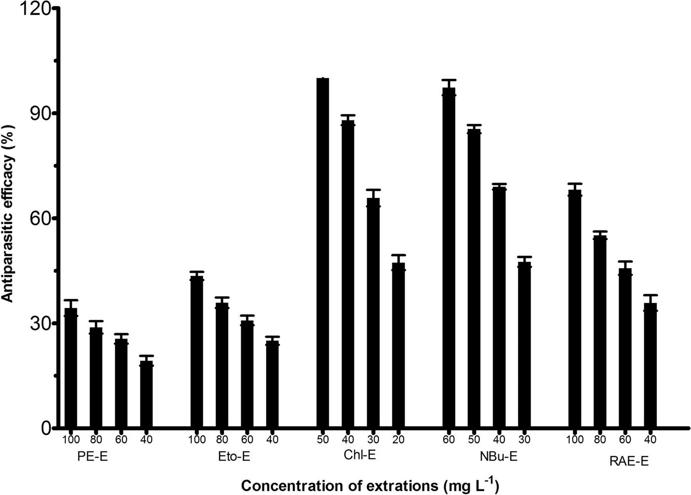
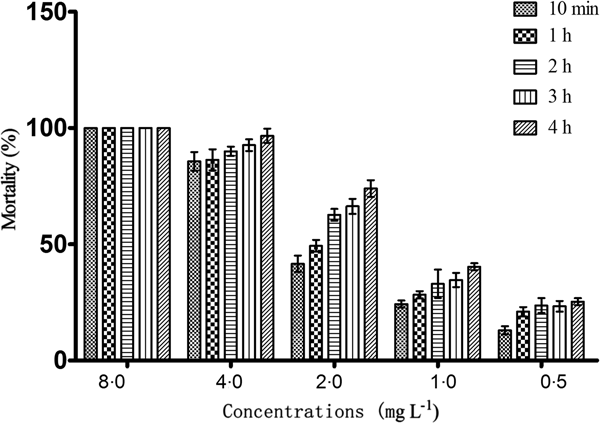
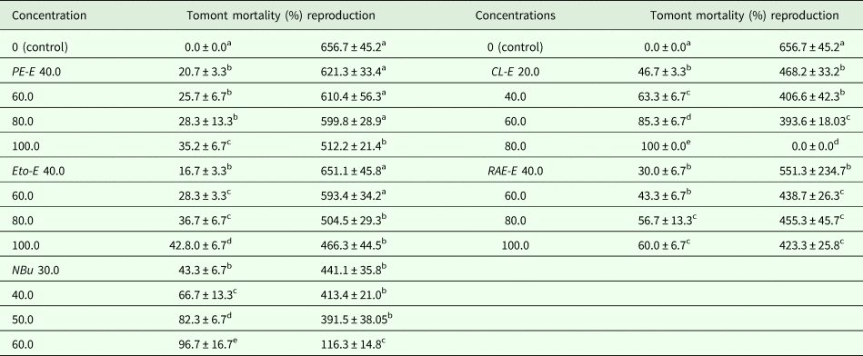
Five extractions of extracellular products from S. albus JSY-2 were submitted to in vitro test; the obtained results showed that Chl-E was the highest in antiparasitic efficacy among the five extractions, and the antiparasitic efficacy of the Chl-E against I. multifiliis theronts in three duplicated trials was 100% at a concentration of 50.0 mg L−1 (Fig. 1). In vitro test also revealed that Chl-E could kill all the tomonts when the concentration reached 50.0 mg L−1 (Table 1), so it was then subject to further separation. SAL was separated from Fr. C by bioactivity-guide isolation and was then subjected to in vitro test to determine the active concentrations. The results of SAL against I. multifiliis theronts were depicted in Fig. 2. As shown in Fig. 2, SAL concentrations vs I. multifiliis theront mortality demonstrated a distinct dose–response and a time–response relationship. The 10 min, 1, 2, 3 and 4 h EC50 [95% confidence interval (CI)] of the compound against theronts of I. multifiliis were 2.12 (2.22–2.02), 1.93 (1.98–1.88), 1.42 (1.47–1.37), 1.35 (1.41–1.31) and 1.11 (1.21–1.01) mg L−1, respectively.
Anti-parasitic efficacy of extractions of extracellular products of S. albus JSY-2 against I. multifiliis theronts after 4 h exposure.

Anti-parasitic efficacy of SAL against I. multifiliis theronts after 4 h exposure.

Ichthyophthirius multifiliis tomont survival and reproduction after 6 h exposure to the extractions of S. albus JSY-2

The reproduction was represented as the number of theronts released by each live tomont. Each value is expressed as mean ± s.d. of three replicates, and within a column, values followed by the different letters are significantly different (P < 0.05, only compared with control).
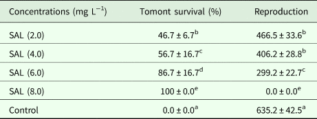
The results of in vitro trial on the effect of SAL against tomonts were listed in Table 2. All tomonts were dead when exposed to SAL at 8.0 mg L−1, and no theronts were released. The mortality of tomonts exposed to 6.0, 4.0 and 2.0 mg L−1 were 86.7, 56.7 and 46.7%, respectively. Tested compound led to an obvious dose-dependent lethal effect against tomonts.
Ichthyophthirius multifiliis tomont survival and reproduction after 6 h exposure to SAL

The reproduction was represented as the number of theronts released by each live tomont. Each value is expressed as mean ± s.d. of three replicates, and within a column, values followed by the different letters are significantly different (P < 0.05, only compared with control).
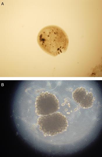
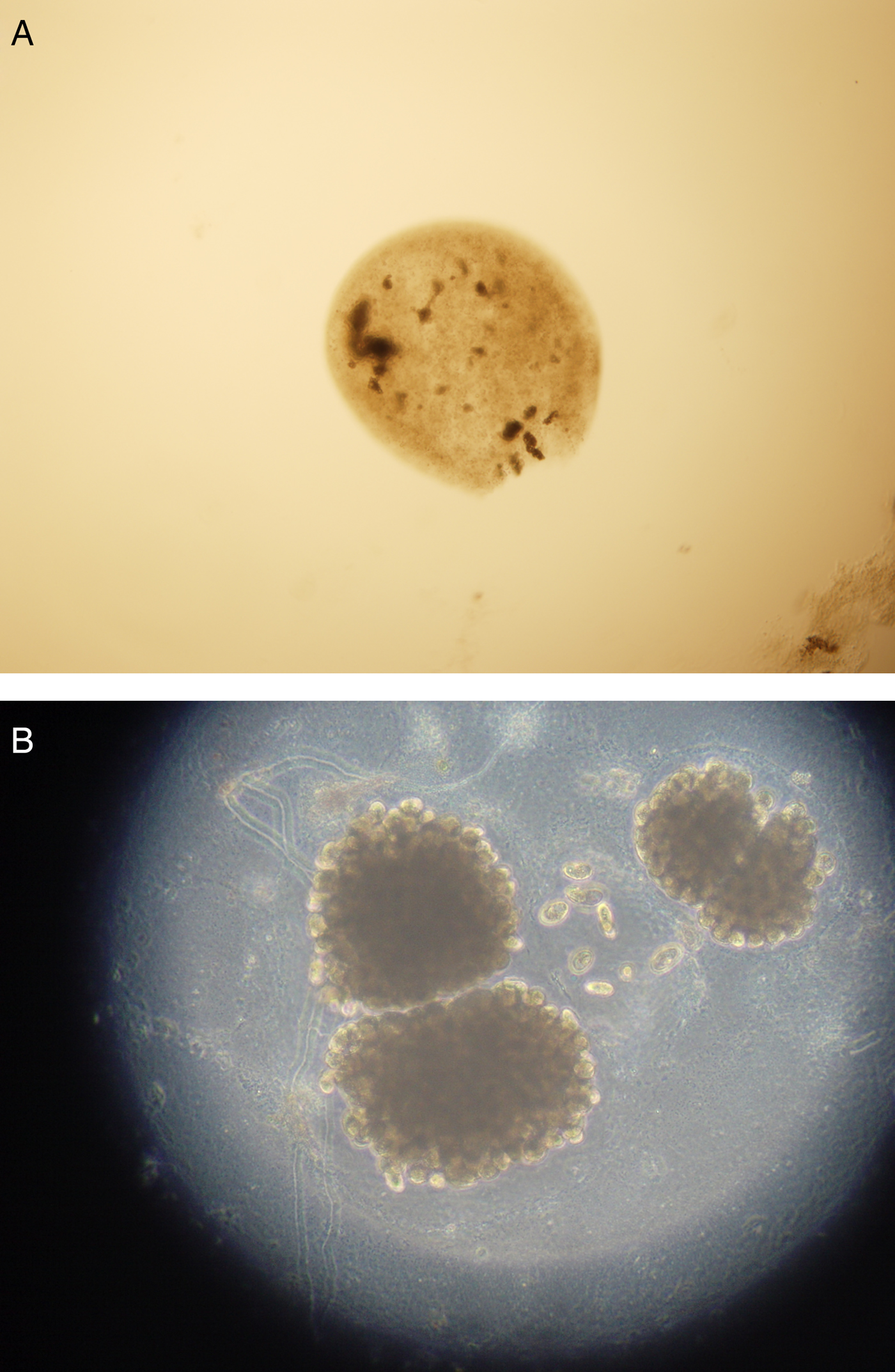
A noticeable effect on I. multifiliis tomonts was observed in treatment groups: the outer cell membrane of tomonts was destroyed, macronucleus was invisible, cell division were stopped and the cytoplasm of the tomonts was characterized by vacuoles (Fig. 3). In contrast, tomonts in the control groups were not affected during the observations in all trials after 6 h exposure.
Morphology of I. multifiliis tomonts.
In vivo efficacy of SAL against Ich
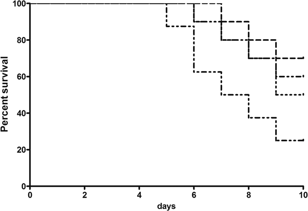
After 3 days infection, white spot (I. multi fi liis) can be easily found in the control group, on day 5, mortality was first recorded due to I. multifiliis infection in the control group. In total, 80.0% fish were died at 10 days in the control group after starting the trail (Table 3). In contrast, I. multi fi liis can be easily observed on day 4 in SAL-treated groups, and the number of parasites was significantly lower than that of the control group (Table 3). The first mortality was recorded on day 7, 30.0% mortality was recorded at 10 days after the exposure (8.0 mg L−1 SAL) (Fig. 4), among the remaining fish, the average number of I. multi fi liis trophonts on the gills and fins was 368.8. Furthermore, SAL at a concentration of 8.0 mg L−1 significantly decreased I. multifiliis tomont survival and reproduction when the tomonts were collected from infected fish bathed for 5 days, whereas there was no significant difference in tomont survival and reproduction between the fish in the control groups and the fish treated with 2.0 and 4.0 mg L−1 SAL.
Survival rate of infected E. ilishaeformis after treated with SAL.

In vivo efficacy of SAL against Ich at 10 days

The reproduction was represented as number of theronts released by each live tomont. Each value is expressed as mean ± s.d. of three replicates, and within a column, values followed by the different letters are significantly different (P < 0.05).
Acute toxicity of SAL
The fish tolerated SAL at a concentration of 30.0 mg L−1 for 48 h without visible effects, but exposure to 35.0 mg L−1 resulted in an increased opercular movement and erratic behaviour of fish within 4 h; all fish died in tanks treated with 60.0 mg L−1 of SAL; the linear equation was y = 3.0929x–79.607 (R 2 = 0.9894), which was derived from the regression analysis of probit mortality of E. ilishaeformis in test solution bioassay; the calculated LD50 of SAL was 32.9 mg L−1 with the 95% CI of 32.2–33.6 mg L−1. There was no fish mortality occurred in the control groups during the experiments.
Discussions
The ciliate I. multifiliis is one of the most pathogenic parasites of fish, and it constitutes a major disease problem in aquaculture (McCallum, Reference Mccallum1985, Reference Mccallum1986; Dickerson and Dawe, Reference Dickerson, Dawe and Woo1995; Bisharyan et al., Reference Bisharyan, Chen, Hossain, Papoyan and Clark2003). Currently, most countries have few effective chemical treatments that can be used to control I. multifiliis (Sudová et al., Reference Sudová, David, Wienke and Meinelt2010). More and more efforts have been spent on searching for more effective anti-I. multifiliis drugs. Microbial secondary metabolites are chemically complex and diverse; there is a tremendous potential to identify novel organisms with various biological properties. In the present study, anti-I. multifiliis compounds were separated from S. albus JSY-2 by bioactivity-guided isolation and identified as SAL. The in vivo tests revealed that bath treatments with SAL at the concentration of 8.0 mg L−1 for three times were significantly effective against I. multifiliis. To the best of our knowledge, it seems to be the first report demonstrating the protection efficacy of SAL from parasite infection for fish. This result extended the general knowledge about the antiparasitic activity of SAL to control fish parasite. It also highlights a new way to explore novel active compounds from bacterial extracellular products to combat fish disease.
SAL, a polyether ionophore antibiotic isolated from S. albus, was originally used to eliminate bacteria, fungi and parasites (Miyazaki et al., Reference Miyazaki, Shibuya, Sugawara, Kawaguchi and Hirsoe1974). Moreover, SAL improves the efficacy of gemcitabine in pancreatic cancer (Zhang et al., Reference Zhang, Liang, Zhou, Chen, Chen, Zhang, Kang and Zhao2011), and sensitizes breast cancer cells to the effects of doxorubicin and etoposide treatment by increasing DNA damage (Kim et al., Reference Kim, Chae, Kim, Kim, Kang, Kim and Yoon2011). Dominant mechanism of SAL activity involves the formation of lipid-soluble complexes with the monovalent cations (particularly K+), thereby promoting cation transport across membranes and increasing the osmotic pressure in the coccidian (Mitani et al., Reference Mitani, Yamanishi and Miyazaki1975; Conway et al., Reference Conway, Johnson, Guyonnet, Long and Smothers1993). SAL acts in different biological membranes, including cytoplasmic and mitochondrial membranes, leading to mitochondrial injury which inhibiting mitochondrial oxidative phosphorylation and ultimately cell death (Mitani et al., Reference Mitani, Yamanishi, Miyazaki and Otake1976; Pressman, Reference Pressman1976). Gao et al. (Reference Gao, Zheng, Ruan, Ji, Lin, Guo and Jiang2018) reported that SAL induces primary chicken cardiomyocytes death via mitochondria-mediated apoptosis. SAL-induced mitochondrial dysfunction was accompanied by the significant decrease of mitochondrial membrane potential and the severe ultrastructure damage. As is well known, mitochondria are the key players that control and regulate apoptosis; mitochondrial dysfunction produced by toxic stress may lead to both apoptotic and necrotic cell death (Lemasters et al., Reference Lemasters, Qian, Bradham, Brenner, Cascio, Trost, Nishimura, Nieminen and Herman1999). A direct action on mitochondria might be involved in the eradication of the parasites. This may be the main mechanism responsible for the anti-I. multifiliis activity of SAL. However, the detailed mechanism of action regarding the antiparasitic activity of SAL should be further addressed.
The life stages of I. multifiliis include an infective theront, a parasitic trophont and a reproductive tomont (Matthews, Reference Matthews2005; Dickerson, Reference Dickerson and Woo2006). It is difficult to kill the parasitic trophont on fish by antiprotozoal drugs, so theronts and tomonts are the alternative choice. Theronts and tomonts are the two free-living stages of the I. multifilliis life cycle, rapidly eliminating either of them will prevent the reproductive cycle and prevent its invasion of fish. The in vitro tests from the present study showed that all theronts could be killed when the concentration of SAL reached 2.0 mg L−1 at 4 h. The exposure duration of SAL was much shorter than potassium ferrate, which eliminates all theronts at a concentration of 24 mg L−1 (Ling et al., Reference Ling, Wang, Wang and Gong2011). Our results also revealed that SAL could be 100% effective against I. multifiliis encysted tomonts at the concentration of 8.0 mg L−1, thus SAL could effectively eradicate theronts and encysted tomonts in water.
Under practical fish farming conditions, it would be necessary to apply drugs repeatedly because tomocysts release new infective theronts continuously over the course of several days. Li and Buchmann (Reference Li and Buchmann2001) reported that the mean time between tomocyst formation and the release of theronts was found to be 18 h at 25 °C, so SAL was administrated at every 2 days in the present study. In vivo test showed that fish treated with SAL at the concentration of 8.0 mg L−1 carried significantly fewer parasites than the control. The observed reduction of parasites in the test groups could be attributed to the effects of the SAL because similar reduction in parasite burden was not observed in the control groups. These results suggest that continuous immerse with SAL at the concentration of 8.0 mg L−1 at every 2 days for three times was suitable for controlling I. multifiliis infection. Even if the parasites were not completely eliminated in the in vivo test, a substantial reduction of the parasite burden, as shown in this study, led to the recovery of the fish and this could seem as immunized fish with live I. multifiliis. As we all known, fish could obtain high protective immunity against I. multifiliis after immunization (Clark and Dickerson, Reference Clark and Dickerson1997). This could be seen as a better alternative for aquaculture and the environment than the use of some chemicals.
In conclusion, using the strategy of bioactivity-guided isolation monitoring the chromatographic separation, a compound showing promising anti-I. multifiliis activity was obtained from S. albus JSY-2 and elucidated as SAL. It can be chosen as a lead compound for the development of new antiparasitic agents against I. multifiliis. Also, further studies are required for field evaluations in the practical system and the mechanism of the antiparasitic activity remains to be performed.
Author ORCIDs
Jia-Yun Yao http://orcid.org/0000-0001-6134-1018
Financial support
The research was supported by the Commonness and Commonwealth Technology Application project program of Zhejiang province (LGN18C190005, 2017F30034), Zhejiang Key R&D Projects (2018C02033), S & T Major Project of Breeding New Varieties of Agriculture (Aquatic Products) in Zhejiang province (2016C02055-1) and the key project program of Huzhou City (2017GY08).
Conflict of interest
None.
Ethical Standards
All the animal experiments in our paper were performed in accordance with the NIH Guide for the Care and Use of Laboratory Animals and approved by the Institutional Animal Care and Use Committee of Zhejiang Institute of Freshwater Fisheryies and all efforts were made to minimize suffering.