

INTRODUCTION
Anti-N-methyl-D-aspartate (NMDA) receptor encephalitis is a recently identified autoimmune encephalitis (Dalmau et al. 2007). Though the frequency is unknown, the condition is becoming increasingly recognised; at one tertiary centre anti-NMDA receptor encephalitis accounted for approximately 20% of patients with encephalitis requiring neurological intensive care unit (ICU) admission over four years (Davies et al. 2010).
Initial presentation may often be with psychiatric symptoms and as such the patient is often first seen by a psychiatrist (Dalmau et al. 2008), however, as this case highlights, management may necessitate the involvement of a number of different specialities including neurology, intensive care and obstetrics and gynaecology.
CASE REPORT
A 25-year-old woman was brought by the police to the accident and emergency department having committed an assault resulting in serious injury and having self-harmed by self-laceration. She denied remembering the incident. She appeared perplexed and distressed and complained of recent depressed mood, poor sleep and self-harm. She admitted to some strange experiences in the previous two weeks such as believing she was a character from a television show. She had no past psychiatric history and medical history was unremarkable except for hypothyroidism. She was taken into police custody with a view to further assessment by forensic psychiatric services.
Whilst in prison custody she showed increasingly bizarre behaviour, with poor sleep and diet. There was sexual disinhibition, self harm and violence towards staff. She exhibited psychotic symptoms with evidence of thought disorder, delusions and auditory hallucinations. She later appeared mute, rigid in posture and disorientated. Whilst in custody she had bitten the distal third of her tongue requiring transfer to the accident and emergency department for medical intervention.
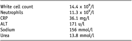
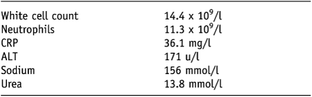
She was subsequently transferred to a psychiatric intensive care unit. A few hours later her physical health deteriorated. She appeared dehydrated and was pyrexial and tachycardic. Laboratory examination (Table 1) revealed a raised white cell count with a raised neutrophil count, a raised C-reactive protein (CRP) and a raised alanine aminotransferase (ALT) suggesting an inflammatory process. Sodium and urea were also raised in keeping with the appearance of clinical dehydration. She was transferred to an acute medical ward and commenced on intravenous fluids, empirical antibiotics and haloperidol in view of her agitation.
Laboratory results
Further deterioration resulted in her transfer to the medical ICU where she was sedated and ventilated. Her vital signs were unstable and she was unconscious for most of the time, even when sedation was lifted. Computerized tomography (CT) and magnetic resonance imaging (MRI) brain scans were normal and an electroencephalogram (EEG) showed no focal abnormality. A lumbar puncture revealed normal cerebrospinal fluid findings and viral serology was negative.
There was input from medical, neurology, intensive care and psychiatric teams. A diagnosis could not be established and she was treated symptomatically with sedatives (clonidine) and intravenous fluids. She was initially treated with further antibiotics and antiviral medication. Abnormal facial grimacing and chewing, posturing of the arms and kicking movements of the legs were noted. She had seven treatments of electroconvulsive therapy (ECT) whilst on ICU for a presumptive diagnosis of malignant catatonia (Box 1). She was started on antipsychotic medication and quetiapine was used due to the low incidence of extrapyramidal side-effects and weak dopamine D2 receptor antagonism. Treatment with benzodiazepines (diazepam) was also initiated. In total she spent a period of six weeks on ICU.

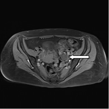
There was some improvement and the patient left ICU and, following a period on a medical ward, was transferred to the psychiatric intensive care unit. After consultation with the neurologist a test for anti-NMDA receptor antibodies was performed and this returned positive. Subsequently an MRI scan of the pelvis revealed a small cyst on the left ovary (Fig. 1). The patient was treated with intravenous immunoglobulin (IVIg) and high dose steroids then underwent a left ovarian cystectomy, histology later confirming an ovarian teratoma containing neural tissue. The patient made a good recovery from this, with no subsequent psychotic or affective symptoms noted, however neuropsychological testing showed residual deficits in the domains of memory and new learning, suggestive of temporal lobe dysfunction, five months after first presentation and one month after removal of the teratoma. She was treated in hospital under the provisions of the Mental Health Act 1983 (as amended in 2007). After improving, she appeared in Court and was placed under a Hospital Order (Section 37) which allowed her treatment to continue. Fourteen months after initial presentation she was discharged.
MRI scan of the pelvis showing left adnexal cyst

DISCUSSION
The NMDA receptor is a ligand gated cation channel consisting of NR1 and NR2 subunits (Waxman & Lynch, 2005). It is thought that antibodies are produced due to the expression of autoantigens (which are heteromers containing NR1 and NR2 subunits of the NMDA receptor) by the tumour, resulting in the encephalitis (Dalmau et al. 2007).
Our patient’s presentation was typical of the characteristic syndrome. In a case series of 100 patients by Dalmau et al. (2008), 91 were women with a median age of 23. 77 presented with prominent psychiatric symptoms and 76 patients had seizures. 88 patients developed decreased consciousness, progressing to a catatonic-like state with akinesia, alternating with agitation. 86 had dyskinesias, of which orofacial dyskinesia was the commonest. 69 had autonomic instability and 66 central hypoventilation. 55 had abnormal findings on MRI brain scan. 59 percent of patients had a tumour, most commonly ovarian teratoma (53 out of 58). Other tumours were identified in 3 female patients: 1 sex-cord stromal tumour, 1 neuroendocrine tumour and 1 teratoma of the mediastinum. In 2 male patients one had an immature teratoma of the testis, the other small cell lung cancer. Notably however 41% of patients did not have a clinically identifiable tumour. At follow-up, 47 patients had made a full recovery, 28 had a mild stable deficit, 18 severe deficits and 7 died.
Treatment includes supportive measures, immunotherapy and tumour resection. Patients whose tumour was diagnosed and treated within the first four months of the onset of neurological disease had better outcomes than those with late or no tumour treatment and those where no tumour was identified (Dalmau et al. 2008).
In our case, the patient had ECT whilst in medical intensive care prior to the diagnosis being established. This did appear to be associated with some improvement and the patient was able to leave intensive care. This is in keeping with a previous case report of an 11-year-old girl with paraneoplastic limbic encephalitis and malignant catatonia related to an ovarian teratoma, who responded to ECT but in whom maximal response only followed removal of the primary tumour (Lee et al. 2006).
CONCLUSIONS
Anti-NMDA receptor encephalitis should be given consideration particularly in patients presenting with sudden onset psychiatric symptoms progressing to rapid deterioration in physical health. As early treatment may prevent mortality and morbidity (Dalmau et al. 2008), increased awareness of the diagnosis may be important in this often treatable disorder.