Coronavirus disease 2019 has affected millions of people worldwide. Cardiac manifestations in adults have included heart failure, cardiogenic shock, arrhythmia, and myocarditis.Reference Shafi, Shaikh, Shirke, Iddawela and Harky1 Acute myocarditis from COVID-19 is thought to be caused by direct viral injury due to myocytes and secondary cardiac injury due to host immune response.Reference Siripanthong, Nazarian and Muser2 In the paediatric population, Multisystem Inflammatory Syndrome in Children was the term coined by the Center for Disease Control and Prevention to describe children who presented with persistent fever, fatigue, and signs/symptoms consistent with multiorgan involvement, elevated inflammatory markers, and current or recent COVID-19 infection. The pathophysiology of heart failure in these patients is unknown, but thought to be due to a post-infective inflammatory syndrome without direct viral injury to the myocytes. This definition overlaps with active COVID-19 infection. Adult studies report malignant ventricular arrhythmias, but there are few paediatric reports of malignant arrhythmias due to COVID-19 myocarditis. Limited paediatric reports suggest that cases of isolated COVID-19 acute fulminant myocarditis are rare, but can be fatal.Reference Fischer, Brillat-Savarin, Ducrocq and Ou3,Reference Beaudry, Dietrick and Lammert4 Herein, we report the case of a child presenting with sustained ventricular tachycardia from acute fulminant myocarditis due to active COVID-19 infection. He required venoarterial extracorporeal membrane oxygenation due to ventricular tachycardia and cardiogenic shock, but completely recovered with an excellent outcome.
Case
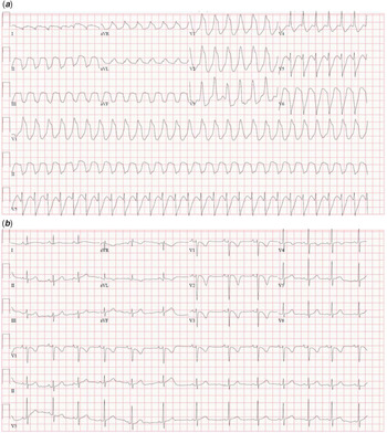
A previously healthy 5-year-old Black, non-Hispanic male presented with 1 day of fatigue and vomiting. He had no other symptoms such as fever, cough, congestion, rash, or diarrhoea. In the emergency department, he was poorly perfused with a heart rate of 170 bpm and rhythm revealed monomorphic ventricular tachycardia (Fig 1a). He was hypotensive with a lactate of 8 mg/dL. Complete metabolic panel showed elevated blood urea nitrogen 37 mg/dL and creatinine 0.91 mg/dL. Electrolytes including magnesium, calcium, phosphorous, and potassium levels were normal. Complete blood count was unremarkable. High-sensitivity troponin I was elevated at 1658 ng/L (normal range 3–7 ng/L) and brain natriuretic peptide was 2298 pg/ml (normal range <101 pg/ml). Chest radiograph showed cardiomegaly and pulmonary oedema. COVID-19 PCR was positive and his serology was negative. The patient had no known exposure to COVID-19, but a family member was a healthcare provider. Initially, he was interactive and talking. Due to hypotension and cardiogenic shock, synchronised cardioversion (1 J/kg) was performed with sedation without conversion. This was repeated (2 J/kg), but he remained in ventricular tachycardia. He became more hypotensive and obtunded and was emergently transferred to the ICU. He was loaded with lidocaine (1 mg/kg) and started on a continuous infusion without improvement. Echocardiogram showed severely decreased biventricular function without chamber dilation (supplementary material – ECHO clip). Central venous and arterial catheters were placed showing profound metabolic acidosis with venous saturation of 22%. He was loaded with amiodarone (5 mg/kg) and his heart rate slowed to 100 bpm, but he remained in ventricular tachycardia. He was intubated and synchronised cardioversion was performed after the amiodarone load, but was unsuccessful; therefore, he was cannulated to VA-ECMO via the right neck.
(a) Initial EKG (top) showed wide QRS morphology concerning for ventricular tachycardia. (b) Bottom EKG showed the patient recovered to sinus rhythm.

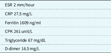
He was treated with intravenous immunoglobulin (2 g/kg) and methylprednisolone (30 mg/kg daily for 3 days). His family declined treatment with Remdesivir. Amiodarone and milrinone infusions were continued during ECMO. His infectious workup (respiratory viral PCR, viral serum PCR/serology for adenovirus, enterovirus, cytomegalovirus, Epstein–Barr virus, and parvovirus and blood and urine cultures) was negative. His inflammatory markers were not severely elevated as seen in MIS-C (Table 1), and his Interleukin-1β and Interleukin 6 were normal. High-sensitivity troponin I peaked at 5948 mg/L and BNP peaked at 3207 pg/ml.
Initial inflammatory laboratory values

He was taken for atrial septostomy due to left atrial hypertension (mean left atrial pressure 28 mmHg) on ECMO. Angiography revealed normal coronary anatomy without stenosis or aneurysm. Endomyocardial biopsy was deferred due to risk of perforation and cardiac magnetic resonance imaging was deferred due to inability to perform on ECMO support and the fact that it would not change acute management. On day 4 of admission, the QRS narrowed and patient converted to sinus rhythm (Fig 1b). Therefore, amiodarone was discontinued. His inflammatory markers decreased and systolic function improved. He was decannulated on admission day 5 and did not require any intervention for the small atrial communication. He was extubated on day 6 and was transitioned from milrinone to enalapril due to mildly reduced function on a repeat echocardiogram. He recovered to his baseline neurocognitive status and was discharged home with outpatient physical therapy.
Discussion
COVID-19 has been associated with malignant arrhythmias (ventricular tachycardia/fibrillation) in adults. Atrial and ventricular arrhythmias occurred in 3–17% of adult COVID-19 patients.Reference Lakkireddy, Chung and Gopinathannair5,Reference Wang, Hu and Hu6 The prevalence of COVID-19 associated arrhythmias in children is still unknown. In the largest descriptive report of children with MIS-C, 80% had cardiovascular involvement and amongst these patients, 12% had arrhythmias.7 Two recent studies published in the paediatric literature described 19–20% of MIS-C patients with various degrees of heart block.Reference Dionne, Mah and Son8,Reference Choi, Fremed and Starc9 In a recent case report, an adolescent presented with MIS-C and had rapid cardiac decompensation, arrhythmia, and died.Reference Fischer, Brillat-Savarin, Ducrocq and Ou3 In MIS-C, we suspect that high levels of pro-inflammatory cytokines affect cardiac output and multiple organ failure contributes to the severity of illness. In comparison to MIS-C, our patient had ventricular tachycardia from acute COVID-19 myocarditis. One paediatric case report describes a child with acute myocarditis with COVID-19 and adenovirus.Reference Beaudry, Dietrick and Lammert4 But, we found only one paediatric report of acute myocarditis due to COVID-19 alone.Reference Lara, Young and Del Toro10 In this report, a 15-year-old boy was diagnosed with acute COVID-19 myocarditis. Unlike our patient, this child had stable haemodynamics without arrhythmia. The mechanism of myocardial injury and inflammation is likely different in children with acute COVID-19 myocarditis compared to MIS-C. We believe our patient’s sustained ventricular tachycardia was secondary to direct injury to the myocardium from COVID-19. Our patient was afebrile, he had no other organs involved, and his inflammatory markers were not impressively elevated as seen in a cytokine storm. With supportive care, he recovered with an excellent outcome.
Conclusion
We present the first case of sustained ventricular tachycardia from acute fulminant myocarditis due to COVID-19 in a paediatric patient. Paediatric case reports of acute COVID-19 myocarditis and arrhythmia are limited, and we report the use of VA-ECMO as a bridge to recovery for this paediatric patient with fulminant myocarditis due to COVID-19. Although rare, this case demonstrates that paediatric acute COVID-19 infection may present with acute myocarditis with malignant arrhythmia and aggressive supportive care can lead to good outcomes.
Acknowledgements
None.
Financial support
This research received no specific grant from any funding agency, commercial, or not-for-profit sectors.
Conflicts of interest
None.
Ethical standards
The authors assert that all procedures contributing to this work comply with the ethical standards of the relevant national guidelines on human experimentation and with the Helsinki declaration and institutional review was exempt by the Central Michigan University Institutional Review Board.