Introduction
Adenosquamous carcinoma is a rare variant of head and neck squamous cell carcinoma (SCC) that usually shows aggressive clinical features. Adenosquamous carcinoma is pathologically characterised by mixed differentiations and distinct areas of both SCC and adenocarcinoma, as defined by the World Health Organization.Reference Cardesa, Zidar, Alos, Barnes, Eveson, Reichart and Sidransky1
Adenosquamous carcinoma of the head and neck was first defined in 1968 by Gerughty et al. in a series of 10 patients, where it was shown to be extremely aggressive and highly malignant, with 80 per cent of the patients having proven metastases.Reference Gerughty, Hennigar and Brown2 Adenosquamous carcinoma has two distinct histologic components. Squamous cell carcinoma usually predominates, and the adenocarcinomatous component can have a tubular or glandular morphology. Immunohistochemical findings are helpful to confirm the diagnosis of adenosquamous carcinoma, as previously reported by Alos et al. Reference Alos, Castillo, Nadal, Caballero, Mallofre and Palacin3
Herein, we report a case of adenosquamous carcinoma of unknown primary origin, and discuss the diagnosis and treatment along with a review of the previous literature.
Case report
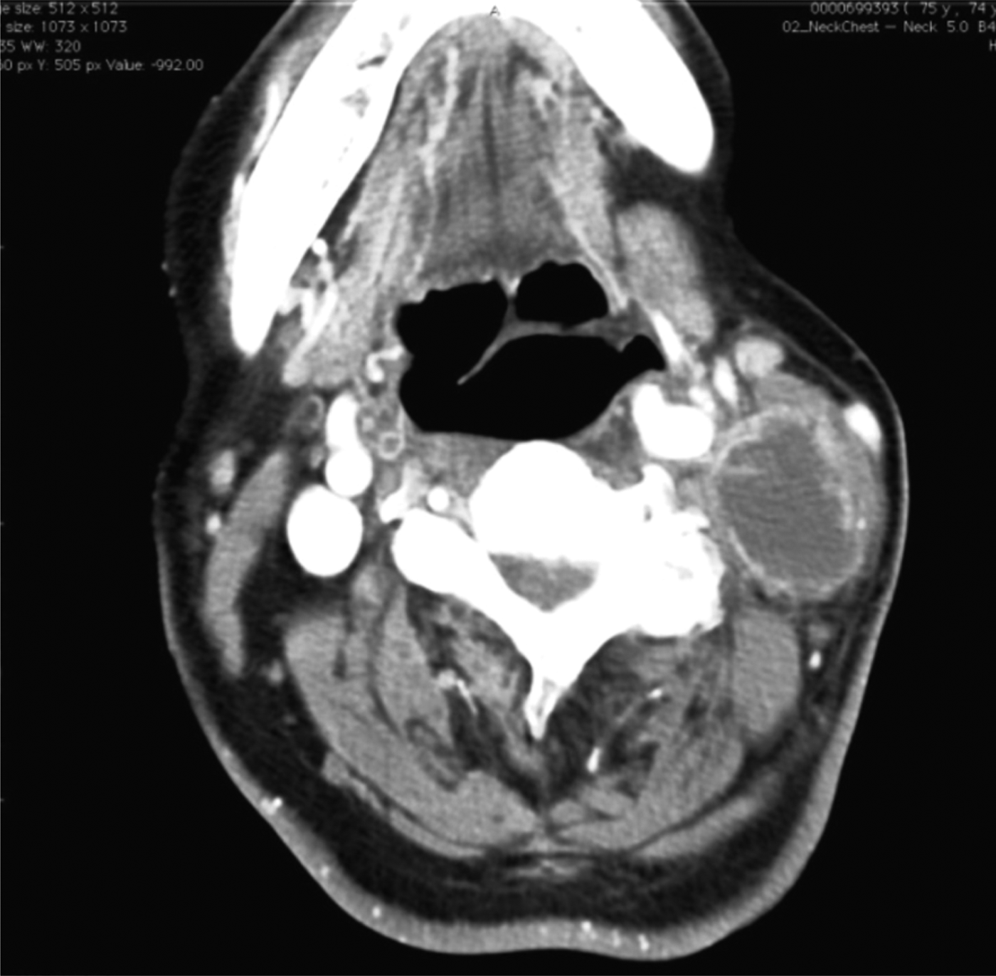
A 74-year-old man, a non-smoker, was presented to our hospital on the third day after noticing swelling on the left side of his neck. His past medical history included paralysis of the left arm due to a machine-related injury. Physical examination revealed a painless, fixed and elastic hard mass on the left side of the mid-neck area. No other abnormal lesions were detected in the ENT region. Contrast enhanced computed tomography (CT) scans showed a well-defined, ring-enhanced cystic mass, measuring up to 56 mm, while no other significant lymphadenopathy was detected (Figure 1).
Axial view of a computed tomography scan showing a ring-enhanced solitary mass in the left side of the neck.

A fine needle aspiration was performed, and the cytological findings raised a suspicion of SCC. To find the primary lesion, the patient underwent an 18F-fluoro-2-deoxy-D-glucose positron emission topography (FDG-PET) and CT scan. High uptake of FDG was detected only in the left neck, and no other hot spots were found in the whole body (Figure 2).
18F-fluoro-2-deoxy-D-glucose positron emission tomography (FDG-PET)–computed tomography showing a hot spot in the left side of the neck. No other hot spots were detected. A: Anterior, P: posterior, R: right, L: left
Although other examinations, such as gastro-intestinal fibrescopy and magnetic resonance imaging, were performed, no primary lesions were found.
According to the standard treatment for SCC of unknown primary origin, the patient underwent an operation consisting of unilateral neck dissection and left palatine tonsillectomy for biopsy.
Macroscopically, the tumour was congruent with the surrounding tissue, including the sternocleidomastoid muscle, scalene muscle and internal jugular vein (Figure 3). Microscopically, the cystic tumour was lined by SCC with papillary structures (Figure 4a). This tumour consisted of two intermingled components, SCC and adenocarcinoma. The SCC component was predominant and included an intracellular bridge (Figure 4b), whereas the adenocarcinomatous component was focally involved and characterised by small tubular structures (Figure 4c). Positive staining with mucicarmine, which revealed apical and occasionally intracytoplasmic mucin, was seen both in the tubular lumen and cytoplasm (Figure 4d). The tumour was diffusely positive for CK5/6 (Figure 4e) and focally positive for CEA (Figure 4c), whereas staining for CK7, CK20, p16, CDX2 and thyroid transcription factor-1 (TTF-1) were negative (data not shown). There was no evidence of malignancy in the palatine tonsil.
Macroscopic findings: a cystic mass invading the surrounding tissues.
(a) The cystic tumour was lined by squamous cell carcinoma (SCC) with papillary structures (×100). (b) The SCC component, showing clear intracellular bridges (×400). (c) The adenocarcinomatous component, consisting of small tubular structures, and showing positive immunoreactivity for CEA (inset) (×200). (d) Mucicarmine positive secretion both in the tubular lumen (a) and cytoplasm (b) (×400). (e) Diffusely positive immunoreactivity for CK5/6 (×200).
A diagnosis of lymph node metastasis of adenosquamous carcinoma from an unknown primary lesion was made. As adenosquamous carcinoma has been known to show low susceptibility to both radiation and chemotherapy,Reference Gerughty, Hennigar and Brown2 the patient was not given any additional therapy in spite of the extra-nodal involvement.
The patient has been alive without relapse for 18 months, and the site of the primary lesion remains unknown.
Discussion
Adenosquamous carcinoma is a rare entity, not only in the head and neck area, but also in other organs. The commonest sites of occurrence within the head and neck would appear to be the larynx and oral cavity, according to the previous reviews.Reference Keelawat, Liu, Roehm and Barnes4, Reference Masand, El-Mofty and Ma5 Adenosquamous carcinoma is considered to be aggressive and to have a poor clinical outcome. In the latest literature review of 93 cases, Masand et al. reported that 47.4 per cent of cases had regional metastases and 24.7 per cent had distant metastases.Reference Masand, El-Mofty and Ma5 There is nearly unanimous agreement about the first-choice treatment of adenosquamous carcinoma, which is thorough resection with an adequate surgical margin. However, there is no clear consensus on additional post-operative therapies, including radiation and chemotherapy.Reference Keelawat, Liu, Roehm and Barnes4, Reference Yoshimura, Mishima, Obara, Yoshimura and Maruyama6
It is ordinarily difficult to give a histological diagnosis of adenosquamous carcinoma before initial treatment, because the pre-operative diagnosis is generally made from small biopsy specimens that lack either component, particularly the adenocarcinomatous one, which often exists in the deeper portion of the lesion. Yoshimura et al. reported in their retrospective study that the rate of correct diagnosis of adenosquamous carcinoma from initial biopsy specimens was only 31.6 per cent, with an incorrect diagnosis of SCC being made in 7 of 19 patients (36.8 per cent).Reference Yoshimura, Mishima, Obara, Yoshimura and Maruyama6 Therefore, it has been suggested that adenosquamous carcinoma may be more common than the published data would suggestReference Sheahan, Toner and Timon7 and many cases of adenosquamous carcinoma may have been treated inadequately, such as with chemoradiotherapy.
• Adenosquamous carcinoma in the head and neck is a very rare tumour, which is characterised pathologically by the simultaneous presence of distinct areas of adenocarcinoma and squamous cell carcinoma and clinically by a very aggressive course with a poor outcome
• We reported a very rare case of a neck adenosquamous carcinoma with an unknown primary site, which was well-controlled by thorough resection
• Although branchiogenic carcinoma was clinically suspected, a follow up of at least five years will be needed to confirm this diagnosis
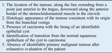
In our case, the cervical lymph node presented as a solitary, cyst-like mass. Morphologically, therefore, we had to consider the possibility of branchiogenic carcinoma. The entity of branchiogenic carcinoma has remained controversial. In many of the previously reported cases of cystic SCC of the neck, the primary site was thought to be the tonsils. Thompson and Heffner analysed 136 cases of cystic SCCs in the neck and concluded that the primary sites were the palatine or lingual tonsils in 87 cases, or 63 per cent of the total cases.Reference Thompson and Heffner8 Therefore, we think that branchiogenic carcinoma should be strictly diagnosed. Martin et al. published a review of 250 cases, including 15 of their own, and proposed a set of conditions that should be satisfied for a diagnosis of branchiogenic carcinoma.Reference Martin, Morfit and Ehrich9 Based on a retrospective review of 67 cases, Khafif et al. proposed that five new criteria be appended to those of Martin et al. Reference Khafif, Prichep and Minkowitz10 (Table I). They reported the fourth criterion, the proof of transition from normal epithelium to carcinoma, seemed to be the most important and indispensable for a proper diagnosis.Reference Khafif, Prichep and Minkowitz10 In our case, the tumour satisfied most criteria, but the zone of transition could not be observed, and thus the evidence was insufficient for a diagnosis of branchiogenic carcinoma. If no further progression is observed in the ensuing years, including recurrences, metastases or detection of primary lesions, then we would consider it clinically acceptable to classify the present case as the first reported case of ‘branchiogenic’ adenosquamous carcinoma. It will thus be necessary to follow this case for at least five years to confirm the diagnosis.
Criteria proposed by khafif et al. For the diagnosis of branchiogenic carcinoma
Acknowledgement
The authors thank KN International for reviewing the English usage in this report.