Introduction
Genetic risk factors, such as the presence of one or both Apolipoprotein-E (APOE) ε4 alleles, have been associated with increased risk for Alzheimer's disease (AD) and late-life cognitive decline (Caselli et al., Reference Caselli, Graff-Radford, Reiman, Weaver, Osborne, Lucas and Thibodeau1999; Farrer et al., Reference Farrer, Cupples, Haines, Hyman, Kukull, Mayeux and van Duijn1997; Saunders et al., Reference Saunders, Hulette, Welsh-Bohmer, Schmechel, Crain, Burke and Rosenberg1996; Swan, Lessov-Schlaggar, Carmelli, Schellenberg, & La Rue, Reference Swan, Lessov-Schlaggar, Carmelli, Schellenberg and La Rue2005). Cross-sectional studies suggest that asymptomatic elders with risk factors for AD (i.e., APOE ε4 carriers and persons with a family history of AD) demonstrate different patterns of brain activation on functional magnetic resonance imaging (fMRI) than elders without risk factors (for a review, see Wierenga & Bondi, Reference Wierenga and Bondi2007). Often, these activation changes occur in brain regions critical for memory processes, such as the hippocampus, posterior cingulate, and lateral posterior temporoparietal regions. Such regions are also the earliest to be affected by AD neuropathology (Bassett et al., Reference Bassett, Yousem, Cristinzio, Kusevic, Yassa, Caffo and Zeger2006; Bondi, Houston, Eyler, & Brown, Reference Bondi, Houston, Eyler and Brown2005; Johnson et al., Reference Johnson, Schmitz, Trivedi, Ries, Torgerson, Carlsson and Sager2006; Seidenberg et al., Reference Seidenberg, Guidotti, Nielson, Woodard, Durgerian, Antuono and Rao2009; Sperling et al., Reference Sperling, Dickerson, Pihlajamaki, Vannini, LaViolette, Vitolo and Johnson2010).
Aberrant patterns of task-activated fMRI may precede the onset of cognitive symptoms and atrophy on structural MRI in elders at-risk for AD (Seidenberg et al., Reference Seidenberg, Guidotti, Nielson, Woodard, Durgerian, Antuono and Rao2009). Several longitudinal studies suggest that task-activated fMRI may be useful for predicting future cognitive decline in intact elders (Bookheimer et al., Reference Bookheimer, Strojwas, Cohen, Saunders, Pericak-Vance, Mazziotta and Small2000; Lind et al., Reference Lind, Ingvar, Persson, Sleegers, Van Broeckhoven, Adolfsson and Nyberg2006; O'Brien et al., Reference O'Brien, O'Keefe, LaViolette, DeLuca, Blacker, Dickerson and Sperling2010; Persson et al., Reference Persson, Nyberg, Lind, Larsson, Nilsson, Ingvar and Buckner2006; Smith et al., Reference Smith, Kryscio, Schmitt, Lovell, Blonder, Rayens and Andersen2005; Woodard et al., Reference Woodard, Seidenberg, Nielson, Smith, Antuono, Durgerian and Rao2010) and in patients with mild cognitive impairment (MCI), the prodromal condition typically preceding the diagnosis of AD (Miller et al., Reference Miller, Fenstermacher, Bates, Blacker, Sperling and Dickerson2008). The majority of these fMRI prediction studies have used episodic memory (EM) tasks (Bookheimer et al., Reference Bookheimer, Strojwas, Cohen, Saunders, Pericak-Vance, Mazziotta and Small2000; Miller et al., Reference Miller, Fenstermacher, Bates, Blacker, Sperling and Dickerson2008; O'Brien et al., Reference O'Brien, O'Keefe, LaViolette, DeLuca, Blacker, Dickerson and Sperling2010; Persson et al., Reference Persson, Nyberg, Lind, Larsson, Nilsson, Ingvar and Buckner2006), which is not surprising because EM dysfunction is among the most prominent of cognitive changes in the 3-year period preceding a diagnosis of AD (Mickes et al., Reference Mickes, Wixted, Fennema-Notestine, Galasko, Bondi, Thal and Salmon2007). EM tasks have been used widely in cross-sectional fMRI studies to contrast cognitively intact individuals at varying risk for developing AD (Trivedi et al., Reference Trivedi, Schmitz, Ries, Hess, Fitzgerald, Atwood and Johnson2008; Wierenga & Bondi, Reference Wierenga and Bondi2007). However, there exist potential methodological problems in using EM tasks during fMRI. EM performance declines with healthy aging and is accelerated in MCI and AD (Bondi & Kaszniak, Reference Bondi and Kaszniak1991). The interpretation of fMRI activation maps is complicated when performance varies across participants. This issue is particularly problematic for blocked trial designs, where the calculated fMRI response is based on the average of both correct and incorrect trials within a block, as the incorporation of incorrect trials may result in activation of unintended, non–memory-related processes. In an event-related fMRI design, it is possible to eliminate error trials from the brain maps, but even this procedure may be invalid if performance is close to chance. In addition, EM tasks are inherently difficult for older adults, resulting in potential activation of brain regions associated with increased effort rather than the memory circuits of interest.
Alternatively, semantic memory (SM) fMRI tasks have also successfully predicted future cognitive decline (Lind et al., Reference Lind, Ingvar, Persson, Sleegers, Van Broeckhoven, Adolfsson and Nyberg2006; Smith et al., Reference Smith, Kryscio, Schmitt, Lovell, Blonder, Rayens and Andersen2005; Woodard et al., Reference Woodard, Seidenberg, Nielson, Smith, Antuono, Durgerian and Rao2010) and may have some advantages over EM tasks. Performance declines on SM tasks are less severe than EM declines in normal aging and MCI (Hodges & Patterson, Reference Hodges and Patterson1995). In addition, SM tasks are typically less effortful for older adults than EM tasks. Finally, brain regions recruited in response to SM tasks overlap with the so-called “default mode network” (DMN), a circuit activated during rest and during semantic processing (Binder, Desai, Graves, & Conant, Reference Binder, Desai, Graves and Conant2009). Regions in this network are susceptible to early AD pathology, including amyloid-beta deposition (Buckner et al., Reference Buckner, Snyder, Shannon, LaRossa, Sachs, Fotenos and Mintun2005; Pihlajamaki & Sperling, Reference Pihlajamaki and Sperling2009; Raichle et al., Reference Raichle, MacLeod, Snyder, Powers, Gusnard and Shulman2001; Rombouts & Scheltens, Reference Rombouts and Scheltens2005). Thus, SM fMRI tasks may be sensitive to the detection of risk for future cognitive decline and AD.
We published a series of fMRI studies using a famous name discrimination task (FNDT) to probe SM networks in at-risk aging and in MCI (Sugarman et al., Reference Sugarman, Woodard, Nielson, Seidenberg, Smith, Durgerian and Rao2012). Seidenberg et al. (Reference Seidenberg, Guidotti, Nielson, Woodard, Durgerian, Antuono and Rao2009) found greater SM activation in individuals at-risk for AD, especially in the posterior cingulate and lateral posterior temporoparietal regions. Increased SM activation in the same regions was also found in MCI patients as compared to healthy older adults, despite equivalent task performance (Woodard et al., Reference Woodard, Seidenberg, Nielson, Antuono, Guidotti, Durgerian and Rao2009). Recently, Woodard et al. (Reference Woodard, Seidenberg, Nielson, Smith, Antuono, Durgerian and Rao2010) demonstrated that SM task activation was more effective in predicting future cognitive decline than hippocampal volumes in elders who were cognitively intact at study entry.
In the current study of cognitively intact healthy elders, we compared the accuracy of EM and SM fMRI brain activation patterns in predicting future cognitive decline after an 18-month retest interval. The FNDT served as the SM task; the EM task involved an old-new recognition task involving famous and unfamiliar names. To equate the brain maps derived from the two tasks, we used an event-related design to eliminate error trials from the image analyses. In addition, a principal components analysis was used to reduce the number of predictors derived from the SM and EM fMRI activation data. We predicted that task performance would be poorer on the EM task relative to the SM task, and that activation during the SM task would be the superior predictor of future cognitive decline.
Method
Participants
Participants were 78 healthy older adults (73% female; mean age = 73 years; SD = 4.9 years; mean education = 14.9 years; SD = 2.7 years). They were drawn from a larger sample of 459 community-dwelling adults who were recruited via newspaper advertisements. Following telephone screening, 92 participants met study inclusion and exclusion criteria. Participants were excluded from the study if they had a history or evidence of: (1) neurological illnesses or conditions including dementia and head trauma with a loss of consciousness greater than 30 min; (2) medical illnesses or conditions that may affect brain function (such as untreated hypertension or insulin-dependent diabetes mellitus); (3) major psychiatric disturbance meeting DSM-IV Axis I criteria; (4) a Mini-Mental State Exam (MMSE) (Folstein, Folstein, & McHugh, Reference Folstein, Folstein and McHugh1975) score less than 28; (5) a Geriatric Depression Scale (Yesavage et al., Reference Yesavage, Brink, Rose, Lum, Huang, Adey and Leirer1982) score greater than 10; (6) substance abuse meeting DSM-IV Axis I criteria; (7) impairments in activities of daily living as determined by the Lawton Instrumental Activities of Daily Living Scale (ADL) (Lawton & Brody, Reference Lawton and Brody1969); (8) taking prescribed psychoactive medications, (9) a Hachinski ischemia score above 4; or (10) left-handedness, as determined by the Edinburgh Handedness Inventory (Oldfield, Reference Oldfield1971). Of the 92 participants who met study criteria, 81 persons agreed to undergo APOE genotyping from blood samples, a neuropsychological evaluation, and an fMRI scanning session. MRI data were not successfully obtained from 3 participants, leaving 78 participants for inclusion.
Family history was defined as a report of a clear clinical diagnosis of AD or a reported history of gradual decline in memory and other cognitive functions, confusion, or judgment problems without a formal diagnosis of AD before death in a first-degree relative. One participant reported a diagnosis of AD in a second-degree relative, with some mild cognitive changes noted in a parent before the parent's death. Because our study examined the influence of AD risk factors on prediction of cognitive decline, approximately half (51.3%) of the participants were purposely selected because they had a positive family history of AD. In addition, 33.3% of the sample carried at least one APOE ε4 allele. All participants underwent neuropsychological evaluation (see below) and were determined to be cognitively intact at baseline. Informed consent was obtained consistent with the Declaration of Helsinki and institutional guidelines established by the Medical College of Wisconsin Human Subjects Review Committee. All participants received financial compensation for their participation.
Neuropsychological Assessment and APOE Genotyping
All participants underwent a baseline and 18-month follow-up neuropsychological battery consisting of the following: MMSE (Folstein et al., Reference Folstein, Folstein and McHugh1975), Mattis Dementia Rating Scale-2 (Jurica, Leitten, & Mattis, Reference Jurica, Leitten and Mattis2001; Mattis, Reference Mattis1988), Rey Auditory Verbal Learning Test (RAVLT) (Rey, Reference Rey1958), Geriatric Depression Scale (Yesavage et al., Reference Yesavage, Brink, Rose, Lum, Huang, Adey and Leirer1982), and ADL (Lawton & Brody, Reference Lawton and Brody1969). Alternate forms of the DRS-2 (Schmidt, Reference Schmidt2004; Schmidt, Mattis, Adams, & Nestor, Reference Schmidt, Mattis, Adams and Nestor2005) and RAVLT (Schmidt, Reference Schmidt1996) were administered during the follow-up examination. Participant APOE genotype was determined using a polymerase chain reaction method described by Saunders et al. (Reference Saunders, Hulette, Welsh-Bohmer, Schmechel, Crain, Burke and Rosenberg1996). DNA was isolated with the Gentra Systems Autopure LS for Large Sample Nucleic Acid Purification.
Definition of Cognitive Decline
Membership into the Declining group (n = 27) was defined as a reduction of at least one standard deviation between baseline and follow-up testing on one or more of the following neuropsychological measures: DRS-2 Total Score (DRS-2 Tot), RAVLT Sum of Trials 1-5 (RAVLT-Tot), or RAVLT Delayed Recall (RAVLT-DR). Residualized change scores were computed for each cognitive measure by predicting retest scores using baseline scores. This procedure adjusts for baseline performance, practice effects, and regression to the mean. Participants with standardized residuals of −1.0 or lower on one or more of the three measures were assigned to the cognitively declining group; the remaining participants were classified as cognitively stable.
fMRI Task
All participants were administered SM and EM tasks while undergoing fMRI in the same scanning session. Corrective lenses were provided to participants as needed. The SM task consisted of the presentation of 30 highly recognizable famous names and 30 unfamiliar names. Stimuli were selected from an original pool of 784 names based on the ability of healthy older adults to correctly classify each name as famous or unfamiliar (see Douville et al., Reference Douville, Woodard, Seidenberg, Miller, Leveroni, Nielson and Rao2005, for details). The 60 names were randomly interspersed with 20 presentations of a centrally placed fixation crosshair to introduce “jitter” into the fMRI time series (inter-stimulus interval = 4 s). Participants made a right index or right middle finger key press for famous or unfamiliar names, respectively. Accuracy and reaction time were recorded. The imaging run began and ended with 12 s of fixation and was 5 min and 44 s in duration.
Following a 20-min delay, participants were administered an EM recognition task. The stimuli consisted of 60 “old” items (the 30 famous and 30 unfamiliar names from the SM task) randomly intermixed with 60 “new” items (30 famous and 30 unfamiliar names). Participants were asked to indicate by button press if the name was old (right index finger) or new (right middle finger). As in the SM task, name stimuli were presented for 4 s and presented in a pseudorandom order with 40 presentations of a centrally placed fixation crosshair to introduce “jitter.” The EM task was split into two runs that began and ended with 12 s of fixation and were each 5 min and 44 s in duration.
Image Acquisition
Whole-brain, event-related fMRI was conducted on a General Electric (Waukesha, WI) Signa Excite 3.0 Tesla short bore scanner equipped with a quad split quadrature transmit/receive head coil. Echoplanar images were collected using an echoplanar pulse sequence (TE = 25 ms; flip angle = 77 degrees; field of view [FOV] = 24 mm; matrix size = 64 × 64). Thirty-six contiguous axial 4-mm-thick slices were selected to provide coverage of the entire brain (voxel size = 3.75 × 3.75 × 4 mm). The inter-scan interval (TR) was 2 s. High-resolution, three-dimensional spoiled gradient-recalled at steady-state (SPGR) anatomic images were acquired (TE = 3.9 ms; TR = 9.5 ms; inversion recovery [IR] preparation time = 450 ms; flip angle = 12 degrees; number of excitations [NEX] = 2; slice thickness = 1.0 mm; FOV = 24 cm; resolution = 256 × 224). Foam padding was used to reduce head movement within the coil.
Image Analysis
Functional images were generated with the Analysis of Functional Neuroimages (AFNI) software package (Cox, Reference Cox1996). Each image time series was shifted to the beginning of the TR and then spatially registered to reduce the effects of head motion using a rigid body iterative linear least squares method. A deconvolution analysis was used to extract a hemodynamic response function (HRF) for famous and unfamiliar names (SM task) and for novel and previously seen names (EM task) from the time-series. Correct and incorrect trials were modeled separately, and only correct trials were used in the second-level analyses. HRFs were modeled for the 0- to 16-s period post-stimulus onset. Motion parameters were incorporated into the model as nuisance regressors. The HRFs were also transposed so that the value of the HRF at trial onset was zero. Area under the curve (AUC) was calculated by summing the hemodynamic responses at time points 4, 6, and 8 s post-trial onset. Individual anatomical and functional scans were transformed into standard stereotaxic Talairach space (Talairach & Tournoux, Reference Talairach and Tournoux1988). To compensate for normal variation in anatomy across participants, functional images were blurred using a 6-mm Gaussian full-width half-maximum filter.
Spatial Extent of Activation Analysis
A voxel-wise analysis was used to determine differences in spatial extent of activation between the stable and declining groups. SM task activation was defined as regions where the AUC for famous names was significantly different from that of unfamiliar names. EM task activation was defined as regions where the AUC for previously shown names was significantly greater than the AUC for novel names. For all voxel-wise analyses, the individual voxel probability threshold was p < .005 with a minimum cluster of 0.731 mL.The statistical threshold was derived from 3,000 Monte Carlo simulations (Forman et al., Reference Forman, Cohen, Fitzgerald, Eddy, Mintun and Noll1995) and was equivalent to a whole brain family-wise error threshold of p < .05.
Functional Region of Interest Analysis
Using voxel-wise t tests, SM and EM activations were calculated separately combining all 78 participants. Significant cluster volumes were used to create functional regions of interest (fROIs) for each task. The average AUC of all voxels within each fROI was then calculated for each participant. For each task, the data from all fROIs were entered into a principal components analysis (PCA) to further reduce the number of predictors for the logistic regression analysis (see Woodard et al., Reference Woodard, Seidenberg, Nielson, Smith, Antuono, Durgerian and Rao2010, for details).
Data Analysis
Logistic regression analyses were conducted to examine the relative accuracy of SM versus EM in predicting cognitive decline. Our previous research (Woodard et al., Reference Woodard, Seidenberg, Nielson, Smith, Antuono, Durgerian and Rao2010) demonstrated that the combination of APOE ε4 status and SM activation outperformed other predictive models that included combinations of hippocampal volume, a family history of AD, and demographic variables. Therefore, we limited our predictors to only task activation and APOE ε4 status to maintain a reasonable number of subjects-to-variables and prevent overfitting the model. We tested four models: APOE ε4 status alone (Model 1), APOE ε4 and SM activation (Model 2), APOE ε4 and EM activation (Model 3), and APOE ε4 and both SM and EM activation (Model 4). Nagelkerke R2 values and the concordance (C) indexes determined the relative fit of each model in predicting participants’ future cognitive decline. Nagelkerke R2 indicates the importance of the predictors in each model relative to a perfectly fitting null model (Nagelkerke, Reference Nagelkerke1991). The C index represents the area under the receiver operating characteristic curve and indicates the proportion of all possible pairs of Stable and Declining subjects in which the Declining subject in the pair had a higher predicted probability of decline than the Stable subject (Harrell, Reference Harrell2001; Woodard et al., Reference Woodard, Seidenberg, Nielson, Smith, Antuono, Durgerian and Rao2010). Therefore, greater C index values indicate greater prediction accuracy for a model. For each logistic regression, values of Nagelkerke R2 and C were validated with a bootstrapping analysis using 5000 resamples (Harrell, Reference Harrell2001). Through bootstrapping, holding data out for cross-validation was not required, and each phase of model development was revalidated using repeated resampling from the entire sample (Harrell, Reference Harrell2001; Woodard et al., Reference Woodard, Seidenberg, Nielson, Smith, Antuono, Durgerian and Rao2010). The four logistic regression models were compared on the basis of their Bayesian Information Criterion (BIC) values, which account for the number of parameters in each model. Lower BIC values suggest a better model for optimally predicting cognitive decline. BIC values can be compared statistically to determine whether one model fits the data significantly better than the other model.
Results
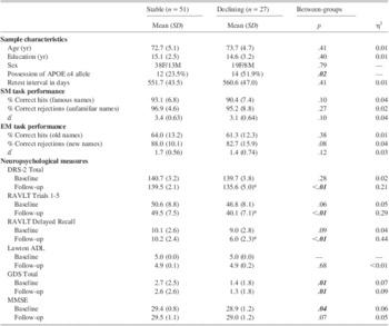
On baseline measures, there were no significant differences between the Stable and Declining groups on the DRS-2 Tot, RALT-Tot, RAVLT-DR, or ADL after controlling for multiple comparisons (Bonferroni adjust alpha level = .0125; 0.04/4 tests; see Table 1). At an 18-month follow-up, 27 of 78 participants (34.6%) demonstrated a reduction in neuropsychological performance of at least 1 SD on one or more of the specified neuropsychological measures, indicating cognitive decline. Of these 27, 22 declined on one measure only (6 = DRS-2 Tot, 8 = RAVLT-Tot, 8 = RAVLT-DR). Four participants declined on two measures (3 = RAVLT-Tot & RAVLT-DR, 1 = DRS-2 Tot & RAVLT-Tot), and one participant declined on all three measures. A 2 (Group) × 2 (Testing Session) ANOVA (analysis of variance) revealed a significant interaction for all three neuropsychological measures (RAVLT-Tot, F(1,76) = 14.95; p < .001, partial η2 = .164; RAVLT-DR, F(1,76) = 34.9; p < .001, η2 = .315; DRS-2 tot, F(1,76) = 11.99; p < .001; η2 = .136). Two (7.6%) participants met Petersen criteria for a diagnosis of MCI (Petersen, Reference Petersen2000). No participants reported impairment in ADLs at baseline or follow-up. The presence of one or both APOE ε4 alleles was significantly greater in the declining group (p = 0.02). There were no significant group differences in age, education, sex, test–retest interval, or fMRI task performance between the two groups (see Table 1).
Sample characteristics and fMRI behavioral data for stable and declining groups

Note. APOE = Apolipoprotein E; F = female; M = male; SM = semantic memory; EM = episodic memory; DRS-2 = Mattis Dementia Rating Scale-2; RAVLT = Rey Auditory Verbal Learning Test; ADL = Activities of Daily Living; GDS = Geriatric Depression Scale; MMSE = Mini-Mental State Exam.
*Significant decrease (p < .05) from baseline to follow-up.
Baseline fMRI
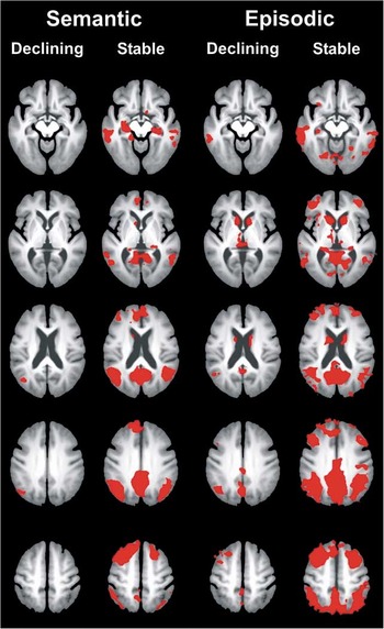
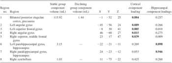
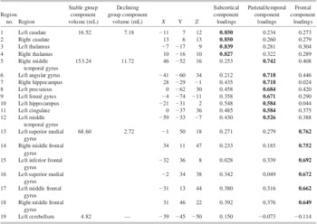
The Declining group demonstrated less overall activation on both the SM and EM tasks as compared to the Stable group (see Figure 1). As described previously in Woodard et al. (Reference Woodard, Seidenberg, Nielson, Smith, Antuono, Durgerian and Rao2010), a voxelwise analysis of the SM task resulted in eight fROIs (see Table 2). The EM voxelwise analysis resulted in 19 fROIs (see Table 3).
Comparison of semantic and episodic task activation between the declining and stable groups. The declining group demonstrated less activation on both the semantic (famous > unfamiliar names) and episodic (previously seen > novel names) memory tasks compared to the non-declining group.

PCA components derived from semantic memory fROIs for stable versus declining groups (from Woodard et al., Reference Woodard, Seidenberg, Nielson, Smith, Antuono, Durgerian and Rao2010)

PCA = principal components analysis; fROIs = functional regions of interest.
PCA components derived from episodic memory fROIs for stable versus declining groups

PCA = principal components analysis; fROIs = functional regions of interest.
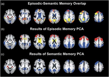
A PCA conducted on the eight SM fROIs resulted in two components: “cortical” and “hippocampal” (see Table 2; Figure 1). The cortical component included significant loadings of fROIs in the bilateral posterior cingulate, left and right angular gyrus, left superior frontal gyrus, and the right superior middle frontal gyrus. The hippocampal component included significant loadings for both the left and right parahippocampal/hippocampal fROIs.
Three components resulted from the PCA of the 19 EM fROIs: “subcortical,” “frontal,” and “parietal/temporal” (see Table 3; Figure 2). The subcortical component consisted of the left and right caudate and left and right thalamic fROIs. The frontal component included the left superior medial gyrus, the left and right middle frontal gyrus, and left inferior frontal gyrus fROIs. The parietal/temporal component included left and right middle temporal gyrus, left angular gyrus, left and right hippocampus, left lingual gyrus, left cingulate, and left precuneus fROIs.
Semantic and episodic activation functional region of interest (fROI) analysis. A: The top row displays the overlap of semantic and episodic activation (yellow), regions that activated only during the semantic task (red), and regions that activated only during the episodic task (blue). A principal components analysis (PCA) of task-related activation for each condition resulted in the following factor loadings: B: The episodic model consisted of subcortical (yellow), frontal (red), and parietal/temporal (blue) components; C: The semantic activation model consisted of cortical (blue) and hippocampal (red) components.

Cerebellar activation was observed during both the SM and EM tasks. However, this activation did not demonstrate significant loadings in either the SM or EM PCA and was thus excluded from logistic regression analyses.
Logistic Regression Analysis
APOE ε4 allele status was found to be a significant predictor of cognitive decline in our previous study using the SM task (Woodard et al., Reference Woodard, Seidenberg, Nielson, Smith, Antuono, Durgerian and Rao2010) and was, therefore, included in the logistic regression analyses comparing SM and EM task activation models in the current study.
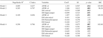
Four logistic regression models were evaluated (see Table 4). Model 1 (APOE ε4 alone) was the poorest fitting model (Nagelkerke R2 = .106; C = .642). Model 2 (SM task+APOE) fit the data much better (Nagelkerke R2 = .285; C = .787), with significant contributions from APOE (p = .003) and both cortical (p = .01) and hippocampal (p = .03) fMRI components. Model 3 (EM task+APOE) was not a significantly better fit than Model 2 (Nagelkerke R2 = .212; C = .711) with none of the EM fMRI components significantly predicting decline. The significance of APOE ε4 as a predictor was reduced to a marginally significant level (p = .051) in Model 3, likely due to a significant relationship between frontal activation during the episodic task and APOE ε4 inheritance; the frontal component became significant when ε4 was removed from the model (p = .039). Model 4 (APOE, SM, and EM) demonstrated the greatest predictive accuracy of all four models (R2 = .422; C = .837). However, an examination of BIC values indicated that Model 4 most likely overfit the data due to the large number of variables relative to number of participants (see below). Of interest, APOE ε4 and both SM fMRI components were significant predictors in this model, but the only significant EM predictor was the parietal-temporal component.
Results of the logistic regressions for the relative accuracy of semantic and episodic memory activation in predicting cognitive decline

Note. BIC = Bayesian Information Criterion; APOE = Apolipoprotein E; SM = semantic memory; EM = episodic memory.
Nagelkerke R2 and C Index values have been corrected for optimism using bootstrapping (see text for additional description).
When comparing the models, BIC values favored the SM model over the combined SM+EM model (p < .036) and the APOE ε4 only model (p < .001) (Table 4). The APOE ε4 model demonstrated a significantly lower BIC than the EM model (p < .034). Thus, the SM+APOE model fit the data significantly better and more parsimoniously than either the EM+APOE model or the APOE only model.
Discussion
In our previous study, we demonstrated that two factors, baseline semantic memory fMRI activation and APOE ε4 allele status, predicted future cognitive decline in healthy participants after an 18-month retest interval (Woodard et al., Reference Woodard, Seidenberg, Nielson, Smith, Antuono, Durgerian and Rao2010). The current study extends these findings by focusing on the type of fMRI activation task used to predict cognitive decline. The current results indicate that, in combination with APOE ε4 status, fMRI brain activation patterns derived from an SM activation task were superior to an EM activation task for predicting cognitive decline after an 18-month interval. These results suggest the type of activation task used in fMRI studies may play an important role in accurately identifying healthy elders at risk for developing future cognitive decline.
Previous research reported that EM fMRI activation may also be effective in predicting future cognitive decline (Bookheimer et al., Reference Bookheimer, Strojwas, Cohen, Saunders, Pericak-Vance, Mazziotta and Small2000; O'Brien et al., 2010). In contrast, we found that when SM and EM predictors were combined in the same model, the EM components did not increase the overall predictive ability of the model relative to APOE status alone. Only the parietal-temporal EM component significantly contributed to prediction of cognitive decline. Whereas there was extensive overlap in fMRI activation between the SM and EM tasks (Figure 2), the greater sensitivity of the SM task may be due to its activation of a more spatially localized network than the multiple activation networks associated with the EM task (Figure 2; Tables 2 and 3). Specifically, the SM task (famous name discrimination) has been previously shown to activate AD-vulnerable regions such as the hippocampus and parahippocampal gyrus (Douville et al., Reference Douville, Woodard, Seidenberg, Miller, Leveroni, Nielson and Rao2005) and the posterior cingulate (Woodard et al., Reference Woodard, Seidenberg, Nielson, Miller, Franczak, Antuono and Rao2007). These regions overlap with the resting state default mode network (DMN), which includes the posterior cingulate, medial prefrontal cortex, medial temporal, and angular gyrus (Raichle et al., Reference Raichle, MacLeod, Snyder, Powers, Gusnard and Shulman2001). Recent studies have indicated that the DMN is disrupted in patients with a diagnosis of MCI or AD (Pihlajamaki & Sperling, Reference Pihlajamaki and Sperling2009; Rombouts & Scheltens, Reference Rombouts and Scheltens2005). In a recent review, Binder and colleagues (2009) point out that the DMN overlaps with regions involved in task-activated fMRI experiments involving semantic processing. Because it appears to selectively activate brain regions associated with the DMN, task-activated fMRI that engages the semantic processing may be more sensitive to future cognitive decline than episodic memory tasks.
The more diffuse pattern of activation observed with the EM task may be associated with task difficulty. Importantly, EM task accuracy was worse than SM task accuracy, which is not surprising because episodic memory declines with age (Cansino, Reference Cansino2009), while semantic memory abilities remain relatively intact (Nilsson, Reference Nilsson2003). The EM task activated more regions than the semantic task, especially those associated with effort and error detection (e.g., frontostriatal, anterior cingulate). Thus, SM activation may be superior to EM activation for predicting cognitive decline due to its ability to effectively stress AD-vulnerable regions, while minimizing activation of networks associated with task performance and effort.
Our study found that a greater magnitude of fMRI activation at baseline was associated with preserved cognitive performance regardless of task (Figure 1). The study of Bookheimer et al. (Reference Bookheimer, Strojwas, Cohen, Saunders, Pericak-Vance, Mazziotta and Small2000) found the opposite results, namely that increased baseline activation was associated with future cognitive decline. Our findings, however, are consistent with those of a more recent study conducted by Lind et al. (Reference Lind, Ingvar, Persson, Sleegers, Van Broeckhoven, Adolfsson and Nyberg2006) demonstrating that decreased activity predicted future cognitive decline. The precise reasons for the divergent results are unclear. It should be noted, however, that greater task-induced activation is consistently observed in asymptomatic APOE ε4-positive individuals relative to non-carriers (Bondi et al., Reference Bondi, Houston, Eyler and Brown2005; Kukolja, Thiel, Eggermann, Zerres, & Fink, Reference Kukolja, Thiel, Eggermann, Zerres and Fink2010; Seidenberg et al., Reference Seidenberg, Guidotti, Nielson, Woodard, Durgerian, Antuono and Rao2009; Trivedi et al., Reference Trivedi, Schmitz, Ries, Hess, Fitzgerald, Atwood and Johnson2008). In addition, increased activation in fMRI studies is frequently observed in older compared to younger subjects (Cabeza, Reference Cabeza2002; Nielson et al., Reference Nielson, Douville, Seidenberg, Woodard, Miller, Franczak and Rao2006; Nielson, Langenecker, & Garavan, Reference Nielson, Langenecker and Garavan2002).
Whether increased activation is helpful or harmful for the individual is an important question. Park and Reuter-Lorenz (Reference Park and Reuter-Lorenz2009) proposed the Scaffolding Theory of Aging and Cognition (STAC) to suggest that increased activation in cognitively intact elders represents a compensatory response to neural changes associated with normal aging and/or the impact of emerging disease processes. The increased activity serves to preserve intact levels of cognitive performance. Conversely, once cognitive symptoms emerge, brain activity diminishes since the “scaffold” is no longer successful in preserving normal performance levels (Han, Bangen, & Bondi, Reference Han, Bangen and Bondi2009). In the context of this study, cognitively intact elders with the ε4 gene would be expected to experience more brain activity than cognitively intact non-carriers presumably because at least some at-risk individuals have begun to experience the early stages of AD-related pathology. In support of this hypothesis, longitudinal fMRI studies suggest a progression of hyperactivation to hypoactivation over the course of AD (cf. Machulda et al., Reference Machulda, Ward, Borowski, Gunter, Cha, O'Brien and Jack2003; O'Brien et al., Reference O'Brien, O'Keefe, LaViolette, DeLuca, Blacker, Dickerson and Sperling2010).
The broader literature has demonstrated that comprehensive neuropsychological testing is predictive of impending cognitive decline (cf. Twamley, Ropacki, & Bondi, Reference Twamley, Ropacki and Bondi2006). Thus, some might question the value of using fMRI for prognostic purposes. The goal of the current study was to use functional MRI as a surrogate for such testing because recent studies suggest it might reveal specific patterns that could eventually serve as better or earlier predictors of cognitive decline. In the current study, the neuropsychological measures used, specifically the DRS-2 and the RAVLT, served as criterion variables for assessing cognitive decline, thereby serving to determine group membership. Thus, they could not be used also as predictors of cognitive performance in our models and no other measures were available. However, the role of cognitive performance in predicting decline could be examined in this study by using the semantic and episodic tasks given in the scanner. When including the d′ score for semantic and episodic performance in the model with the other significant predictors (i.e., adding these behavioral factors to Model 2), the bootstrapped C index was .80 compared with .79 without them, and neither of the cognitive performance factors offered significant prediction (semantic d′ p = .16; episodic d′ p = .77). While behavioral performance may provide adequate prediction of impending decline, in this context where it was examined directly in conjunction with fMRI activation, fMRI was superior. One limitation of this interpretation is that the semantic task (FNDT) was specifically designed to produce >90% accuracy in performance. The value of the design was to limit task difficulty contributions to activation, but a consequence of it is a limited ability to discern behavioral differences between groups.
There are several other limitations of the current study worth noting. Participant performance on the EM task was poorer than performance on the SM task. Although only correct trials were used in the brain imaging analysis, an easier EM task may have resulted in improved sensitivity for predicting cognitive decline. Similarly, EM task performance was dependent upon encoding during the semantic task. During the EM task, participants in both groups sometimes judged novel famous names as previously seen due to familiarity effects with these stimuli, adding to task difficulty. Furthermore, both the SM and EM tasks used in this study relied on retrieval in a forced-choice recognition format. Activation maps based on encoding evoke different brain systems and may possess different degrees of accuracy in predicting cognitive decline (Bondi, Salmon, Glasako, Thomas, & Thal, Reference Bondi, Salmon, Glasako, Thomas and Thal1999; Wolk & Dickerson, 2011). Finally, results of the current study require replication using larger sample sizes and a longer follow-up interval to draw more definitive conclusions on the ability of fMRI task activation to predict conversion to MCI or AD.
In summary, our findings suggest that fMRI activation during a semantic memory task is more accurate in predicting future cognitive decline in asymptomatic older adults than activation during an episodic memory task. Future studies are required to determine the relative sensitivity of task-activated fMRI in comparison to other biomarkers (structural MRI, resting state functional connectivity MRI, amyloid positron emission tomography scanning, cerebrospinal fluid and blood analyses) in identifying individuals at-risk for future cognitive decline or the development of MCI/AD. Results of this study suggest that semantic memory activation in combination with APOE ε4 status holds promise for identifying asymptomatic at-risk individuals for inclusion in primary prevention randomized clinical trials of interventions designed to prevent or delay cognitive decline.
Acknowledgments
This project was supported by NIH grant, R01 AG022304, awarded to SMR, the Medical College of Wisconsin General Clinical Research Center (M01 RR00058), and the Medical College of Wisconsin Advancing a Healthier Wisconsin Program. The content is solely the responsibility of the authors and does not necessarily represent the official views of the National Institute on Aging or the National Institutes of Health. The information in this manuscript has never been published either electronically or in print. The authors report no conflicts of interest.