Acute rheumatic fever remains one of the most common causes of acquired cardiac morbidity and mortality worldwide. Major and minor Jones criteria are used in the diagnosis of the disease. Reference Gewitz, Baltimore and Tani1 It is well known that acute rheumatic fever affects the cardiac conduction system and often causes first-degree atrioventricular block, one of Jones’s minor criteria. Complete atrioventricular block, supraventricular tachycardia, and atrial/ventricular ectopic beats are other rarely reported rhythm disturbances. While complete atrioventricular block was initially observed in our patient, atrioventricular block and then sinus rhythm were detected at different levels (2nd degree type 2, type 1, and 1st degree atrioventricular block) after anti-inflammatory treatment. It is emphasised that atrioventricular block at different levels, including 3rd-degree complete atrioventricular block, is a rare condition during the same attack and should be kept in mind with this case.
Case report
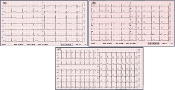
An 11-year-old male patient applied to an external health centre with complaints of knee pain and limited movement that started two days ago and was referred to us after laboratory tests revealed elevated acute phase reactants and complete atrioventricular block on electrocardiographic. In his medical history, it was learned that he had an upper respiratory tract infection approximately 1 month ago. On physical examination, there was a 2/6 systolic murmur and bilateral pain and limited movement in the knees. The electrocardiographic at the time of admission showed complete atrioventricular block. It was learned that he had a cow’s milk allergy in his medical history and there was no rheumatological disease in the family. Laboratory studies revealed C-reactive protein as 173 mg/dl (normal range: <5 mg/dl), anti-streptolysin O as 786 IU/ml (normal range: <250 IU/ml), and erythrocyte sedimentation rate as 66 mm/hour. Echocardiography revealed mild mitral and aortic valve insufficiency. Chest radiography showed a cardiothoracic ratio of 50%. With these findings and the presence of evidence supporting group A beta-haemolytic streptococcal infection, the case was diagnosed with acute rheumatic fever if it met two major and two minor criteria. The patient was admitted to the paediatric cardiology ward. Benzathine penicillin G (IM) was administered. The patient, whose electrocardiography revealed complete atrioventricular block, was started on anti-inflammatory treatment with prednisolone at a maximum dose of 60 mg/day. On the second day of treatment, it was observed that the 3rd-degree atrioventricular block on the electrocardiographic was replaced by 2nd-degree type 1 and type 2 atrioventricular block. Twenty-four-hour rhythm Holter recordings showed predominantly 2nd degree type 1 and type 2 and intermittent complete atrioventricular block during the night hours (Figure 1a–c). On the 4th day of treatment, it was observed that acute phase reactants regressed, the clinical picture improved, and there was no pathology in the electrocardiographic except for 1st degree atrioventricular block (PR: 165 ms) (Figure 2a–c). In the examinations performed on the 10th day of his hospitalisation, the patient had negative acute phase reactants, no clinical complaints, sinus rhythm on electrocardiographic and no atrioventricular block (PR distance within normal limits for age) (Figure 2a–c). He was discharged after continuing his paediatric cardiology outpatient clinic follow-ups. The steroid dose was gradually reduced after 2 weeks and discontinued in 4 weeks. Ibuprofen treatment (15 mg/kg/day) was started in the last week when steroid treatment was reduced. After two weeks, ibuprofen treatment was tapered to 4 weeks and then discontinued. Atrioventricular block was not observed again during the follow-up of the patient, who continued with depot treatment for twenty-one days.
Examples of patient’s 24-hour rhythm Holter recording. In ( a ), 3rd degree complete AV block on 24-hour rhythm Holter recording. In ( b,c ), 2nd degree AV block.

Electrocardiogram images of the case. In ( a ), baseline ECG consistent with complete AV block at the time of admission. In ( b ), 1st degree AV block on ECG on day 4 of steroid treatment. In ( c ), ECG in sinus rhythm before discharge.

Discussion
Acute rheumatic fever is an immune-mediated nonsuppurative complication of group A streptococcal (GAS) pharyngitis. Reference Karthikeyan and Guilherme2 It remains the most common cause of acquired heart disease in some countries around the world. The most common conduction disorder associated with the disease is first-degree atrioventricular block, which is also one of the minor criteria. Reference Karacan, Işıkay, Olgun and Ceviz3 In addition to first-degree atrioventricular block, advanced atrioventricular block, premature atrial contractions, ventricular extrasystoles, and ventricular/supraventricular tachycardias have been observed. Reference Zalzstein, Maor, Zucker and Katz4 Yakut et al. reported a rare case of acute rheumatic fever in which three different degrees of atrioventricular block were observed in the same patient. Reference Yakut, Eybek, Erolu and Karacan5 In the study by Agnew et al., transient atrioventricular conduction abnormalities were detected in 8.5% of patients with acute rheumatic fever, and the rate of second- and third-degree atrioventricular block was determined as 2.5%. Additionally, nodal rhythm was observed in 6% of patients in this study. Reference Agnew, Wilson, Skinner and Nicholson6 In the literature, cases of Mobitz type II and third-degree atrioventricular block developing in the same acute rheumatic fever attack have been rarely reported. Reference Zalzstein, Maor, Zucker and Katz4 Karacan et al. showed that analysis of 24-h rhythm Holter recordings showed that more arrhythmias were observed in acute rheumatic fever patients. Reference Karacan, Işıkay, Olgun and Ceviz3 When our patient’s arrival rhythm Holter recordings were evaluated, atrioventricular block was initially observed at different degrees (2nd degree type 1 and type 2 and complete atrioventricular block), similar to the literature, and with the treatment process, it was observed that first the 1st degree atrioventricular block and then completely returned to normal.
Although the mechanism of atrioventricular block in acute rheumatic fever is not fully understood, it is thought to be caused by increased vagal tone and immunologic effects on the atrioventricular node. Conduction abnormalities associated with acute rheumatic fever are usually self-limited and resolve within a few weeks after initiation of nonsteroidal anti-inflammatory therapy. Reference Carano, Bo, Tchana, Vecchione, Fantoni and Agnetti7 A good response to corticosteroids has been reported in some studies. Reference Reddy, Chun and Yamamoto8,Reference Kibar, Erdem and Oflaz9 This suggests that corticosteroids may be a good option for the treatment of advanced atrioventricular block due to acute rheumatic fever. In our case, in whom steroid treatment was initiated, it was observed that complete atrioventricular block turned into 2nd-degree atrioventricular block at the end of the 2nd day and into 1st-degree atrioventricular block at the end of the 4th day along with the treatment process. There are studies in the literature suggesting that specific treatment, such as temporary pacemaker implantation, should only be considered when complete atrioventricular block leads to an Adams-Stokes attack, Reference Carano, Bo, Tchana, Vecchione, Fantoni and Agnetti7 while cases of permanent pacemaker implanted cases with atrioventricular block causing syncope and haemodynamic instability have also been reported. Reference Argun, Baykan, Özyurt, Pamukçu, Üzüm and Narin10
In our study, we presented a patient whose complaint was joint pain and who was diagnosed with acute rheumatic fever after examinations, who had complete atrioventricular block on the electrocardiographic at admission and different degrees of atrioventricular block on the Holter recordings before treatment. It should be kept in mind that although first-degree atrioventricular block is the most common accompaniment to acute rheumatic fever, other types of atrioventricular blocks may also be observed during the disease. atrioventricular blocks developing secondary to acute rheumatic fever return to normal with anti-inflammatory treatment. In patients with complete atrioventricular block, aetiology should be thoroughly investigated before pacemaker implantation, and patients should be evaluated holistically with history, physical examination, and all laboratory tests.