
Introduction
Two-dimensional (2D) transition metal dichalcogenides (TMDs) have received attention as potential materials for fabricating ultrathin electronic devices, owing to their inherent semiconductor band gap which is absent in the graphene.[ Reference Yin, Li, Li, Jiang, Shi, Sun, Lu, Zhang, Chen and Zhang 1 – Reference Kim, Konar, Hwang, Lee, Lee, Yang, Jung, Kim, Yoo, Choi, Jin, Lee, Jena, Choi and Kim 3 ] However, the atmospheric stability of semiconducting TMDs is a crucial parameter, which could significantly affect the performance of TMD-based devices. Previously reported work emphasized the degradation of TMDs under specific conditions such as high-temperature annealing, elevated humidity or exposure to electron beam irradiation.[ Reference Wu, Li, Yin, Li, Liu, Cao, Zhang and Zhang 4 – Reference Late, Liu, Matte, Dravid and Rao 6 ] Compared with other TMDs, MoS2 is considered to be fairly stable under ambient conditions. However, recent studies on MoS2 electronic devices have suggested that atmospheric conditions may significantly affect device properties.[ Reference Lee, Cui, Kim, Arefe, Zhang, Lee, Ye, Watanabe, Taniguchi, Kim and Hone 7 , Reference Qiu, Pan, Yao, Li, Shi and Wang 8 ] Recently, Gao et al. presented a study on the ambient stability of MoS2 and WS2 layers grown by chemical vapor deposition (CVD).[ Reference Gao, Li, Tan, Chow, Lu and Koratkar 9 ] They observed cracks develop on the surface of ultra-thin MoS2 and WS2 flakes over the 1-year period of study. These changes were attributed primarily to the sulfur vacancies, which are inherently present in TMD films grown by CVD.
Mechanically exfoliated flakes have always been considered of superior quality in comparison with CVD grown layers.[ Reference Liu, Wong and Chi 10 , Reference Zhou, Zou, Najmaei, Liu, Shi, Kong, Lou, Ajayan, Yakobson and Idrobo 11 ] The pristine quality and crystallinity of exfoliated films are maintained because they are exfoliated directly from the parent crystal. In this work, the long-term stability of mechanically exfoliated MoS2 flakes is compared for samples stored in the air and under vacuum. Optical microscopy and contrast difference measurements were used to identify any changes in lateral dimensions and thickness of MoS2 flakes. The effect of storage conditions was observed on both ultra-thin and bulkier MoS2 flakes, which have a thickness ranging up to several hundreds of nanometers. Scanning electron microscopy (SEM) and non-contact mode atomic force microscopy (AFM) were used to analyze the surface of the flakes on the different samples. Any changes in quality and crystallinity of the flakes were studied by carrying out Raman spectroscopy and energy-dispersive X-ray spectroscopy (EDX) on representative flakes.
Experimental methods
Layers of MoS2 were mechanically exfoliated from a commercially available natural molybdenum disulfide crystal (supplied by 2D Semiconductors) using Scotch® tape. Exfoliated flakes were then transferred from Scotch® tape onto a PF-X4 gel-film as obtained from Gel-Pak®, before final transfer onto a thermally oxidized (315 nm) silicon substrate.[ Reference Budania, Baine, Montgomery, McNeill, Mitchell, Modreanu and Hurley 12 ] Samples were also prepared by direct Scotch® tape exfoliation. Details of sample preparation are given in the Supplementary material.
Thin MoS2 flakes were identified on the samples by optical microscopy and optical profile lines.[ Reference Li, Wu, Huang, Lu, Yang, Lu, Xiong and Zhang 13 ] Initially, one sample (the control sample) was stored in a continuously pumped vacuum desiccator and another sample was stored at room temperature in cleanroom conditions better than class 1000 and with a relative humidity of 60%. Over a period of 1 year, optical microscopy of both samples was done at regular intervals to observe any modifications taking place in the physical appearance of the flakes. Additional samples, which were prepared and stored in the air after initial optical imaging, were subsequently transferred to vacuum storage to provide further experimental validation.
Long-term effects of storage conditions
The control sample was kept under vacuum after preparation and was taken out only for short time periods to image the flakes. Over a period of 1 year, there was no distinguishable change in optical contrast of the ultra-thin flakes. As an example of this, Figs. S1(a) and S1(b) show a comparison between ultra-thin flakes on the day of preparation and after 222 days of vacuum storage. In addition, bulk flakes on the control sample had a uniform appearance without any apparent development of defects on their surface 222 days after sample preparation as shown in Fig. S2.
After sample preparation and initial optical imaging, the flakes on the sample stored in the air were again imaged after 10, 25, and 55 days. The first changes in the flake appearance were observed 55 days after sample preparation, as shown in Figs. S3(a) and S3(b), where a crack has appeared in a thin flake. The effect of storage conditions was also observed on bulk MoS2 flakes. Within 55 days of preparation, small speckles had started to appear on the surface of the bulk flakes on the sample stored in the air as shown in Fig. S4. These speckles were seen to emerge primarily at the edges of the bulk flakes. However, no changes were observed in the lateral dimensions of these bulk flakes.
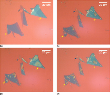
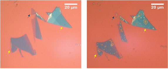
As time progressed, the appearance of both thin and bulk flakes on the sample stored in the air continued to evolve. Figures 1(a)–1(d) show optical microscope images of thin MoS2 flakes on this sample on the day of preparation, after 110, 170, and 205 days, respectively. After 110 days, the ultra-thin MoS2 flake pointed out by the blue arrow in Fig. 1(a) had almost disappeared from the sample, as seen in Fig. 1(b). Defects which appeared on the surface of other thin flakes shifted in size and position as pointed out by the yellow arrows. It was observed that the lateral dimensions of the bilayer MoS2 flake shown in Fig. S3 had reduced by 50% over a period of 1 year as shown in Figs. S5 and S6.
Optical microscope images showing the change in size and appearance of thin MoS2 flakes on a sample stored in the air: (a) on the day of sample preparation, (b) after 110 days, (c) after 170 days, and (d) after 205 days. Arrows are used for identification of the same region.

Because of the marked difference between flakes stored in vacuum and flakes stored in the air, it was suspected that the observed flake degradation could be the result of progressive absorption of water molecules from the atmosphere. Therefore, two sets of samples which had been initially stored in the air, and which had already developed speckled flakes, were annealed in vacuum at 200 °C for 2 h in order to desorb any water. The flakes on these samples were optically imaged just before and after this anneal. One set of samples was then stored in the air and the other was stored under vacuum. The flakes on these samples were imaged at regular intervals.
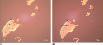
Figure 2(a) shows an optical image of some speckled flakes on one of these samples before annealing. After vacuum annealing, most of the speckles had either disappeared from the surface of the flakes or had become smaller, as shown in Fig. 2(b). Figures S7(a) and S7(b) show optical images of the same flakes after 5 and 133 days of subsequent vacuum storage, respectively. There were no distinguishable changes in the position and size of the speckles after storing the samples under vacuum. No new speckles had appeared on the surface of the flakes. However, speckles reappeared on the surface of flakes on samples that were stored in the air after vacuum annealing, as shown in Fig. S8. The emergence of speckles was observed on all the samples irrespective of the method of mechanical exfoliation. Figures S9 and S10 show optical images of speckles developed on MoS2 flakes on samples prepared by Scotch® tape exfoliation and the gel-film transfer method, respectively. The reduction in speckles on the surface of decomposing flakes achieved by annealing emphasizes that the speckles are most likely caused by adsorbed water molecules on the surface of MoS2 flakes. It is believed that, after annealing, storage under vacuum prevents further interaction of water molecules with the flakes. On the other hand, when the annealed samples were stored in the air again, the speckles re-emerged on the surface of the MoS2 flakes due to fresh absorption of ambient moisture.
Optical microscope images of speckled MoS2 flakes: (a) before annealing and (b) just after vacuum annealing. Arrows are used for identification of the same region.

Investigation of degradation mechanism
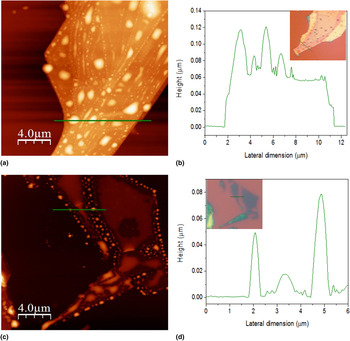
The speckles on the surface of both thin and bulk flakes were further analyzed by SEM as shown in Fig. S11. The decomposing thin MoS2 flake on the sample stored for 240 days in the air was lined with small beads along the edges of the flake as shown in Fig. S11(b). These beads trace the original boundary of the flake. The speckles on the surface of bulk MoS2 flakes on the same sample were scattered across the flake rather than being concentrated at the edges, as shown in Fig. S11(c). The bulk flake is composed of many MoS2 layers stacked on top of each other, so these speckles may form at surface steps on the bulk flake, thus covering the whole surface of the flake. However, by SEM analysis it could not be confirmed if the speckles on the surface of the bulk flakes were holes or raised features on the surface of the flake. Therefore, the decomposed flakes were analyzed by non-contact mode AFM. Figure 3 shows the AFM images of decomposing bulk and thin flakes on the sample stored in the air. In the AFM scan, the speckles on the surface of the bulk MoS2 flake were shown to be raised features as seen in Fig. 3(b). Similarly, the beads along the boundary of the thin MoS2 flake were protruding above the surface as seen in Fig. 3(d). The height of these speckles and beads ranged up to ~0.1 µm, and their lateral dimensions up to about 1 µm. It can be noted from Fig. 3(d) that the thickness of the beads surrounding the decomposing thin MoS2 flake was actually much greater than the thickness of the flake itself.
(a) AFM image of bulk MoS2 flake on the sample stored in the air, (b) corresponding profile along the green scan line, (c) AFM image of the thin MoS2 flake on the sample stored in the air, and (d) corresponding profile along the green scan line. Insets are optical images of the measured flakes.

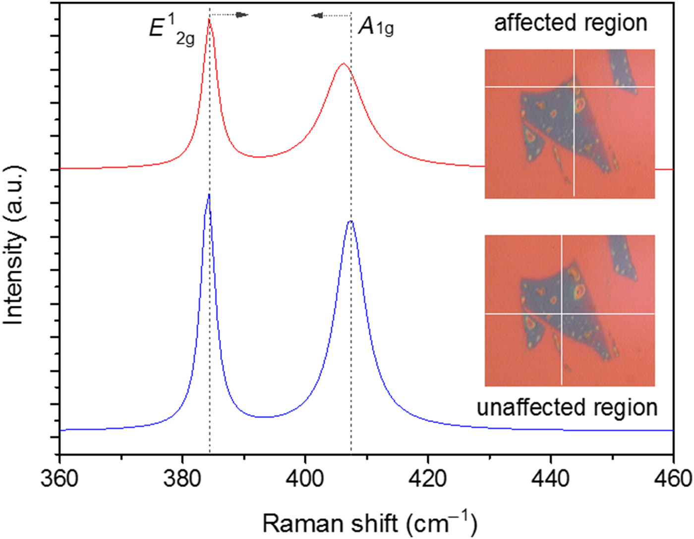
Raman analysis was done on decomposing thin and bulk flakes on the sample stored in the air for 100 days. Figure 4 shows the Raman spectra taken from a speckled region (red curve) and from an unaffected region (blue curve) on a thin MoS2 flake. On the unaffected region of this flake, the significant MoS2 Raman in-plane (E 1 2g) and out-of-plane (A 1g) peaks were observed at 384 and 407.3 cm−1, respectively. These values are consistent with the reported in-plane and out-of-plane peak values for exfoliated MoS2 flakes.[ Reference Li, Zhang, Yap, Tay, Edwin, Olivier and Baillargeat 14 , Reference Lee, Yan, Brus, Heinz, Hone and Ryu 15 ] However, the out-of-plane (A 1g) peak underwent significant change on the speckled region of the flake. The in-plane (E 1 2g) and out-of-plane (A 1g) peak values for the affected region were observed at 384.4 and 406.1 cm−1, respectively. There was a red shift in the A 1g peak value. The FWHM (full-width half-maximum) for the A 1g peak increased from 5.84 to 8.06 cm−1 and it remained almost constant at 2.8 cm−1 for the E 1 2g peak. On the affected region of the flake, the intensity ratio of the in-plane and out-of-plane peaks increased from 1.12 to 1.41. The severely broadened A 1g peak and alteration in the intensity ratios emphasize the change of the MoS2 flake from a crystalline to an amorphous nature.[ Reference Houben, Luysberg, Hapke, Carius, Finger and Wagner 16 , Reference Sahoo, Gaur, Ahmadi, Guinel and Katiyar 17 ] Figure S12 shows the Raman spectra that were taken from an affected and unaffected regions on a bulk MoS2 flake on the same sample. There was no distinguishable shift in the MoS2 Raman peak values in the two different regions. The E 1 2g and A 1g peak values were observed at 383.5 and 408.8 cm−1, respectively. The change in the out-of-plane Raman vibrations is more likely to be noticeable in a thin MoS2 flake in comparison to a thick flake, which is composed of numerous layers of MoS2.
Raman spectra from affected and unaffected regions of a thin MoS2 flake on the sample stored in the air. Insets are optical images of the measured flakes. The measurement was done at the intersection point of the markers on the inset images.

The decomposition of MoS2 flakes on the sample stored in the air was further confirmed by EDX spectroscopy. A JEOL JSM-6500F field-emission scanning electron microanalyzer with an accelerating voltage of 10 kV was used for this analysis. Figures S13(a) and S14(a) show the unaffected and affected regions of a bulk MoS2 flake where EDX data were collected. The atomic percentage of S and Mo atoms on the unaffected region was 18.61 and 7.33%, respectively. Since the flakes were deposited on a 315 nm silicon-dioxide layer, Si and O atoms were also detected. The affected region of the bulk flake was depleted in both S and Mo atoms as the atomic percentages reduced to 5.93 and 3.46%, respectively. The ratio of S-to-Mo atoms reduced from 2.54 in the unaffected region to 1.71 in the affected region. This indicates that the speckled regions are rich in Mo atoms in comparison with S atoms. In the affected region, the atomic percentage of C increased significantly from 7.27 to 31.44%. Figures S13(b) and S14(b) give details of the atomic percentages detected by EDX analysis at the unaffected and the affected sites, respectively. It is believed that the higher concentration of Mo atoms at the speckled sites on the flake is due to oxidation of MoS2 flakes by environmental oxygen and water molecules. Oxidation of MoS2 is thought to lead to the formation of volatile sulfur dioxide and less volatile molybdenum-oxide compounds. It is recently reported by Zhang et al. that the water molecules can remain adhered to the surface of oxidized MoS2 in the form of MoO3.H2O.[ Reference Zhang, Jia, Yang and Song 18 ]
The first signs of these dome-shaped speckles on the thin and the bulk MoS2 flakes were mainly observed along the edges and/or at varying step heights, respectively. This is in accordance with previous reports, which suggest that edge sites are predominantly more reactive than the basal surfaces of the 2D materials because they are created by cleaving the 2D crystal perpendicular to the basal plane, which results in breaking of covalent bonds.[ Reference Fleischauer and Lince 19 , Reference Lince and Frantz 20 ] The decomposition due to oxidation of 2D crystals is not limited to edge sites as the vacancies in the basal plane also contribute toward oxidation of TMDs.[ Reference Liu, Han and Zhao 21 ] Mirabelli et al. reported the appearance of similar domed features on the surface of mechanically exfoliated HfSe2.[ Reference Mirabelli, McGeough, Schmidt, McCarthy, Monaghan, Povey, McCarthy, Gity, Nagle, Hughes, Cafolla, Hurley and Duffy 22 ] In their case, the domes were Se-rich blisters formed due to oxidation of HfSe2 into HfO2. Similarly, the speckles in the present experiment were found to be deficient in S atoms and rich in Mo atoms. Formation of bubbles, irrespective of flake thickness, has also been reported on black phosphorus.[ Reference Wood, Wells, Jariwala, Chen, Cho, Sangwan, Liu, Lauhon, Marks and Hersam 23 ] It was reported that, due to prolonged ambient exposure, the bubbles constantly changed in their size. We observed a similar behavior for speckles on relatively stable mechanically exfoliated MoS2 flakes.
Conclusions
The long-term stability of mechanically exfoliated MoS2 flakes has been investigated. A comparison was made between flakes on samples stored in the air and under vacuum. Significant changes were observed in the lateral dimensions and appearance of MoS2 flakes stored in the air. However, flakes on the control sample did not undergo any physical change. Over the period of study, small speckles appeared on the surface of flakes stored in the air. Vacuum annealing reduced the size and number of speckles, which indicated that they were most likely caused by water absorption on the flake surface. This was followed by thinning and eventual decomposition of flakes.
By SEM analysis, it was found that the decomposition of thin flakes begins along the flake boundary, forming beads of decomposed material along its edges. AFM analysis confirmed that these beads and speckles on partially decomposed flakes were raised dome-shaped features. Due to exposure to oxygen and water molecules, the crystallinity of the flakes was also changed as observed by Raman spectroscopy. The speckles were found to be rich in Mo atoms by EDX spectroscopy. This suggested that the speckles were oxidized MoS2 sites which had a higher concentration of Mo atoms due to evaporation of sulfur dioxide and formation of MoO3. It is believed that ambient moisture is held on the surface of the flake due to the hygroscopic nature of MoO3.
Although there are previous reports showing substantial degradation in the electrical properties of MoS2-based devices due to ambient exposure, there is a lack of detailed analysis of decomposition of mechanically exfoliated TMD flakes. The present work shows the step-by-step degradation mechanism for mechanically exfoliated MoS2 flakes, which are otherwise expected to be stable in comparison with other films deposited by various CVD techniques. Degradation of thin MoS2 flakes can be prevented by storing samples under vacuum. However, for practical device fabrication, careful encapsulation of MoS2 flakes is recommended to prevent the ingress of water molecules and subsequent device degradation.
Supplementary material
The supplementary material for this article can be found at https://doi.org/10.1557/mrc.2017.105
Acknowledgments
The authors acknowledge financial funding from the US-Ireland Research and Development Partnership Programme (project USI 057). Stephen McFarland, unit manager for electron microscopy unit at School of Mathematics and Physics, Queen's University, Belfast is acknowledged for carrying out EDX analysis of MoS2 flakes.