Introduction
Wallace's line is a famous zoogeographical boundary separating Oriental and Australian realms (Lomolino et al., Reference Lomolino, Riddle, Whittaker and Brown2010). The endemic non-volant eutherian mammals in the region east of this line are considered to have originated in the west region, especially Sundaland, then dispersed across the line to the east, and diversified (Musser, Reference Musser and Whitmore1987). As a key island in the region east of Wallace's line, Sulawesi has a unique mammalian fauna that is not only particularly rich in species, but also features a high level of endemism. Many researchers have given much attention to the rodents of Sulawesi, due to their unique biogeography and distribution (Amoria et al., Reference Amoria, Gippoliti and Helgen2008; Maryanto et al., Reference Maryanto, Prijono and Yani2009).
Species of the nematode genus Syphacia Seurat, 1916 occur in the rodent families Cricetidae and Muridae (Hugot, Reference Hugot1988). From the area east of Wallace's line, 15 fully described species have been recorded: S. abertoni Weaver & Smales, Reference Weaver and Smales2006, S. australasiensis Smales, Reference Smales2004, S. boodjamullaensis Weaver & Smales, Reference Weaver and Smales2010, S. brevicaudata Weaver & Smales, Reference Weaver and Smales2008, S. carnarvonensis Weaver & Smales, Reference Weaver and Smales2010, S. coccymyos Smales, Reference Smales2011, S. darwini Hugot & Quentin, Reference Hugot and Quentin1985, S. helidonensis Weaver & Smales, Reference Weaver and Smales2010, S. longaecauda Smales, Reference Smales2001, S. lorentzimyos Smales, Reference Smales2010, S. mamelontenuis Smales, Reference Smales2010, S. muris (Yamaguti, 1935), S. pseudomyos Weaver & Smales, Reference Weaver and Smales2008, S. rifaii Dewi & Hasegawa, Reference Dewi and Hasegawa2010, S. sulawesiensis Hasegawa & Tarore, Reference Hasegawa and Tarore1996 (Hugot & Quentin, Reference Hugot and Quentin1985; Hasegawa & Tarore, Reference Hasegawa and Tarore1996; Smales, Reference Smales2001, Reference Smales2004, Reference Smales2010, Reference Smales2011; Weaver & Smales, Reference Weaver and Smales2006, Reference Weaver and Smales2008, Reference Weaver and Smales2010; Dewi & Hasegawa, Reference Dewi and Hasegawa2010). All of them are assigned to the subgenus Syphacia Seurat, 1916, but exhibit considerable diversity in morphology. Except for the cosmopolitan species S. muris, they are endemic species parasitic in endemic murids, suggesting co-speciation with hosts in their localities.
For Sulawesi, two endemic Syphacia species were hitherto recorded: S. sulawesiensis from Rattus xanthurus in north Sulawesi, and S. rifaii from Bunomys chrysocomus and B. prolatus in central Sulawesi (Hasegawa & Tarore, Reference Hasegawa and Tarore1996; Dewi & Hasegawa, Reference Dewi and Hasegawa2010). Syphacia muris has also been recorded from Rattus rattus and R. xanthurus in north Sulawesi (Hasegawa & Tarore, Reference Hasegawa and Tarore1996). During investigative research on the biodiversity of south-east Sulawesi, under project collaboration with the Indonesian–International Cooperative Biodiversity Groups (ICBG) Project Mekongga, two new Syphacia were discovered from two endemic Sulawesian murids. Their morphology is described herein, along with a phylogenetic discussion on Syphacia of Sulawesi and adjacent regions.
Materials and methods
Collection and examination of rodents
Samples were collected from the ICBG project area, Masembo Watershed, Mekongga Mountains, south-east Sulawesi, as a part of making an inventory of animals and plants. Rodents were captured using small cage traps, 28 × 12 × 12 cm, baited with peanut butter, and then euthanized using ether alcohol. Mammal specimens were kept as vouchers (either as skins and/or skeletons, or as whole preserved specimens stored in ethyl alcohol) and deposited at the Museum Zoologicum Bogoriense, Bogor, Indonesia (MZB) and the Museum of Wildlife and Fish Biology at the University of California, Davis, USA (MWFB). Determination of the rodent species was made by A.S. Achmadi (MZB) and K. Rowe (Victoria Museum, Australia). Viscera were removed in the field, fixed in 10% formalin and then examined for helminths under a stereomicroscope.
Morphological examination of nematodes
Recovered nematodes were re-fixed and stored in 70% ethanol. Later, worms were examined under a compound Olympus BH-2 series microscope (Tokyo, Japan) with a drawing tube and a JEOL JSM5310LV scanning electron microscope (Akishima, Tokyo, Japan) (SEM) at an accelerating voltage of 20 kV. For light microscopy, the specimens were cleared in glycerine. Measurements were made with an ocular micrometer. For SEM studies, the specimens were post-fixed in glutaraldehyde, dehydrated through an ethanol series and vacuum-dried using TAITEC VC-96N (TAITEC, Koshigaya, Saitama, Japan), at least for 30 min. Dried specimens were then coated with gold at 5–8 mA for 5 min. Measurements are given in micrometres unless otherwise stated, and presented as the range, followed by the mean in parentheses. Additional specimens collected in the survey carried out in south Sulawesi in 1992 (Hasegawa et al., Reference Hasegawa, Miyata and Syafruddin1999) were also examined. All Syphacia specimens were deposited in MZB and the National Museum of Nature and Science (NSMT), Tokyo, Japan.
Syphacia (Syphacia) taeromyos sp. n.
Type host. Taeromys celebensis (Gray, 1867) (long-tailed Taeromys) (Rodentia: Muridae).
Site of infection. Caecum.
Type locality. Masembo Watershed, Mekongga Mountains, south-east Sulawesi, Sulawesi, Indonesia (121°22′10″E, 03°63′33″S; elevation 1899 m).
Date of collection. 4 July 2011.
Specimens deposited. Holotype male and allotype female (MZB Na 602), five male and three female paratypes (MZB Na 603 and NSMT-As 3904).
Etymology. Species epithet is derived from the generic name of the host rodent, Taeromys.
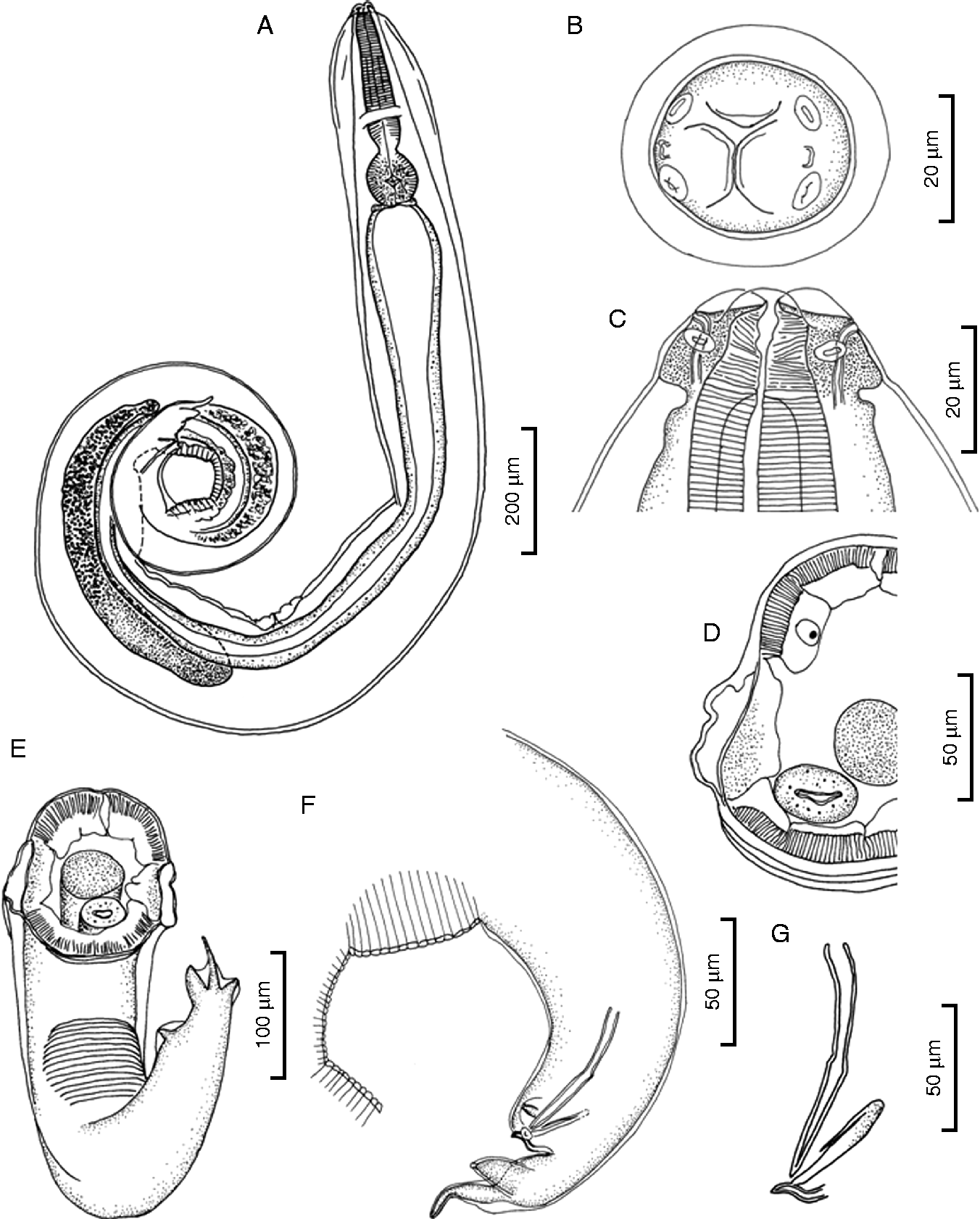
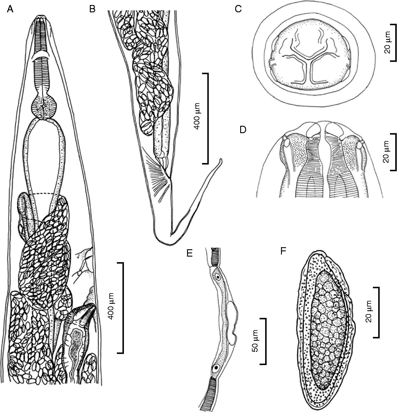
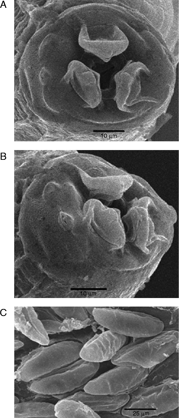
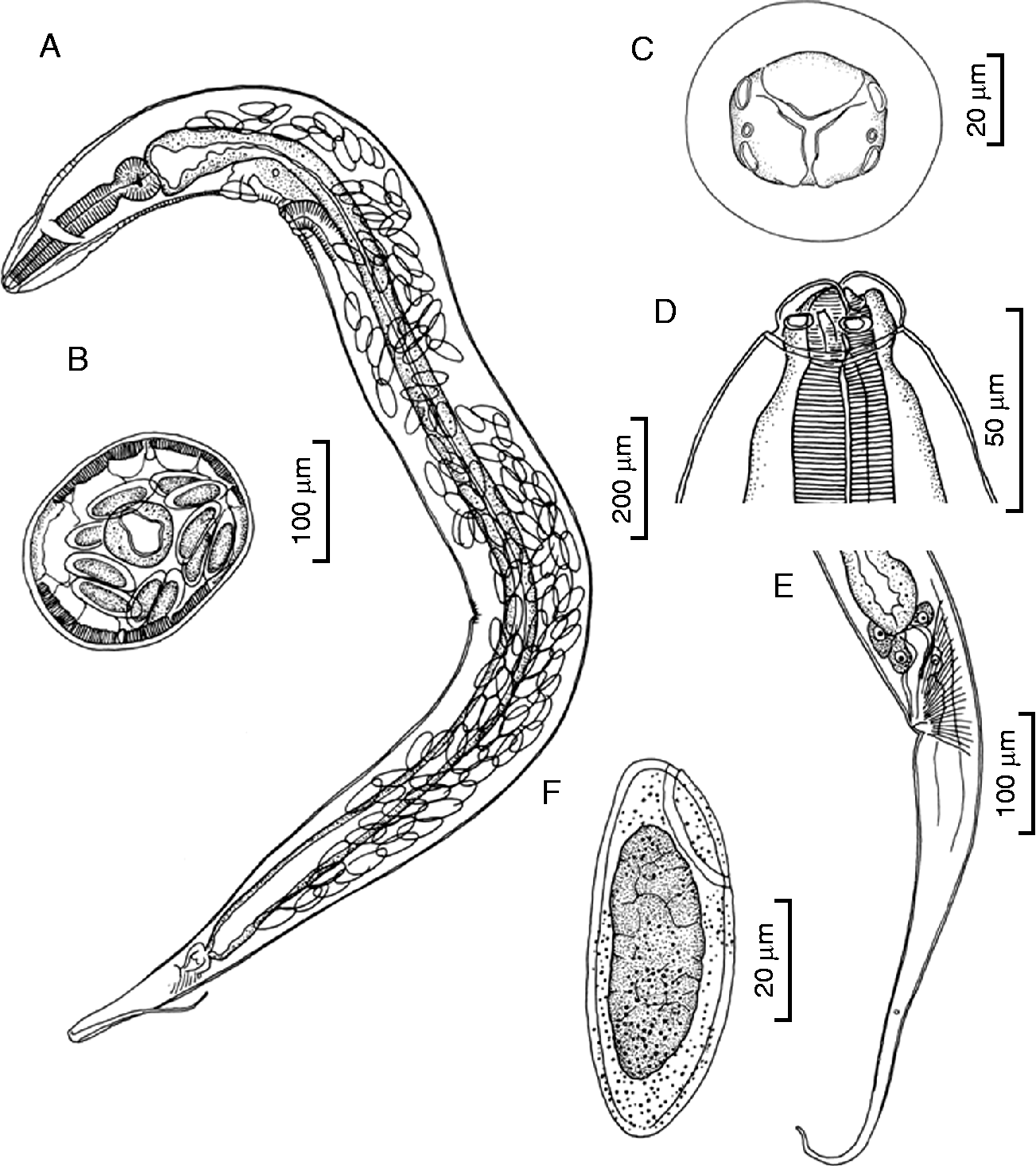
Description. Small nematodes with transverse cuticular striations; cephalic vesicle developed, continuous to body cuticle posteriorly; cephalic plate round, only slightly extended laterally; mouth opening surrounded by three protruded lips, one dorsal and two subventral. Four cephalic papillae, large, arranged almost square; amphids opening sublaterally, with porous patches laterally (figs 1B, ); oesophagus of typical oxyuroid form with valved bulb; cervical alae absent; deirids not seen.
Male of Syphacia (Syphacia) taeromyos sp. n. from Taeromys celebensis in south-east Sulawesi, Indonesia: (A) holotype, lateral view; (B) cephalic end, apical view; (C) cephalic end, ventral view; (D) midbody in cross-section; (E) posterior end, ventral view; (F) posterior end, lateral view; (G) spicule and gubernaculum, lateral view.

Female of Syphacia (Syphacia) taeromyos sp. n. from Taeromys celebensis in south-east Sulawesi, Indonesia: (A) anterior portion, lateral view; (B) posterior portion, lateral view; (C) cephalic end, apical view; (D) cephalic end, ventral view; (E) lateral field, midbody in cross-section; (F) egg.
SEM of Syphacia (Syphacia) taeromyos sp. n. from Taeromys celebensis in south-east Sulawesi, Indonesia: (A) cephalic end, apical view; (B) cephalic end, lateral view; (C) eggs.
Male (holotype and six paratypes). Posterior body bent ventrally (fig. 1A); length 1.95–2.17 (2.09) mm, width 144–165 (153); distance between amphids 23 (n= 1); cephalic vesicle 104–134 (119) wide; lateral alae vesicular, moderately developed (figs 1D, E); total oesophagus including pharynx, corpus and bulb 235–261 (248) long: pharynx 9–13 (12) long and 12–15 (13.5) wide (fig. 1C), corpus 163–177 (173) long and 32–41 (36) wide, isthmus 17–21 (19) wide at narrowest level, bulb 59–69 (64) long by 63–72 (66) wide; nerve ring at posterior part of oesophageal corpus, 146–157 (152), and excretory pore far posterior to oesophago-intestinal junction, 821–958 (919) from cephalic end, respectively; three mamelons with prominent transverse furrows present at ventral surface of posterior body: anterior mamelon 119–134 (127) long, middle mamelon 74–96 (82) long and posterior mamelon 61–72 (66) long; distance from cephalic end to anterior edges of anterior, middle and posterior mamelons 1.19–1.36 (1.28), 1.43–1.58 (1.52) and 1.65–1.83 (1.77) mm, respectively; spicule single, thin, needle-shaped, slightly constricted at middle, relatively short, 62–77 (68) long (i.e. 3.0–3.7 (3.3)% of total body length (TBL)); gubernaculum rod-like, 28–34 (31) long; accessory piece of gubernaculum relatively thin, unornamented (fig. 1G); caudal papillae present in three pairs, two pairs small, near cloaca and one posterior pair large, protruding posterolaterally (fig. 1F); tail including short terminal process 64–71 (67) long (i.e. 2.95–3.4 (3.2)% of TBL).
Female (allotype and four paratypes). Body relatively stout; length 4.85–5.85 (5.28) mm, width 390–476 (419); distance between amphidial pores 41–43 (n= 2); small flat vesicular lateral alae present (fig. 2E); total oesophagus including pharynx, corpus and bulb 422–462 (445) long: pharynx 16–22 (19) long and 17–18 wide (n= 2) (fig. 2D), corpus 302–395 (334) long and 58–70 (65) wide, isthmus 26–32 (30.4) wide at narrowest level, bulb 99–114 (108) long by 96–119 (109) wide; nerve ring at middle of oesophageal corpus, 179–202 (187), excretory pore 1.07–1.15 (1.11) mm from cephalic end (fig. 2A); vulva not protruding, 1.19–1.31 (1.27) mm (i.e. 22.4–24.4 (23.8)% of TBL) from cephalic end; vagina and ovejector directed posteriorly; distance between excretory pore and vulva 119–133 (155) (i.e. 2–4 (2.9)% of TBL); eggs long, elliptical, asymmetrical with one side flattened, operculated, shell surface uneven and densely pitted, containing morula-stage embryo in uterus, 57–62 (59) × 21–24 (23) (figs 2F and 3C); uterus winding, extending anteriorly to oesophageal bulb and ending posteriorly near anus; tail conical with pointed end, relatively long, 693–886 (804) (i.e. 13.5–18.1 (14.7)% of TBL) (fig. 2B).
Remarks. The protruded lips and much longer (more than 1.5 times) anterior mamelon in relation to other mamelons are characteristic of the present species, being readily distinguished from other congeners found in the Indonesia–Australian regions (Hugot & Quentin, Reference Hugot and Quentin1985; Hasegawa & Tarore, Reference Hasegawa and Tarore1996; Smales, Reference Smales2001, Reference Smales2004, Reference Smales2010, Reference Smales2011; Weaver & Smales, Reference Weaver and Smales2006, Reference Weaver and Smales2008, Reference Weaver and Smales2010; Dewi & Hasegawa, Reference Dewi and Hasegawa2010). By having a round cephalic plate without dorsoventral constrictions laterally, S. taeromyos resembles S. australasiensis, S. darwini, S. longaecauda, S. lorentzimyos, S. mamelontenuis, S. rifaii and S. sulawesiensis (Hugot & Quentin, Reference Hugot and Quentin1985; Smales, Reference Smales2001, Reference Smales2010, Reference Smales2011; Weaver & Smales, Reference Weaver and Smales2008 Dewi & Hasegawa, Reference Dewi and Hasegawa2010). However, S. australasiensis possesses cephalic papillae on lateral projections, a vulva with knob in the female and lacks lateral alae in the male; S. darwini has only two mamelons, S. lorentzimyos possesses cervical alae, S. longaecauda has a much greater anteriorly positioned excretory pore at the level of the proximal intestine in the male, and S. mamelontenuis lacks lateral alae, all being readily distinguished from the present species (Hugot & Quentin, Reference Hugot and Quentin1985; Smales, Reference Smales2001, Reference Smales2004, Reference Smales2010; Weaver & Smales, Reference Weaver and Smales2006). The relative distance between the excretory pore and vulva of S. taeromyos is comparable with that of S. sulawesiensis (2.3–3.8%), but shorter than that in S. rifaii (4.8–8.4%) (Hasegawa & Tarore, Reference Hasegawa and Tarore1996; Dewi & Hasegawa, Reference Dewi and Hasegawa2010). The uneven eggshell surface of S. taeromyos resembles that in S. rifaii, but egg size is smaller (68–70 × 23–29 in the latter) (Dewi & Hasegawa, Reference Dewi and Hasegawa2010).
Syphacia (Syphacia) paruromyos sp. n.
Type host. Paruromys dominator (Thomas, 1921) (Rodentia: Muridae).
Site of infection. Caecum.
Type locality. Masembo Watershed, Mekongga Mountains, south-east Sulawesi, Sulawesi, Indonesia (121°19′43″E, 03°63′66″S; elevation 1515 m).
Other locality. Indonesia: south Sulawesi, Lambanan.
Date of collection. June 2011 (Mekongga); July 1992 (Lambanan).
Specimens deposited. Holotype male and allotype female (MZB Na 604), six male and nine female paratypes (MZB Na 605); other specimens from Mekongga (MZB Na 612; NSMT-As 3905); three males and five females from Lambanan (NSMT-As 3906).
Etymology. Species epithet is derived from the generic name of the host murid, Paruromys.
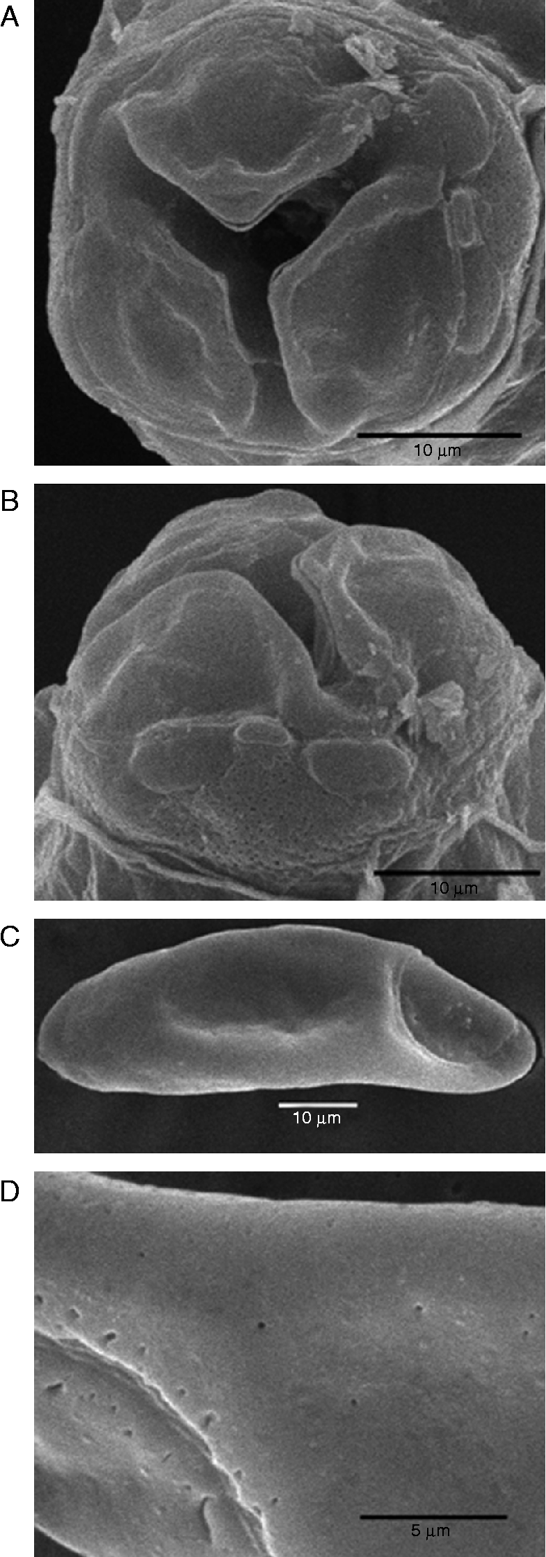
Description. Small nematodes, cuticle with faint striations; cephalic portion set-off from body by constriction, especially clearly in female (fig. 5D); cephalic plate round, without lateral extension (figs 4B, 6A); mouth with three developed lips, one dorsal and two subventral; four large papillae present; two at each lateral side; amphids with porous patches laterally (fig. 6B); oesophagus of typical oxyuroid form with valved bulb; cephalic vesicle present; cervical alae absent; deirids not seen.
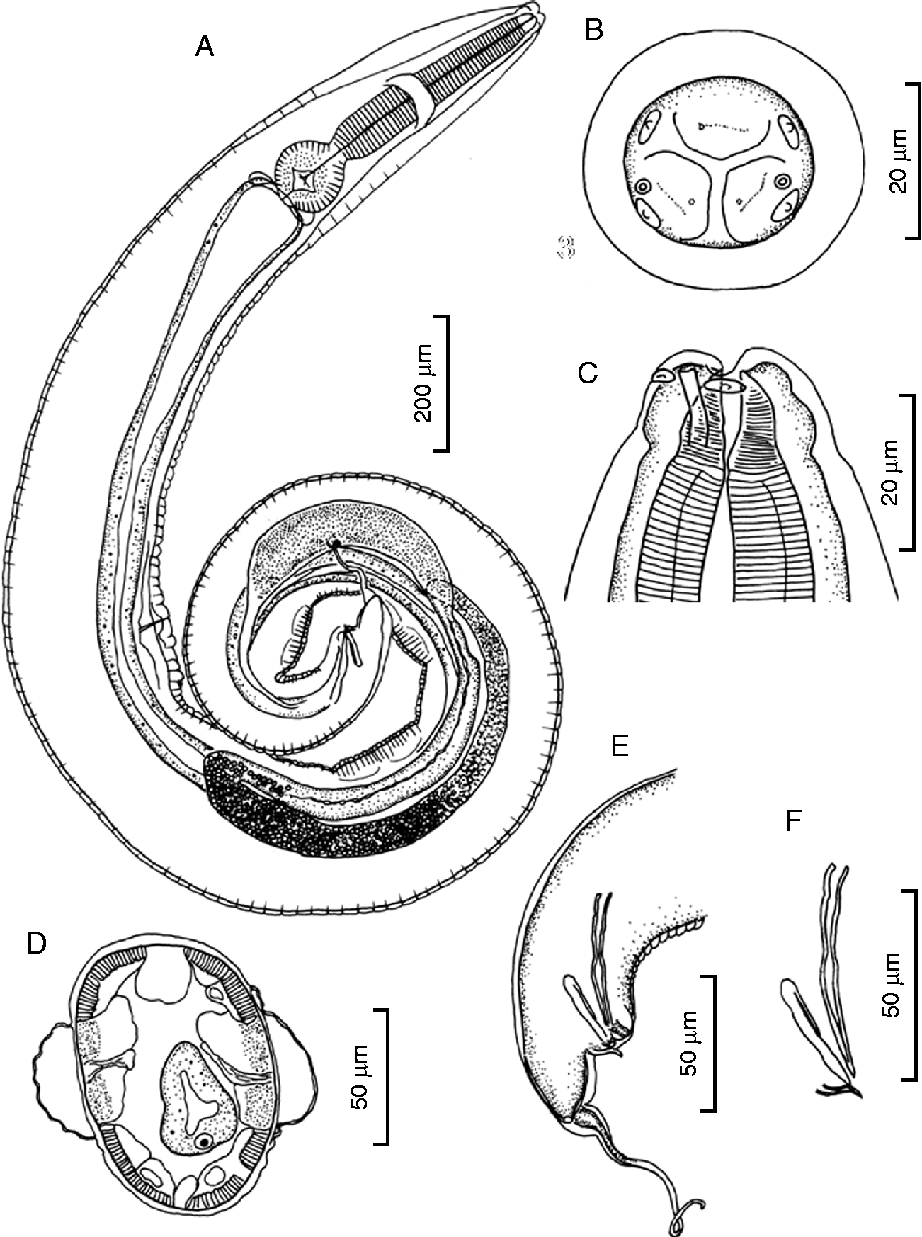
Male of Syphacia (Syphacia) paruromyos sp. n. from Paruromys dominator in south-east Sulawesi, Indonesia: (A) holotype, lateral view; (B) cephalic end, apical view; (C) cephalic end, sublateral view; (D) midbody in cross-section; (E) posterior end, lateral view; (F) spicule and gubernaculum, lateral view.
Female of Syphacia (Syphacia) paruromyos sp. n. from Paruromys dominator in south-east Sulawesi, Indonesia: (A) paratype, lateral view; (B) midbody in cross-section; (C) cephalic end, apical view; (D) cephalic end, ventral view; (E) posterior portion, lateral view; (F) egg.
SEM of female Syphacia (Syphacia) paruromyos sp. n. from Paruromys dominator in south-east Sulawesi, Indonesia: (A) cephalic end, apical view; (B) cephalic end, lateral view; (C) egg, lateral view; (D) enlarged view of eggshell surface.
Male (holotype and six paratypes from P. dominator of Mangolo). Posterior body bent ventrally (fig. 4A). Length 1.37–1.65 (1.49) mm, maximum width 114–133 (123); lateral alae large, vesicular (fig. 4D); distance between amphids 18 (n= 1); cephalic vesicle 67–89 (78) wide; total oesophagus 219–245 (229) long: pharynx 13–16 (14) long and 14–16 (15) wide, corpus 157–176 (164) long and 28–33 (30) wide, isthmus 15–18 (17) wide at narrowest level, and bulb 48–56 (51) long by 47–59 (53) wide; nerve ring slightly posterior to midlevel of oesophageal corpus, 104–115 (109), and excretory pore far posterior to oesophago-intestinal junction, 544–618 (568) from cephalic end; three mamelons developed at ventral posterior body; anterior mamelon 66–83 (72) long, middle mamelon 54–64 (58) long and posterior mamelon 45–61 (53) long; distance from cephalic end to anterior edges of anterior, middle and posterior mamelons 0.76–0.88 (0.81) mm, 0.90–1.09 (0.97) mm and 1.06–1.32 (1.15) mm, respectively; spicule single, relatively short, thin, needle-shaped, slightly constricted basal to middle, 53–59 (56) long (i.e. 3.5–4.1 (3.8)% of TBL); gubernaculum, 27–35 (30) long with thin, unornamented accessory piece of 11–14 (13) long (fig. 4F); caudal papillae present in three pairs, two pairs small, near cloaca and one pair, large, protruding posterolaterally; tail 112–134 (124) long (i.e. 6.8–9.3 (8.4)% of TBL), with whip-like appendage of 71–102 (82) long (fig. 4E).
Female (allotype and nine paratypes from P. dominator of Mangolo). Body relatively stout (fig. 5A); length 2.65–2.98 (2.80) mm, width 201–256 (223); distance between amphids 32 (n= 1); lateral alae absent (fig. 5B); cephalic vesicle 112–127 (119) wide; total oesophagus 299–346 (326) long: pharynx 15–17 (16) long and 17–19 (18) wide, corpus 217–257 (237) long and 45–53 (50) wide, isthmus 24–30 (26) wide at narrowest level, and bulb 67–81 (72) long by 71–90 (83) wide; nerve ring at midlevel of oesophageal corpus, 115–143 (130), excretory pore 396–584 (491), from cephalic end; vulva not protruding, 566–699 (650) from cephalic end (i.e. 21.9–26.4 (23.4)% of TBL); vagina and ovejector directed posteriorly; distance between excretory pore and vulva 126–188 (147) (i.e. 4.4–7.1 (5.3)% of TBL); eggs elliptical, asymmetrical with one side flattened, operculated, shell surface pitted, embryonated in uteri, 64–72 (68) × 24–29 (26) (figs 5F, 6C, D); uterus extending anteriorly to the oesophageal bulb and ending posteriorly near anus; tail relatively long, tapering to pointed end, 384–449 (408) long (i.e. 12.8–17.4 (14.6)% of TBL) (fig. 5E).
Measurements of worms from P. dominator of Lambanan. Male (three worms): length 2.04–2.12 (2.07) mm, maximum width 131–138 (134); cephalic vesicle 96–128 (112) wide; total oesophagus 237–253 (243) long: pharynx 14–16 (15) long and 14–16 (15) wide, corpus 166–176 (170) long and 30–34 (32) wide, isthmus 15–22 (18) wide at narrowest level, and bulb 51–58 (55) long by 49–61 (54) wide; nerve ring 136–141 (138), and excretory pore 769–825 (797) from cephalic end; anterior, middle and posterior mamelons 74–108 (90), 54–80 (68) and 45–70 (58) long, respectively; distance from cephalic end to anterior, edges of anterior, middle and posterior mamelons 1.12–1.19 (1.16), 1.33–1.45 (1.39) and 1.63–1.71 (1.67) mm, respectively; spicule 56–59 (58) long (i.e. 2.6–2.9 (2.8)% of TBL); gubernaculum 26–37 (31) long; accessory piece of gubernaculum relatively thin, 10–13 (11) long; tail 138–158 (150) long (i.e. 6.8–7.7 (7.2)% of TBL); whip-like appendage 99–126 (116) long.
Female (five worms). Length 3.16–3.91 (3.58) mm, width 267–293 (280); cephalic vesicle 131–150 (141) wide; total oesophagus 329–352 (338) long: pharynx 19–22 (20) long and 19–21 (21) wide, corpus 224–250 (236) long and 54–61 (57) wide, isthmus 26–32 (30) wide at narrowest level, bulb 74–93 (83) long by 86–102 (95) wide; nerve ring 118–128 (122), and excretory pore 503–650 (587), from cephalic end; vulva 612–871 (783) from cephalic end (i.e. 19.4–23.0 (21.8)% of TBL); distance between excretory pore and vulva 109–240 (196) (i.e. 3.5–6.8 (5.4)% of TBL); eggs 65–72 (69) × 24–29 (28); tail 541–576 (557) (i.e. 14.4–17.1 (15.6)% of TBL).
Remarks. By having a round cephalic plate without dorsoventral constrictions laterally, the present species resembles S. australasiensis, S. darwini, S. longaecauda, S. lorentzimyos, S. mamelontenuis, S. rifaii, S. sulawesiensis and S. taeromyos among the representatives of the subgenus known from the Indonesia-Australian region (Hugot & Quentin, Reference Hugot and Quentin1985; Smales, Reference Smales2001, Reference Smales2004, Reference Smales2010, Reference Smales2011; Weaver & Smales, Reference Weaver and Smales2008; Dewi & Hasegawa, Reference Dewi and Hasegawa2010). The present species is easily distinguished from S. australasiensis, S. darwini, S. lorentzimyos, S. longaecauda and S. mamelontenuis, by the same criteria stated above for S. taeromyos. It is also distinguished from S. rifaii, S. sulawesiensis and S. taeromyos, by having a whip-like appendage of the tail in the male (Hasegawa & Tarore, Reference Hasegawa and Tarore1996; Dewi & Hasegawa, Reference Dewi and Hasegawa2010). The relative distance between the excretory pore and vulva of the present species is close to that of S. rifaii but longer than that in S. sulawesiensis and S. taeromyos; egg size is almost identical with that of S. rifaii, but smaller than that in S. sulawesiensis (77–86 × 24–29) and larger than that in S. taeromyos (Hasegawa & Tarore, Reference Hasegawa and Tarore1996; Dewi & Hasegawa, Reference Dewi and Hasegawa2010).
Discussion
The aboriginal murids of Sulawesi have been assumed to have ancestors on the South-East Asian mainland and on the Sunda shelf islands (Musser, Reference Musser and Whitmore1987; Musser & Durden, Reference Musser and Durden2002). They are composed of so-called new endemics, relatives of which are distributed on the Sunda shelf, and old endemics, of which there are no known living relatives from the Sunda shelf (Musser, Reference Musser and Whitmore1987). There have been arguments on the origin and dispersal of the endemic murids of Sulawesi. Some researchers believe murids migrated via a land bridge connecting Sulawesi to the Sunda shelf in or before the Pleistocene (Audley-Charles, Reference Audley-Charles and Whitmore1981), though there has been no evidence of such a land connection during the Cenozoic era (see Hall, Reference Hall, Hall and Holloway1998). Meanwhile, mammalogists consider dispersal on the sea by drift a possible scenario (Musser, Reference Musser and Whitmore1987). In any case, dispersal to Sulawesi might have exerted a bottleneck effect on the murids and their parasites. Since arriving on Sulawesi, murids have experienced adaptive radiation and formed many endemic species. Sulawesi also provided one of the two routes via which ancestors of endemic murids of the Sahul region (New Guinea and Australia) migrated with their helminths (Smales, Reference Smales, Triunveri and Scalise2012).
In the Sahul region, native murids are also composed of new endemics and old endemics (Flannery, Reference Flannery1995; Archer et al., Reference Archer, Arena, Bassarova, Black, Brammall, Cooke, Creaser, Crosby, Gillespie, Godthelp, Gott, Hand, Kear, Krikmann, Mackness, Muirhead, Musser, Myers, Pledge, Wang and Wroe1999; Rowe et al., Reference Rowe, Reno, Richmond, Adkins and Steppan2008). Among endemic Syphacia species described from the Sahul region, the shape of the cephalic plate is of two types: elongated cephalic plate with dorso-ventral constrictions, and round or oval cephalic plate without dorso-ventral constrictions. Species with the former type have been found only from the old endemic murids of the genera Pseudomys, Zysomys and Coccymys (Weaver & Smales, Reference Weaver and Smales2006, Reference Weaver and Smales2008, Reference Weaver and Smales2010; Smales, Reference Smales2011). Meanwhile, endemic Syphacia with the latter type of cephalic plate are known from both old and new endemic murids (Hugot & Quentin, Reference Hugot and Quentin1985; Smales, Reference Smales2001, Reference Smales2004, Reference Smales2010). If the shape of cephalic plate has a phylogenetic significance, two lineages could be postulated in the endemic Syphacia of the Sahul region.
Murids hitherto known to host endemic Syphacia on Sulawesi, i.e. Bunomys, Paruromys, Taeromys and Rattus, are all assigned to new endemics (Musser, Reference Musser and Whitmore1987). All four endemic Syphacia species hitherto described from Sulawesi share common characteristics, such as a round cephalic plate in both sexes and vesicular lateral alae in the males (Hasegawa & Tarore, Reference Hasegawa and Tarore1996; Dewi & Hasegawa, Reference Dewi and Hasegawa2010). It is likely that they belong to one lineage that has evolved with murids during colonization of this island. If Syphacia species also parasitize old endemic murids of Sulawesi, it is expected that they possess different morphological characteristics. Further surveys are necessary to demonstrate evolutionary processes, and the use of molecular phylogenetic analysis based on DNA sequences will contribute to our understanding of the natural history of the Syphacia–host murid relationship in this area.
Acknowledgements
We would like to express our sincere thanks to the ICBG Project Mekongga team, especially to A.T. Hitch (University of California, Davis) and A.S. Achmadi (Zoology Division, RC Biologi-LIPI) for providing the vertebrate materials examined for this study. A.S. Achmadi (MZB) and K. Rowe (Victoria Museum, Australia) provided identification of host murids. We want to thank Y. Apriyanti for her assistance with preparing the material for the SEM observation. We also wish to thank the Indonesian–ICBG Project Mekongga Leaders, R. Ubaidillah and E.E. Widjaja (LIPI), A. Engilis Jr (University of California, Davis) and S. Chidlow (Oita University) for the English revision. This study was partly supported by Grant #U01TW008160 from the Fogarty International Center, the Office of Dietary Supplements, the National Science Foundation and the Department of Energy; the USDA Agricultural Food Research Initiative of the National Institute of Food and Agriculture, US Department of Agriculture, Grant #35621-04750; the project ‘Peningkatan Kemampuan Peneliti dan Perekayasa (PKPP) 2012’, sponsored by the State Ministry of Research and Technology of Indonesia; and a grant-in-aid (C) 23570120 from the Japan Society for Promoting Science. The content is solely the responsibility of the authors and does not necessarily represent the official views of the Fogarty International Center or the National Institutes of Health, the Office of Dietary Supplements, the National Science Foundation, the Department of Energy or the Department of Agriculture.