Introduction
Kawasaki disease, also known as mucocutaneous lymph node syndrome, is an acute inflammatory vasculitis, and coronary artery aneurysm is its most serious complication. It is the leading cause of acquired heart disease in children under 5 years of age.Reference Zeinali, Frishman and Aronow1 In recent years, the incidence of Kawasaki disease has increased, with racial and regional differences. In China, the incidence among children under 5 years of age is approximately 56–110 per 100,000. The onset of Kawasaki disease may be related to environmental, genetic, infectious, and immune factors, but the specific cause has not been fully clarified.Reference Jone, Tremoulet and Choueiter2 Approximately 20 to 25% of untreated children and 3 to 5% of treated children develop cardiac sequelae related to coronary artery aneurysm, and some children show resistance to intravenous immunoglobulin. Initial intensive treatment does not significantly reduce the overall incidence of coronary artery aneurysm.Reference Agrafiotou, Sapountzi, Margoni and Fotis3 Coronary artery aneurysm may lead to further complications, including coronary artery thrombosis, aneurysm rupture, coronary artery stenosis, myocardial infarction, and even death. Some studies have also found that in some children with coronary artery aneurysm, even if the aneurysm diameter normalises within two years, the morphology of the coronary arteries continues to change.Reference Shimizu, Pérez Pomares and Loy4 This suggests that once the coronary artery wall is affected, endothelial dysfunction persists, and intimal thickening and vascular regeneration may continue for several years. Therefore, long-term use of antiplatelet or anticoagulant therapy is necessary. Additionally, the risk of coronary atherosclerosis and other cardiovascular diseases in adults who had Kawasaki disease as children remains relatively high, which significantly impacts their quality of life and may even pose a life-threatening risk.Reference Elias, Brothers and Hogarty5,Reference Tsuda and Hashimoto6
The basic pathological change in Kawasaki disease is nonspecific vasculitis, predominantly affecting medium and small arteries. Inflammatory injury and dysfunction of vascular endothelial cells are central to the pathogenesis of coronary artery aneurysm.Reference Paolini, Guida and Calvaruso7 Coronary endothelial cells are located in the intima of the coronary arteries, serving as the interface between circulating inflammatory cells, vascular mediators, and the outer membrane. These cells are the primary targets of the early inflammatory attack in Kawasaki disease. Coronary angiitis typically begins 6 to 8 days after the onset of Kawasaki disease, with inflammation rapidly involving all layers of the artery, leading to severe damage to the arterial wall structure.Reference Kumrah, Goyal, Rawat and Singh8 Following inflammatory damage to endothelial cells, the immune system is further activated, resulting in abnormal T cell activation. The cytokines produced by T-cells exacerbate endothelial cell injury.Reference Wang, Qian and Liu9 Abnormal T cell activation and endothelial cell damage are key factors in coronary vascular dysfunction. Current research into the mechanisms of endothelial cell damage has identified several proinflammatory cytokines, such as interleukin-1 beta (IL-1β) and tumor necrosis factor-alpha, along with oxidative stress and inflammatory activation genes such as NOD1 and NLRP3. However, these studies have not yet addressed the underlying cause of endothelial cell damage, a question that remains urgent and requires further exploration.Reference Lin, Wang and Luo10,Reference Yao, Lai and Li11 Studies have shown that multiple triggering factors, in combination with genetic and epigenetic mechanisms, can contribute to endothelial cell injury and dysfunction.Reference Kumrah, Vignesh, Rawat and Singh12
In recent years, studies have shown that various types of extrachromosomal circular DNAs are widely present in eukaryotic cells and play a significant role in genetic processes. Their involvement in the mechanisms underlying vascular endothelial cell apoptosis has garnered increasing attention.Reference Shi, Yang, Qiao, Yu and Zhang13 Therefore, from the perspective of extrachromosomal circular DNAs, in-depth research into the aetiology and pathogenesis of Kawasaki disease complicated by coronary artery aneurysm is crucial for reversing endothelial cell dysfunction and identifying new targets for the prevention and treatment of coronary artery aneurysm.
Methods
Patients and samples
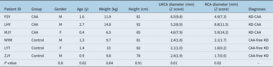
The key aspects of the study design, including the research question, primary outcome measures, and statistical analysis plan, were prepared and registered before data collection commenced. This study was approved by the Ethics Committee of The First Affiliated Hospital of Guangxi Medical University [No.2025-E1076], and parental consent was obtained prior to all research procedures. Three patients with Kawasaki disease complicated by coronary artery aneurysm group and three patients with Kawasaki disease without coronary artery aneurysm (control group) were enrolled in the sequencing study. All patients were treated in 2024. Kawasaki disease was a clinical syndrome, diagnosed primarily based on clinical manifestations and laboratory tests, while excluding other febrile illnesses. A coronary artery aneurysm was diagnosed via echocardiography, with a Z score corrected by body surface area of ≥2.5. The characteristics of the six patients who provided samples for the sequencing study are summarised in Table 1. Whole blood samples were collected during the non-acute phase after intravenous immunoglobulin therapy, and stored at −80°C until DNA extraction.
Baseline characteristics of 6 patients who provided samples for analysis

Table 1. Long description
Table summarizes the baseline characteristics of 6 Kawasaki disease patients who provided blood samples for eccDNA analysis, stratified by coronary artery aneurysm status. The CAA group (n=3) consisted of patients FSY (1.6 years, male), LHY (2.7 years, male), and MJY (0.4 years, female). The non-CAA control group (n=3) consisted of patients WYM (1.3 years, male), LYT (1.4 years, female), and ZJY (0.9 years, male). Demographic comparisons between the CAA and control groups showed no significant differences: gender (p=0.80), age (p=0.62), weight (p=0.64), and height (p=0.91), indicating that the two groups were well-matched for baseline characteristics. However, coronary artery diameters differed significantly between groups. For the left main coronary artery (LMCA), the CAA group had a mean diameter of 5.4 mm (Z-scores ranging from 6.9 to 9.8) compared to 2.3 mm (Z-scores 1.0 to 1.9) in the control group (p=0.01). For the right coronary artery (RCA), the CAA group had a mean diameter of 6.5 mm (Z-scores ranging from 7.3 to 14.2) compared to 1.8 mm (Z-scores 0.2 to 1.7) in the control group (p=0.02). These findings confirm that the CAA group had significantly dilated coronary arteries while remaining comparable to the control group in all demographic parameters.
CAA = coronary artery aneurysm; LMCA = left main coronary artery; RCA = right coronary artery.
extrachromosomal circular DNA library preparation and sequencing
Genomic DNA was isolated from whole blood using the QIAamp DNA Mini Kit (Qiagen, #51306). DNA quantity and quality were assessed by NanoDrop and agarose gel electrophoresis. Mitochondrial DNA was linearised with PacI, and linear chromosomal DNA was removed using Plasmid-Safe ATP-dependent DNase. The enriched circular DNA was amplified by rolling circle amplification with phi29 DNA polymerase (#M0269L, NEB) at 30°C for 14 h and inactivated at 65°C for 10 min. Amplified extrachromosomal circular DNAs were fragmented to an average size of approximately 400 bp using a Bioruptor (Diagenode). Sequencing libraries were prepared using the NEBNext Ultra II DNA Library Prep Kit for Illumina (NEB, #E7645L), quality-checked using an Agilent 2100 Bioanalyzer, and sequenced on the Illumina NovaSeq 6000 platform with the NovaSeq 6000 S4 Reagent Kit (300 cycles).
Bioinformatic analysis of extrachromosomal circular DNA profiles
Raw reads were trimmed using fastp (v0.23.2) and aligned to the human reference genome (UCSC hg38) using BWA (v0.7.17). Putative extrachromosomal circular DNAs were identified using Circle-Map (v1.1.4), with at least two split reads required for detection. Differential extrachromosomal circular DNAs between the coronary artery aneurysm and control groups were defined using |log2 fold change| ≥1 and P < 0.05. extrachromosomal circular DNAs were annotated based on overlapping genes using GENCODE (v42).
Functional enrichment analysis
To investigate the potential biological relevance of differential extrachromosomal circular DNAs, functional enrichment analyses were performed on genes associated with significantly upregulated extrachromosomal circular DNAs. Gene Ontology (GO)
enrichment analysis was conducted to characterise enriched biological process, molecular function, and cellular component terms. Kyoto Encyclopedia of Genes and Genomes (KEGG) pathway enrichment analysis was further performed to identify significantly enriched biological pathways related to extrachromosomal circular DNA-associated genes.
Immune infiltration analysis
Immune infiltration characteristics were assessed using the single-sample gene set enrichment analysis algorithm. Based on publicly available gene sets for immune cells and immune-related pathways, immune infiltration features were quantified for each sample group.
Statistical analysis
All statistical analyses were performed using SPSS 25.0 (IBM) and R software. Differences in clinical or demographic variables between groups were assessed using Student’s t-test or Fisher’s exact test, as appropriate. A two-sided P value of <0.05 was considered statistically significant. Given the limited sample size, the enrichment and immune infiltration analyses were considered exploratory.
Results
Differential expression and length distribution of extrachromosomal circular DNAs in the coronary artery aneurysm and control groups
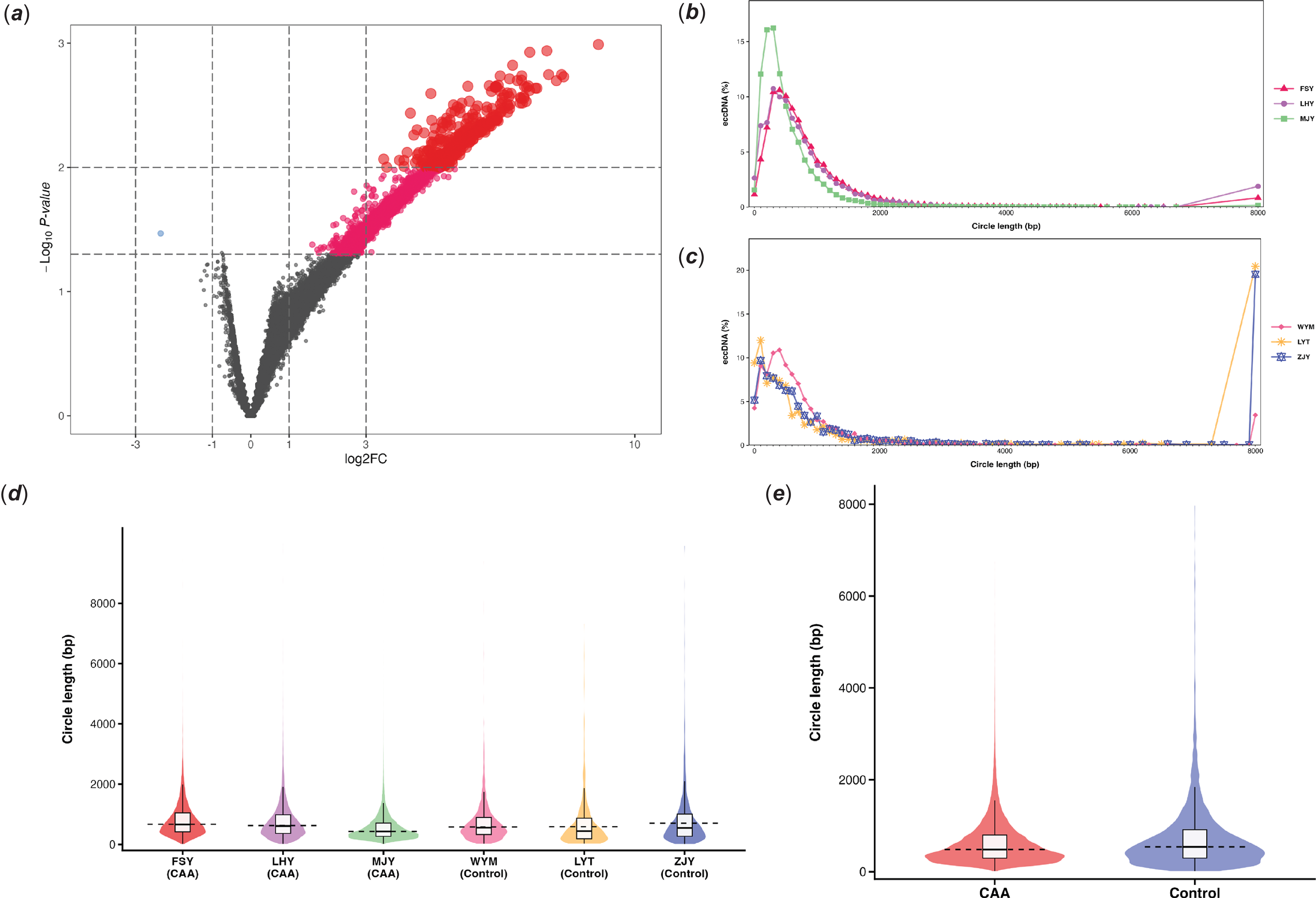
We systematically compared eccDNA profiles between the coronary artery aneurysm and control groups using eccDNA sequencing. The volcano plot (Figure 1a) showed 4,790 significantly differential extrachromosomal circular DNAs between the two groups, 4,789 of which were upregulated (log2FC ≥ 1, P < 0.05). We further compared the length distribution of extrachromosomal circular DNAs between the two groups (Figure 1b–c). The analysis demonstrated that the length distribution of extrachromosomal circular DNAs in the coronary artery aneurysm group was more concentrated compared to the control group. More than half of the extrachromosomal circular DNAs in both groups ranged from 100 to 1,000 bp in length. In the coronary artery aneurysm group, the length distribution showed a dominant peak at 400–500 bp, while the control group exhibited multiple peaks across the 100–1000 bp range. Both groups exhibited a peak around 8000 bp, with this peak being more pronounced in the control group. Furthermore, the coronary artery aneurysm group exhibited a more compact and uniform distribution of eccDNA lengths (Figure 1d–1e), whereas the control group displayed a broader and more dispersed distribution, with a greater proportion of longer extrachromosomal circular DNAs.
Length distribution of eccDNAs in CAA and control groups. (a) Volcano plot illustrating differentially expressed eccDNAs between CAA and control groups (fold change ≥2 and P <0.05). (b–c) Line charts depicting the percentage length distribution of eccDNAs in the CAA group (B) and control group (C). (d–e) Violin plots comparing eccDNA length distribution across individual samples (D) and between the combined CAA and control groups (E).

Figure 1. Long description
Figure 1 presents the differential expression and length distribution characteristics of eccDNAs in the CAA and control groups across five panels (A–E). Panel A is a volcano plot showing 4,790 significantly differentially expressed eccDNAs between the two groups (log2FC ≥ 1, P < 0.05), of which 4,789 were upregulated (red) and only one was downregulated (blue) in the CAA group; gray points indicate no significant difference. Panels B and C are line charts displaying the percentage length distribution of eccDNAs in the CAA group (Panel B) and control group (Panel C), respectively. In both groups, over half of the eccDNAs ranged from 100 to 1,000 bp in length, but with different distribution patterns: the CAA group showed a dominant peak at 400–500 bp, while the control group exhibited multiple peaks across the 100–1,000 bp range; both groups shared a peak at approximately 8,000 bp, which was more pronounced in the control group. Panels D and E are violin plots comparing eccDNA length distribution across individual samples (Panel D, 3 CAA and 3 control samples) and between the combined CAA and control groups (Panel E). The CAA group exhibited a more compact and uniform distribution, whereas the control group displayed a broader and more dispersed distribution with a greater proportion of longer eccDNAs.
Chromosomal distribution and functional enrichment of extrachromosomal circular DNAs
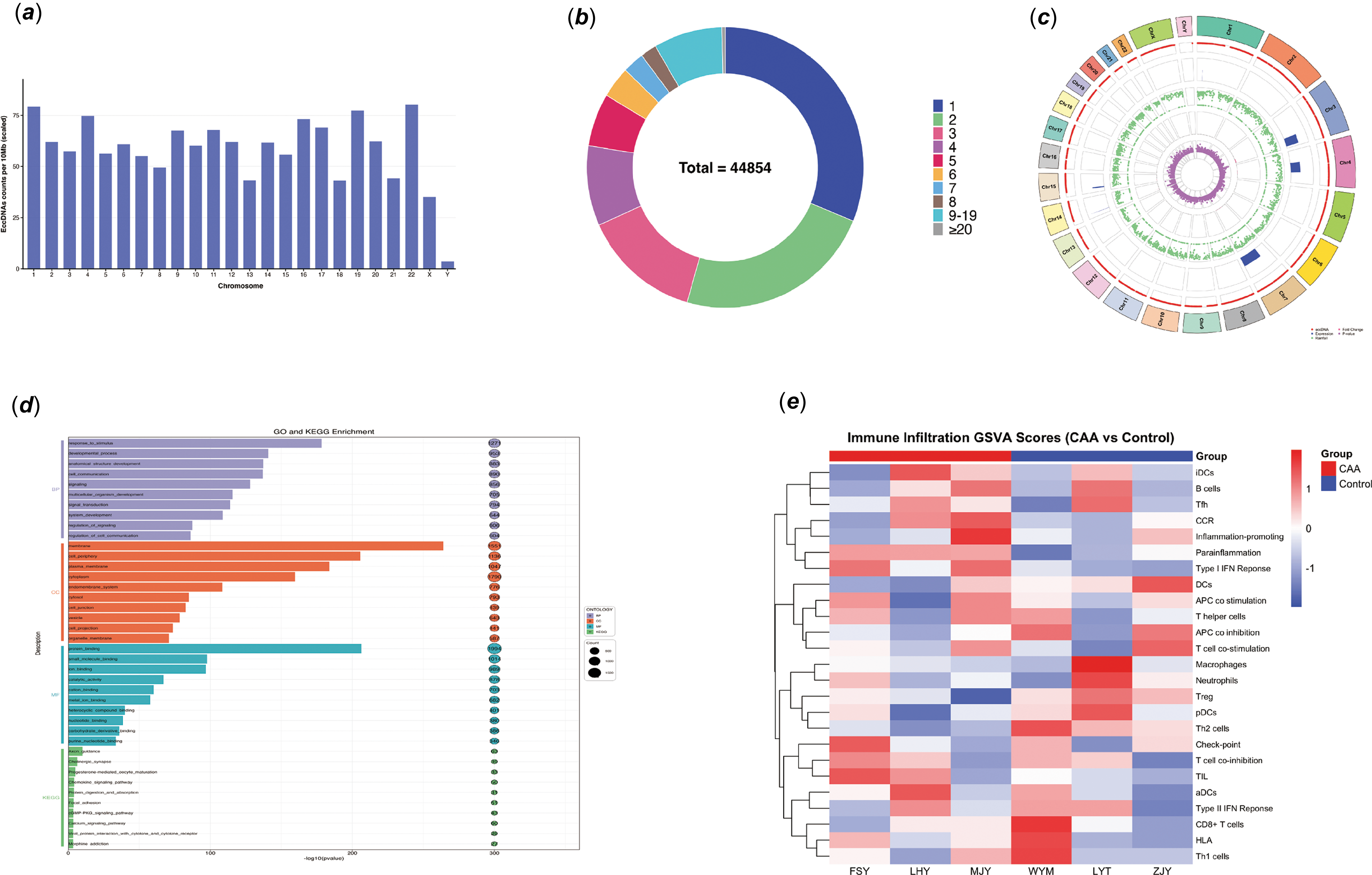
We further investigated the chromosomal distribution of extrachromosomal circular DNAs (Figure 2a). The frequency of extrachromosomal circular DNAs shedding per 10 Mb differed across chromosomes, with increased shedding on chromosomes 1, 19, and 22, and minimal shedding on the Y chromosome. Given that eccDNA production can vary by gene, we quantified the number of extrachromosomal circular DNAs produced by different genes (Figure 2b). Most genes generated three or fewer extrachromosomal circular DNAs, indicating that eccDNA formation may be selective. To further analyse the chromosomal origin of significantly upregulated extrachromosomal circular DNAs (log2FC ≥ 1, P < 0.05) in the coronary artery aneurysm group, we visualised the data using a Circos plot (Figure 2c). We observed that no significant extrachromosomal circular DNAs were detected in the 120–140 Mb region of chromosome 1, the 40–60 Mb region of chromosome 9, or the 0–20 Mb regions of chromosomes 13, 14, and 15. Additionally, almost no significantly differentially expressed extrachromosomal circular DNAs were detected on the Y chromosome. By comparing the distribution of the upregulated extrachromosomal circular DNAs across chromosomes, we found that they were predominantly located on chromosomes 3, 4, and 7. The distribution of the extrachromosomal circular DNAs, as shown in the rainfall plot, indicates a tendency for aggregation at specific chromosomal locations.
Landscape of eccDNA distribution, expression, functional enrichment, and immune signatures in CAA and control groups. (a) The frequency of eccDNAs per 10 Mb across each chromosome. (b) Pie chart representing the proportion of genes (n = 44,854) generating varying numbers of eccDNAs. (c) Circos plot displaying chromosomal distribution and expression levels of the upregulated eccDNAs in the CAA group. From outer to inner rings: chromosome ideograms (chr1–Y); eccDNAs genomic positions; basemean expression levels of corresponding genes; rainfall plot indicating eccDNAs clustering density (y-axis: minimum inter-region distance); fold change of genes; and associated P-values. (d) KEGG pathway enrichment and GO functional analysis of genes associated with CAA-related upregulated eccDNAs. (e) Heatmap of immune infiltration GSVA scores comparing CAA and control groups.

Figure 2. Long description
Figure 2 provides a comprehensive landscape of eccDNA chromosomal distribution, gene proportion, expression profile, functional enrichment, and immune signatures across five panels (A–E). Panel A is a bar chart showing the frequency of eccDNAs per 10 Mb across chromosomes, with chromosomes 1, 19, and 22 showing higher frequencies and the Y chromosome showing the lowest frequency. Panel B is a pie chart demonstrating that most genes generated three or fewer eccDNAs, suggesting that eccDNA formation is selective. Panel C is a Circos plot displaying the chromosomal distribution and expression characteristics of upregulated eccDNAs (log2FC ≥ 1, P < 0.05) in the CAA group, with concentric rings representing chromosome ideograms (chr1-Y), genomic positions of eccDNAs, basemean expression levels, rainfall plot (y-axis: minimum inter-region distance, indicating clustering density), fold changes, and P-values from outer to inner. Notably, no significant eccDNAs were detected in the 120–140 Mb region of chromosome 1, the 40–60 Mb region of chromosome 9, or the 0–20 Mb regions of chromosomes 13, 14, and 15; almost no significantly differentially expressed eccDNAs were detected on the Y chromosome. Upregulated eccDNAs were predominantly concentrated on chromosomes 3, 4, and 7. The rainfall plot revealed a tendency for eccDNA aggregation at specific chromosomal loci. Panel D is a dot plot showing GO and KEGG enrichment analyses of genes associated with upregulated eccDNAs in the CAA group; a total of 3,546 biological processes, 523 cellular components, 790 molecular functions, and 101 KEGG pathways were identified, with significant enrichment in terms related to response to stimulus, developmental processes, cell communication, cell periphery, cytoplasm, endomembrane system, protein binding, small molecule binding, catalytic activity, as well as the chemokine signaling pathway, cGMP–PKG signaling pathway, and calcium signaling pathway. Panel E is a heatmap showing immune infiltration ssGSEA scores comparing CAA and control groups; the CAA group exhibited higher scores for iDCs, B cells, Tfh cells, and type I interferon response, while the control group showed higher scores for DCs, Tregs, pDCs, Th2 cells, and immune checkpoint-related signatures, indicating distinct immune infiltration patterns between the two groups.
Gene Ontology and Kyoto Encyclopedia of Genes and Genomes\ enrichment analyses were performed on genes associated with the upregulated extrachromosomal circular DNAs. We identified 3,546 BPs, 523 CCs, 790 MFs, and 101 KEGG pathways. Genes associated with the upregulated extrachromosomal circular DNAs were enriched in terms related to response to stimulus, developmental processes, cell communication, cell periphery, cytoplasm, endomembrane system, protein binding, small molecule binding, and catalytic activity. The most significantly enriched KEGG pathways included the chemokine signalling pathway, cGMP–PKG signalling pathway, and calcium signalling pathway (Figure 2d).
Immune infiltration differences between coronary artery aneurysm and control groups
Immune infiltration was assessed using the single-sample gene set enrichment analysis algorithm. Single-sample gene set enrichment analysis analysis revealed that the coronary artery aneurysm group exhibited higher scores for several immune-related signatures, including immature Dendritic cells, B cells, T follicular helper cells, and type I interferon response, whereas the control group showed higher scores for Dendritic cells, regulatory T cells, plasmacytoid dendritic cells, T helper 2 cells, and immune checkpoint signatures (Figure 2e).
Discussion
Extrachromosomal circular DNAs are a type of non-mitochondrial, non-plasmid circular DNA molecules derived from linear chromosomal genomic DNA and involved in both physiological and pathological processes. It is widely accepted that eccDNA formation is associated with DNA damage repair. While most broken DNA fragments of chromosomes are repaired or removed by the DNA repair system, the retained or damaged fragments may reassemble into circular DNA structures, which replicate and segregate randomly during cell division.Reference Yang, Jia and Ge14 Extrachromosomal circular DNAs exhibit diverse biological functions. They regulate various cellular processes, including enhancing gene transcription and translation, modulating genome replication to induce cell death, stimulating immune responses as cytokine, and mediating intercellular communication. In cancer, extrachromosomal circular DNAs frequently carry oncogenes, contributing to genetic heterogeneity and promoting tumour progression.Reference Zheng, Li and Wang15 Studies have shown that extrachromosomal circular DNAs play a role in gouty arthritis and are strongly associated with stress responses, as well as immune and inflammatory responses, including the T-cell receptor and JAK-STAT signalling pathways.Reference Pang, Pan and Lin16 In 2021, research demonstrated that apoptosis promotes the generation of extrachromosomal circular DNAs in both human and mouse cells. Similar to chromosomal cleavage, apoptosis induces DNA breaks, generating numerous DNA fragments that contribute to eccDNA formation, thus highlighting the close relationship between extrachromosomal circular DNA generation and the apoptosis mechanism.Reference Wang, Wang and Djekidel17 However, the role of extrachromosomal circular DNAs in children with Kawasaki disease complicated by coronary artery aneurysm, as well as the mechanisms regulating the development of coronary artery aneurysm, are not yet well understood and require urgent in-depth exploration.
This study revealed that extrachromosomal circular DNAs in thecoronary artery aneurysm group tended to be more aggregated and shorter in length, which may suggest that specific DNA fragmentation or recombination events during coronary aneurysm development contribute to the formation of a distinct extrachromosomal circular DNA population. A study on dilated cardiomyopathy showed that a similar accumulation of extrachromosomal circular DNAs smaller than 1000 bp is present in the heart tissue of patients with heart failure, providing a precedent for investigating extrachromosomal circular DNAs in other cardiovascular pathological conditions.Reference Lin, Dai, Li, Zhao and Wang18
Our research found that extrachromosomal circular DNAs were shed more from autosomes and less from the Y chromosome, which is basically consistent with current findings on extrachromosomal circular DNA. Eextrachromosomal circular DNA is typically a sign of genomic instability, resulting from chromosome fragmentation, recombination, or abnormal repair.Reference Møller, Mohiyuddin and Prada-Luengo19,Reference Yan, Mischel and Chang20 Although Kawasaki disease complicated by coronary artery aneurysms are significantly more common in boys than in girls,Reference Burns21 Extrachromosomal circular DNAs were less frequently detected on the Y chromosome, presenting an apparent ‘paradox’.This may suggest that the Y chromosome itself is not the primary site of damage. The heightened risk in boys could stem from other factors, such as immune response intensity, sex hormone effects, or autosomal susceptibility genes, rather than genomic instability of the Y chromosome itself.Reference Dunn, Perry and Klein22 Kawasaki disease is considered an infection-triggered autoimmune vasculitis targeting the blood vessels. The systemic inflammatory response subsequently activates signalling pathways, including NF-κB, IL-1β, and IL-6, and induces DNA damage. The target genes of these processes are widely distributed across both autosomes and the X chromosome.Reference Baatarjav, Komada and Gunji23 This is consistent with the observation that susceptibility genes for many autoimmune diseases, such as systemic lupus erythematosus, are predominantly located on autosomes.Reference Sakaue, Kanai and Tanigawa24 Future research should systematically analyse which autosomal regions in coronary aneurysm tissues serve as hotspots for extrachromosomal circular DNA. The genes encoded or regulated by these extrachromosomal circular DNAs are likely to be key factors driving vascular injury and aneurysm formation, offering potential new targets for intervention.
In the context of coronary artery aneurysms in Kawasaki disease, our study found that upregulated extrachromosomal circular DNAs mainly originated from chromosomes 3, 4, and 7. One possible explanation is that these chromosomes contain key gene regions that are highly stressed during immune inflammatory storms, such as common fragile sites and immune gene clusters.Reference Gerovska, Fernández Moreno, Zabala and Araúzo-Bravo25,Reference Habibi, Talebi and Bahrami26 These regions may become fragile due to high transcriptional activity and replication stress, eventually giving rise to extrachromosomal circular DNAs. This finding may help localise regions of genomic instability and identify core pathogenic genes involved in coronary artery aneurysm formation, thereby providing clues for the development of biomarkers and therapeutic targets. KEGG pathway and GO analyses revealed that genes associated with the upregulatedextrachromosomal circular DNAs were enriched in the chemokine signalling pathway, cGMP–PKG signalling pathway, and calcium signalling pathway. Therefore, we speculate that extrachromosomal circular DNAs may promote the occurrence of coronary artery aneurysm by regulating the amplification of specific genes, driving the excessive production of chemokines and abnormal interactions between cells, such as immune cells and endothelial cells.Reference Chen, Yang and Zhang27,Reference Liu, Luo and Fan28 Extrachromosomal circular DNAs are likely not only a “scar” of genomic instability but also the “engine” driving the vicious cycle of vascular inflammation in Kawasaki disease. They amplify chemokines and immunomodulatory genes, intensifying inflammatory signals and disrupting physiological anti-inflammatory mechanisms, ultimately causing irreversible damage to the coronary arteries. This hypothesis offers a new perspective on the sex differences observed in Kawasaki disease and may inform the development of gene-targeted therapies.
This study further conducted an immune infiltration analysis. Single-sample gene set enrichment analysis algorithm suggested that the coronary artery aneurysm group showed a predominance of immune features associated with more specific immune responses, while the control group showed a greater tendency for broader immune regulatory processes, suggesting that the nature of the immune response may vary. This interpretation aligns with previous reviews highlighting the heterogeneity of Kawasaki disease immunopathology.Reference Noval Rivas and Arditi29 It is worth noting that the reference database used for immune characteristic analysis in this study was mainly constructed based on tumour transcriptome data. Whether it is fully applicable to the background of Kawasaki disease, an autoimmune vasculitis, still needs further verification.
In conclusion, this study provides the first characterisation of extrachromosomal circular DNA alterations in Kawasaki disease complicated by coronary artery aneurysm and its specific chromosomal distribution. Our findings suggest that extrachromosomal circular DNAs may contribute to coronary artery injury by modulating vascular inflammation, immune responses, and interactions between immune cells and endothelial cells. These results broaden our understanding of the molecular mechanisms underlying coronary artery aneurysm in Kawasaki disease and may facilitate the development of extrachromosomal circular DNA-based biomarkers and targeted therapeutic strategies in the future. Several limitations should be acknowledged in this study. The relatively small sample size may reduce statistical power, while the single-centre design and single time-point sampling after intravenous immunoglobulin treatment may limit the generalisability of the findings and prevent evaluation of temporal changes in circulating extrachromosomal circular DNA profiles during disease progression. The limited sample size is partly attributable to the rarity of Kawasaki disease complicated by coronary artery aneurysm and the challenges associated with establishing sufficiently large clinical cohorts. In addition, pathway enrichment and immune infiltration analyses were based on computational inference and should be interpreted as hypothesis-generating rather than direct evidence of biological mechanisms. As this study represents the first investigation of extrachromosomal circular DNA alterations in Kawasaki disease complicated by coronary artery aneurysm, independent validation cohorts were not available within the scope of this study. Future multicentre studies with larger cohorts, longitudinal sampling, and further experimental investigations will be important to validate these findings and clarify the biological significance of extrachromosomal circular DNAs.
Availability of data and material
The raw data were uploaded to Gene Expression Omnibus (GSE318432). The data supporting this article’s conclusions will be made available by the authors without unnecessary reservations to qualified researchers.
Acknowledgements
The authors sincerely thank all participating patients and their families for their invaluable contributions to this study.
Author contributions
(I) Conception and design: Yanlin Wu; Danyan Su; (II) Administrative support: Danyan Su; (III) Provision of study materials or patients: Danyan Su; Yusheng Pang; (IV) Collection and assembly of data: Yanlin Wu; (V) Data analysis and interpretation: Yanlin Wu; Danyan Su; (VI) Manuscript writing: All authors; (VII) Final approval of manuscript: All authors.
Financial support
None.
Competing interests
All authors have completed the ICMJE uniform disclosure form. The authors declare no conflict of interest.
Ethical standard
The authors are accountable for all aspects of the work in ensuring that questions related to its accuracy and integrity are appropriately investigated and resolved. The study was approved by the Ethics Committee of The First Affiliated Hospital of Guangxi Medical University (No. 2025-E1076): the registration number of the ethics board. Written informed consent was obtained from the parents of all individual participants. The study conformed to the provisions of the Declaration of Helsinki and its subsequent amendments.