Left ventricular pseudoaneurysm is a very rare but well-known complication after infective endocarditis.Reference Itoda, Komae, Yamamoto and Takeda 1 – Reference Sachdeva and Imamura 3 Its occurrence is unpredictable and usually detected by routine follow-up echocardiography. Our patient developed left ventricular pseudoaneurysm 4 weeks after aortic valve replacement. We present CT scan images, which might have, retrospectively, presumed the development of left ventricular pseudoaneurysm.
Case report
A 12-year-old boy was admitted with hyperthermia, asthenia, and vomiting. He presented with purpura localised in both hands and feet. Blood samples showed inflammatory response with elevated white cells count and hepatic cytolysis. He had no neurological symptoms. Cardiac murmurs and transthoracic cardiac echography were normal.
Four days after admission, repetitive transthoracic cardiac echography showed significant aortic leak with vegetations appended to the non-coronary cusp.
Antibiotherapy was initiated and adjusted once hemoculture revealed sensible Staphylococcus aureus infection.
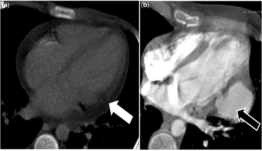
Six days after admission, whole-body CT scan was performed to stage the patient, for which a late post-contrast acquisition was added, which showed lack of enhancement in the postero-lateral wall of the left ventricle, suggestive of ischemic lesions in circumflex supplying area (Fig 1a). Electrocardiogram remained normal but troponin was slightly elevated. Despite adequate antibiotherapy, infectious syndrome was uncontrolled and the patient was getting worse.
(a) CT scan, late post-contrast acquisition, axial oblique 4-chamber view showing lack of enhancement in the postero-lateral wall of the left ventricle (white arrow) suggestive of ischemic lesions. (b) CT scan, arterial phase acquisition, axial oblique 4-chamber view demonstrating the pseudoaneurysmal evolution exactly at the same level of previously detected myocardial ischemic injury (black arrow).

He was brought to the operating room at day 10 for mechanical aortic valve replacement (19 mm). Post-operative course was complicated by cerebral bleeding requiring surgical drainage. The patient was discharged home 3 weeks later in very good clinical condition, with good cardiac function and no neurological sequelae.
One week later, routine outpatient transthoracic cardiac echography follow-up showed a large cavity (36 × 39 × 43 mm) with flow, adjacent to left ventricle communicating by a small neck (12 mm) at the postero-lateral wall. Neither wall motion abnormalities of left ventricle nor mechanical aortic valve dysfunction was seen.
CT scan confirmed the pseudoaneurysmal evolution exactly at the same level of previously detected myocardial ischemic injury (Fig 1b).
Given its potential spontaneous rupture, the patient was brought back in emergency to the operating room for pseudoaneurysm resection and closure. The orifice was closed using a Dacron patch.
Discussion
Pseudoaneurysm is a rare but potentially fatal complication of infective endocarditis.Reference Itoda, Komae, Yamamoto and Takeda 1 Most of the time, reported cases refer to pseudoaneurysm developed close to annulus abscess.Reference Sartipy, Ivert and Ugander 2 , Reference Sachdeva and Imamura 3 In those cases, the etiology of myocardial damage is more likely connected to abscesses spreading to muscular area. In our case, troponin elevation and necrotic/ischemic myocardium area on CT emphasise the hypothesis of coronary embolisation from valvular vegetation. Left ventricular pseudoaneurysm development occurred 4 weeks later exactly in the same area. This suggests the relationship between myocardial injury and pseudoaneurysm growth.
In our reported case, timing of left ventricular pseudoaneurysm occurrence after infective endocarditis seems to be around 4 to 5 weeks post-surgery. This emphasises the need of close follow-up during this period, especially when myocardial images show lack of enhancement. Early CT arterial phase can miss this diagnosis. CT late acquisition should be recommended. Late-enhancement contrast MRI might be discussed.Reference Brodoefel, Klumpp and Reimann 4 , Reference Brodoefel, Klumpp and Reimann 5
Conclusion
For patient with valvular endocarditis and vegetation, myocardial embolisation must be actively detected. The close follow-up of ischemic territory is mandatory so as not to miss its potential complication. This case report highlights the necessity of systematic follow-up to detect the complications of endocarditis that can be asymptomatic.
Acknowledgements
None.
Financial Support
This research received no specific grant from any funding agency, commercial or not-for-profit sectors.
Conflict of Interest
None.
Ethical Standard
Permission was granted by the parents to publish the case report.