I. INTRODUCTION
A great deal of research has focused in the past 60 years on understanding and describing the short- and long-term behavior of metals and alloys under irradiation. This in-depth understanding of the behavior of alloys subjected to harsh environments (neutron fluxes, corrosive surroundings, high temperatures, etc.) was spurred on by the development of electricity generating nuclear systems such as Candu, pressurized water reactors, and fast breeder reactors which all include metallic structural materials such as the fuel cladding and pressure vessel. The focus on materials' related issues in relation to electricity generation has gradually turned toward the permanent disposal of nuclear waste, the proliferation of nuclear weapon technologies and materials, and the sustainability of a once-through nuclear fuel cycle. In addition, the effective utilization of nuclear power will require continued improvements in nuclear technology, particularly in relation to safety and efficiency. There are many areas that require the performance of ceramics to be understood and improved. To name but a few of the relevant ceramics and corresponding applications: boron carbide used as neutron absorbing material, silicon and metal carbides as cladding material for future nuclear plants and accident tolerant concepts, oxide fuels for high burn up applications (UO2), mixed plutonium and minor actinide oxides and carbides for fourth generation nuclear plant (GENIV) fuels, inert matrices for storage of nuclear wastes (pyrochlores, spinels, ZrO2, MgO), SiC fiber-reinforced SiC composites and other ceramics used as first-wall materials for fusion reactors. Reference Hopkins and Price1
Fundamentally, differences between ceramics and metals are due to differences in the nature of the bonds between atoms. Metals and metallic alloys exhibit no band gap and the ionic charges of the nuclei are screened by electrons: they may be described as a sea of electrons facing a background of positive charges. Thermally activated and radiation induced diffusion results from atomic jumps of these neutral interstitials and vacancies. Under irradiation, both neutral vacancies and interstitials are created as Frenkel pairs in metals. These Frenkel pairs may agglomerate to produce extended defects. With regard to metals, the development of ab initio and molecular dynamics (MD) calculations nowadays can give a quantitative description of the formation energies of both point and extended defects such as dislocations. On the strength of these atomic scale descriptions, predictive thermodynamic models are now available to describe the phase stability of alloys, Reference Ducastelle2 including under irradiation. Reference Martin and Bellon3
The complex nature of chemical bonds, partly ionic and partly covalent, even metallic in the case of metal carbides, Reference Gusev, Rampel and Magerl4 has for long precluded any similar approach for ceramics. Actually, the term “ceramics” (derived from the Greek word “keramos” meaning pottery) is in itself misleading since it covers a broad range of refractory materials from insulators to metallic solids. These peculiar bonds produce materials which in certain cases exhibit a band gap responsible for the formation of complex charged defects. Iono-covalent ceramics are either insulators exhibiting large band gaps such as zirconia, spinels, and pyrochlores, or semiconductors with small band gaps such as SiC, B4C, or UO2. However, this is not an unequivocal criterion for distinguishing metals from ceramics. For instance, metal nitrides and carbides considered as ceramics do not possess a band gap. However, these ceramics exhibit a weak electron density at their Fermi level. The existence of a band gap has far reaching implications. Defects with charged and neutral states may be created at some specific energy level in the band gap. Hence, calculating the band gap of a ceramic accurately (in which standard density functional theory (DFT) often fails Reference Freysoldt, Grabowski, Hickel and Neugebauer5 ) is a prerequisite to any predictive approach to the formation energies of these defects and their concentrations. Whereas in metal, impurities have little incidence on defect energetics, in ceramics they modify the position of the Fermi level and hence have a major impact on these energies. Reference Bourgoin and Lannoo6
An additional complication with ceramics stems from the existence of several sublattices that produce the broad diversity of crystalline structures in contrast to essentially cubic and hexagonal structures in metals and alloys. For ionic ceramics, vacancies can be created on both cation and anion sublattices. Pairs or trios of vacancies which satisfy the electroneutrality condition are known as Schottky defects that are often the most stable defects in halides, like NaCl, and oxides, like UO2.
As for the case for metals and alloys, the excess energy due to dopants or charged defects produced under irradiation can be accommodated by the formation of extended or complex defects in ceramics. Examples of electrically neutral dislocation lines or loops are common. Reference Whapham and Sheldon7–Reference Yasunaga, Yasuda, Matsumura and Sonoda9 Complex defects such as Willis clusters in UO2 Reference Willis10 are created in situations where the defect concentration increases, e.g., when deviation from stoichiometry increases, to relax the repulsive forces between anions. Because ceramics are usually refractory materials with high melting points, the temperatures at which they equilibrate with their surroundings are much greater than in the case of metals. Hence, reliable measurements on the formation energies of point defects are difficult to perform. If one adds to this the complex defect landscapes that largely depend on the amount of impurities in the pristine ceramic or the difficulties in characterizing point defect nature and concentration in situ, then the task of constructing a comprehensive macroscopic description of the material is bound to have an extremely uncertain outcome. Nonetheless, multiscale approaches are needed to incorporate atomic scale information into accurate models describing relevant phenomena across multiple length and time scales. Such models with a sound physical basis are only just emerging. Reference Brydon, Schmiedt and Timm11
Despite the difficulties inherent to the existence of a band gap and the challenge it poses to well-established quantum approaches, it is a paradox that this characteristic actually provides experimental benefits as it enables probing these materials at the atomic scale using powerful spectroscopic techniques such as Raman and luminescence spectroscopy. These techniques offer a unique opportunity to track local symmetries and charged defects but unfortunately cannot be applied to determine the nature and concentration of defects in metal carbides and graphite. Coupling these spectroscopic techniques with synchrotron (x-ray) and neutron sources provides structural information at atomic and nanometric scales simultaneously. These recent experimental advances in association with cutting edge computational tools have a key role to play in understanding the long-term evolution of ceramics subjected to radiation. In turn they could prove to be a tremendous accelerator for progress in fundamental materials and chemical sciences, thus bridging the gap between basic scientific approaches and the development of advanced nuclear energy systems.
A number of reviews Reference Hobbs, Clinar, Zinkle and Ewing12–Reference Trocellier, Gosset and Simeone15 related to radiation effects in ceramics have been published over the years. The specific aim of the present paper is to emphasize the underlying physics at the atomic and nanometric scales specific to some ceramics subjected to radiation damage. We will provide an overview of some of the more important theoretical and experimental requirements for describing primary radiation damage as well as the long-term behavior of archetypal ceramics used in the nuclear industry, focusing especially on the most recent and promising developments. We focus on a restricted number of materials that exemplify a particular class of phenomena. Crucial issues relating to radiation damage in glasses, Reference Peuget, Delaye and Jegou16 clays, Reference Sorieul, Allard, Morin, Boizot and Calas17–Reference Allard and Calas19 and graphite Reference Bonal and Gosmain20,Reference Billington and Crawford21 must unfortunately remain beyond the scope of this paper.
The first part of this work is devoted to a discussion of primary radiation damage production, pointing out differences and indeed similarities with metallic systems. The second part deals with specific characterization techniques which may be used to track point defects and structural modifications in ceramics. X-ray diffraction techniques are highlighted. Based on a review of microstructural observations, we attempt in the last part of the work to draw a comprehensive picture of the challenges, experimental as well as theoretical, for a control of the long-term microstructural evolution of ceramics.
II. PRIMARY DAMAGE
The main characteristic of radiation damage is the ability of a neutron or charged particle flux to induce multiple collisions in materials at the atomic scale. For crystalline metals and ceramics, neutrons as well as impinging ions and electrons lose their kinetic energy in numerous collisions with target atoms. During these collisions, a large amount of the kinetic energy of the projectile is transferred to atoms initially at rest. Because the energy transfer to these atoms (few tens of kiloelectron volts) is larger than the binding energy in the solid (a few electron volts), atoms will be ejected from their initial positions and proceed to produce in turn more point defects. These collision sequences will last a few tens of picoseconds and drastically increase the amount of defects expected from thermodynamic equilibrium at a given temperature or in a given chemical environment. The nature and concentration of defects produced a few picoseconds following a collision with the high-energy particle (neutron, swift ion, or electron) are termed primary damage and is highly localized in well-defined areas called displacement cascades. The inhomogeneous distribution of primary defects (Frenkel pairs, antisites and Schottky defects or even dislocation loops) over a few nanometers is the main characteristic of radiation damage.
A. Threshold displacement energy (TDE) in ceramics
The most important physical parameter describing this primary damage in a crystal is the TDE. This physical quantity represents the minimum energy that must be transferred to an atom in the crystal for a stable Frenkel pair to be formed. Such a definition was also extended to ceramics. The TDE is independent of temperature and approximately one order of magnitude greater than the cohesive energy of the crystal. It can be understood as the minimum energy required for an atom to be ejected at a distance larger than the recombination radius. However, these energetic considerations do not take into account the kinetic aspect of the process. No one migration path leads to a stable Frenkel pair which results in a certain degree of scatter in the experimental data, in part attributable to the crystal orientation with respect to impinging ions. By analogy with resistivity measurements performed on metals, Reference King, Merkle and Meshii22 the dependence of the threshold displacement energies upon crystal orientation Reference Vajda23 was further applied with some degree of success to TDE measurements in carbides like TaC. Reference Gosset, Morillo, Allison and De Novion24 However, the TDE can be derived from direct in situ observations/electron irradiations of ceramics in conjunction with various spectroscopic techniques such as optical absorption (OA), positron annihilation (PAS), Rutherford backscattering (RBS), and electron paramagnetic resonance (EPR). From single crystal studies, the TDE may be mapped against crystal orientation. However, few ceramics have been experimentally studied and Table I provides available data Reference Zinkle and Kinoshita14,Reference Pells33 for some relevant oxides and carbides along with some of the calculated values.
Comparison between measured and calculated threshold displacement energies of some ceramics.

From the analysis of these data, there is no obvious dependence of this fundamental quantity neither upon the nature of atomic species of target solids nor crystal structure. As a result, it is doubtful that any correlations may be extracted between TDEs and cohesive energies or crystalline structure. It must be noted that no TDE values have been determined from direct measurements in the case of the following metal carbides and nitrides: ZrN, ZrC, and TiC. Reference Gosset, Dollé, Baldinozzi, Simeone and Thomé48 Spectroscopic methods are liable to provide us with the most accurate TDE values but they are only sensitive to charged defects in the band gap. High-voltage electron microscopy (HVEM) studies remain the most commonly used tools to estimate TDE in ceramics. They are usually derived from the observation of extended defects and this method may not be entirely straightforward or reliable. For instance, in cerium oxide as will be seen later, Reference Yasunaga, Yasuda, Matsumura and Sonoda46 the presence of charged oxygen dislocation loops is assumed and oxygen TDEs derived based essentially on indirect, albeit convincing evidence. Also, it is possible that displacement of the oxygen sublattice occurs but does not necessarily lead to the formation of dislocation loops or stacking faults. This may lead to overestimating this crucial quantity. These caveats, along with the dependence of TDEs upon crystal orientation, may be the cause of the rather large scatter observed in the data reported in Table I.
Complementary to these experimental approaches, numerical simulation techniques such as MD have been extensively used. Using empirical pair potentials, it appeared possible to monitor the formation of stable point defects. This technique was applied to the computation of stable defect configurations in ceramics, mainly in oxides. Reference Williford, Davanathan and Weber26,Reference Meis and Chartier28,Reference Park, Weber and Corrales34,Reference Kittiratanawasin, Smith, Uberuaga and Sickafus35,Reference Xiao, Zhang and Weber45,Reference Crocombette and Galheb49,Reference Moreira, Devanathan, Yu and Weber50 Over recent years, some MD calculations were performed to compute displacement threshold energy in more complex oxides such as pyrochlore Reference Xiao, Gao and Weber51 (Y2Ti2O7) and zircon Reference Moreira, Devanathan, Yu and Weber50 (ZrSiO4). Table I also reports displacement threshold energies computed using MD. However, it must be borne in mind that the charge on cations can vary substantially with position even in simple oxides. In α-Al2O3 with a simple hexagonal structure, it was shown experimentally that the charge associated with Al cations varies from 2.35 in its standard position to 0.65 in an interstitial position. Reference Kotomin and Popov52
The parameterization of the interatomic pair potentials then appears as a key point in these simulations. In the formal charge approximation, the charge associated with cations and anions is fixed and fitted to describe Coulomb interactions adequately. For MgO, the TDEs for Mg were found to be equal to 136 and 90 eV along the [100] and [131] directions respectively, and reaches 64 eV along the [131] direction for O ions.
Reference Park, Weber and Corrales34
Comparison of these values with experimental results reported in Table I is not particularly good. The ratio between the cation and anion displacement threshold energies simulated is twice the experimental values for MgO.
Reference Park, Weber and Corrales34
In alumina, the values of 28 and 54 eV are reported for aluminum and oxygen ions along
 $[10\bar 10]$
(in hexagonal notation), respectively
Reference Williford, Davanathan and Weber26
in disagreement with the corresponding experimental data in Table I. Since pair potentials appear to lack reliability in predicting experimental values, authors have resorted to fitting potentials to reproduce them adequately.
Reference Crocombette and Galheb49
Such an approach does not ensure that fitted potentials with fixed charges for anions and cations can reproduce other properties such as the hierarchy of stable defects. MD simulations using empirical potentials may differ significantly from ab-initio methods in the determination of TDE, as found for SiC.
Reference Lucas and Pizzagalli53
It is important to note that no electron exchange is allowed with empirical interatomic potentials in classical MD calculations. The use of charge variable potentials depending on the local environment Reference Rappe and Goddard54,Reference Streitz and Mintmire55 seems to be an alternative way for a more realistic computation of TDEs in ceramics, or rather a complementary method to resource consuming ab initio calculations. However, ab initio MD simulations were recently performed Reference Xiao, Zhang and Weber45 to take into account the role of charge transfer during the displacement process.
B. Topology of displacement cascades and defect production
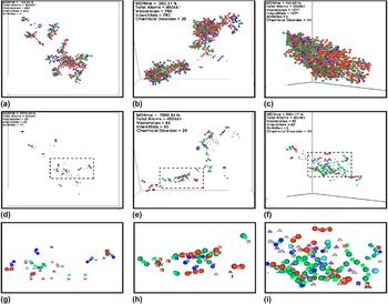
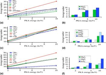
Despite the limitations inherent to MD simulations, the time-dependent distribution of energy and defects produced in a displacement cascade was extensively used to describe the topology of cascades in ceramics. Reference Sickafus56,Reference Chartier, Catillon and Crocombette57 For high-energy primary knock on atoms (PKAs) with a kinetic energy of a few hundreds of kiloelectron volts, the displacement cascades separate into dense subcascades within a few picoseconds following the primary collision as occurs in metals. Reference Stoller and Greenwood58 This is not surprising because the energy of displaced atoms is so large that displacement is hardly affected by the nature of chemical bonds in the solid. When the PKA energy decreases below a few tens of kiloelectron volts, a single cascade is created Reference Martin, Garcia, Van Brutzel, Dorado and Maillard59 in agreement with other investigations. Reference Simeone, Luneville and Serruys60 An example of such an approach was applied at the beginning of the century to discuss the structural stability of spinels and pyrochlores under irradiation. Calculations of defects in spinel structures were performed to investigate the effect of mass and the initial amount of disorder in pristine samples (antisite defects on cation sublattices) on the topology and the amount of primary damage production. Reference Bacorisen61 These authors performed MD simulations on three different spinels MgAl2O4 (M Al = 27) exhibiting no antisite defects on the cation sublattices, MgGa2O4 (M Ga = 70) in which Mg and Ga atoms are present in both sublattices and MgIn2O4 (M In = 114) where half of the In cations lies on the Mg sublattice. The authors also computed average concentrations of antisite defects at the end of simulations. The results are summarized in Fig. 1.
Snapshot of displacement cascades generated by a 5 keV PKA in the three spinels showing the displacement of ions at the end of the collisional phase of the cascade. (a) 150 fs and (d, g) 9975 fs, for a normal MgAl2O4 spinel and Al PKA; (b) 250 fs and (e, h) 7250 fs, for a half-inverse MgGa2O4 spinel and Ga PKA; (c) 400 fs and (f, i) 9001 fs, fully inverse spinel, MgIn2O4 and In PKA (c, f, and i are zoom-ins of the core of the cascades). Blue spheres are associated with Mg interstitials, green spheres are associated with B (Al, Ga, In) interstitials, cubes denote cationic vacancies, and red spheres denote oxygen atoms in perfect sites. Nominal charges for cations and anions were used in the simulation. Reference Bacorisen61

Even if the density of defects in the core of the cascades seems to be more important for In than Mg spinels, no drastic difference is observed on cascade morphology. The amount of oxygen vacancies and interstitials as well as the number of cation antisite defects does not seem to evolve with the degree of antisite defects in pristine spinels. These results are illustrated in Fig. 2. In this example, it appears that the amount of disorder in pristine spinels has little impact on the production of point defects. There is experimental evidence that in yttrium-stabilized zirconia (YSZ), which displays a cubic structure, the amount of native oxygen vacancies (controlled by the yttria content) has no significant effect on the production of F+-type centers involving oxygen atom displacements through elastic collisions, in spite of large recombination volumes. Reference Costantini, Beuneu, Morrison Smith, Devanathan and Weber47 However, very few studies are available to extend this analysis to other ceramics.
Average number of interstitials, vacancies, and antisite/cation disorder defects created at energies of 2, 5, and 10 keV in a (a) normal MgAl2O4, (c) half-inverse MgGa2O4, and (e) inverse MgIn2O4 spinel. The average number and type of cation disorder defects and their coordination are shown in (b), (d), and (f) figures. The superscripts T and O refer to tetrahedral and octahedral coordination, respectively. Reference Bacorisen61

MD simulations based on interatomic pair potentials performed on complex oxides such as spinels, zircon, and pyrochlore clearly show that Frenkel, antisite, and Schottky defects can be created under irradiation in each sublattice. Also, the number of Frenkel pairs and antisite defects follows a power law of the PKA energy with an exponent less than 1. For covalent materials, more sophisticated interatomic potentials as the Tersoff potential Reference Tersoff62 describing oriented bonds were used to simulate displacement cascades. This type of interatomic potential was successfully used to describe cubic and hexagonal SiC. Reference Gao and Weber63,Reference Gao, Weber and Devanathan64 Again simulations indicate Reference Gao, Weber and Devanathan64 that the number of surviving defects (Frenkel pairs, Schottky pairs, antisites, and small clusters) follows a power law of the PKA energy. These results are similar to those encountered in metals. The exponent of the power laws depends on the nature of the ceramic studied and the type of defect.
To understand the effect of crystal structure or the nature of bonds on the production of primary defects, it is common and helpful to calculate the ratio of the surviving Frenkel pairs at the end of a displacement cascade, derived from calculations or experiments, over the calculated (BCA) number of defects. A question naturally arises: in ionic crystals, Does this efficiency depend upon the choice of interatomic potentials? Recent ab initio MD simulations of SiC Reference Gao, Xiao, Zu, Posselt and Weber65 reveal that charge transfer is significant during the displacement of knocked-on Si and C atoms. This charge transfer modifies the energy barrier for the formation and migration of stable defects. However, these calculations remain so far too time consuming to be extended to high-energy PKAs and to compare the amount of Si and C clusters produced to classical MD simulations.
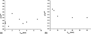
Zinkle first estimated this efficiency in oxides from OA experiments. However, this analysis was based on oxygen vacancies (F centers) measured at low temperature Reference Zinkle and Kinoshita14 (T irr < 80 K) in Al2O3 and MgO. Reference Chen66 He supposed in his analysis that the number of oxygen vacancies is the signature of Frenkel pairs on the anion lattice. Moreover, he assumed that there was no migration of point defects in the displacement cascade at the irradiation temperature. Zinkle showed that there is no clear dependence of this efficiency with the PKA energy in Al2O3. Figure 3 displays the evolution of this efficiency extracted from experiments and calculations for Al2O3 and SiC.
Variation in the ratio of the number of surviving Frenkel pairs over the number of Frenkel pairs estimated by the NRT formula in (a) Al2O3, Reference Zinkle and Kinoshita14 and (b) SiC Reference Gao, Xiao, Zu, Posselt and Weber65 versus the energy of PKA.

As regards the Al2O3 results however, it is possible that oxygen vacancies are also due to the formation of Schottky defects. Some more recent RBS results Reference Schnohr, Wendler, Gartner, Welsch and Ellmer67 relating to Al2O3 seem to contradict the previous analysis of Zinkle and show a distinct effect of PKA energy upon the defect production efficiency. One possible reason for this disagreement may be related to the fact that the number of defects derived from RBS measurements probably cannot be ascribed to single oxygen interstitials only. It is no doubt related to the presence of complex defects such as dislocation loops. Reference Chu, Mayer and Nicolet68 This illustrates the difficulty involved in determining from experiments accurate data for primary defects in ceramics.
The relative independence of this efficiency with the PKA energy in Al2O3 may be understood as the signature of Coulomb interactions between charged stable defects produced in the displacement cascades. It must be noticed that MD simulations performed on MgO with fixed charges for anions and cations Reference Uberuaga69 do not reproduce experimental results. Reference Chen66 The disagreement between simulations and experiments suggests that charge transfer between atoms in MgO during multiple collision events in displacement cascades has a substantial effect. If charge transfer occurs while atoms are at saddle positions, then the apparent energy barrier required to form stable defects may decrease thereby modifying the amount of stable defects. This charge transfer effect has already been observed and discussed in Si Reference Streitz and Mintmire55 and more recently in SiC. Reference Gao, Xiao, Zu, Posselt and Weber65
These results show that both the topology of a displacement cascade as well as the evolution of associated primary defects need to be more clearly understood in ceramics. Variable charge MD simulations or/and ab initio MD simulations Reference Gao, Xiao and Weber70 may provide useful tools for understanding the impact of crystal structure on the topology of displacement cascades and may help explain discrepancies between experimental results and MD simulations performed using empirical interatomic potentials.
C. Impact of inelastic collisions on primary damage
The production of stable defects due to elastic collisions can be described since the corresponding mechanisms at the atomic scale are relatively well defined and well known. On the other hand, the production of defects by inelastic collisions in materials, i.e., the slowing down of high-energy PKAs by multiple collisions with electrons in matter, results from indirect and complex mechanisms is not fully understood. Reference Itoh and Stoneham71 The concept of displacement cascades can no longer be used to describe the changes of materials irradiated with such swift heavy ions, i.e., with a kinetic energy above a few megaelectron volts per unit of atomic mass such as fission products in UO2. For insulator materials with wide band gaps, inelastic collisions produced by these swift heavy ions, whether followed by self-trapped exciton formation as in SiO2 Reference Devine72,Reference Itoh73 or not, induce defect production in halides, silicates, and carbonates as reported from numerous experimental studies. Reference Clinard, Hobbs, Johnson and Orlov74 The analysis of these experimental results enabled Clinard and Hobbs to outline a few favorable conditions for the creation of defects via inelastic interactions Reference Clinard, Hobbs, Johnson and Orlov74 :
-
(i) The electronic excitations, like polarons or excitons, must be localized over a few atomic sites.
-
(ii) The available energy in the excitation must be comparable to the displacement threshold energy.
-
(iii) The lifetime of the excitation must be of the order of the lattice vibration (>0.1 ps) to allow coupling with the motion of atoms.
-
(iv) Non-radiative transitions must prevail over radiative transition, i.e., luminescence.
Obviously, metals do not satisfy conditions (i), (ii), or (iii) and so are in the main insensitive to electronic excitations. In small gap semiconductors, the energy given to electrons during inelastic collisions is much smaller than the TDE. Even if collective excitations created by the electric field of the impinging ion involve a greater energy, these excitations are not localized. This implies that the first condition is not fulfilled. These materials seem to be insensitive to inelastic excitations. However, athermal melting of silicon by laser beams may be accounted for by the lattice instability following a critical amount of the above band-gap electronic excitations Reference Stampfli and Bennemann75 as expected from the Coulomb explosion mechanism.
In insulators, the energy of electronic excitation is about 10 eV. As a result, inelastic collisions can create defects in ionic compounds with small TDEs. Two distinct processes may lead to the creation of defects: the self-trapped exciton formation like in halides and silica Reference Tanimura, Tanaka and Itho76 and the creation of a deep hole associated with Auger de-excitation. Reference Knotek and Feibelman77 In alkali halides, Reference Itoh and Stoneham71,Reference Itho and Tanimura78,Reference Williams and Song79 self-trapped excitons associated with the electronic excitation dissociate into a hole and an electron leading to the formation of stable color centers. Reference Kayamuna80 Excitons are also responsible for the creation of oxygen Frenkel pairs in pure SiO2. Reference Tsai and Griscom81,Reference Hosono, Kawasoe and Matsunami82 Ab initio calculations reveal a coherent scenario for this mechanism. Reference Roma, Limoge and Baroni83 Such a mechanism is possibly responsible for the phase separation and the oxygen bubble formation observed in silicate glasses. Reference Peuget, Delaye and Jegou16
On the other hand, deep holes Reference Knotek and Feibelman77 produced in the cation sublattice formed by metals of columns IV and V of the Mendeleev table vanish leading to the formation of positive or neutral oxygen atoms that are able either to desorb or create oxygen clusters. Such a mechanism explains the reduction of many oxides observed when subjected to electronic excitation irradiations. Anionic desorption from alkali halides and alkaline-earth oxide surfaces arises from bi-exciton production by electronic excitations with laser beams. Reference Xiong, Joly, Beck and hess84
Defect creation is not the only effect induced by an inelastic energy transfer in ceramics. It is well known Reference Itoh and Stoneham71,Reference Itoh73 that inelastic effects largely modify the mobility of defects enhancing it under certain circumstances. The seminal work by Bourgoin and Lannoo clearly showed that this inelastic energy transfer largely affects the diffusion of point defect in semiconductors. Reference Streitz and Mintmire55,Reference Stievenard, Boddaert, Bourgoin and Bardeleden85,Reference Seebauer and Kratzer86 Similar effects such as a-thermal point-defect recombination have also been reported for SiC from calculations and experiments. Reference Weber, Zhang and Wang87 In oxides, a large amount of experimental data is available. It appears that inelastic energy transfers induce an annealing of radiation damage in oxides like Al2O3 and MgO. Reference Krefft88 Until now, a clear description of the physical mechanisms responsible for the large modification of the microstructure, like the formation of colloids in MgO irradiated with electrons Reference Clement and Hodgson89 or the conflicting role of the electronic excitation in the nucleation or growth of loops in Al2O3/MgO system Reference Devanathan, Sickafus, Weber and Nastasi90 is unavailable. It must be noted that the impact of inelastic energy transfers on the diffusion of defects strongly seems to depend on the amount of impurities in the insulator. Reference Itoh73
Moreover, one must bear in mind that for swift heavy ions with a high inelastic stopping power (>10 keV/nm), the intense electric field produced by the impinging ion creates a high density coherent excitation of electrons that are able to produce some damage even in pure metal, Reference Lesueur and Dunlop91 via some poorly elucidated mechanism. No attempts are made up to now to point out the impact of such an intense electric field on phase transition in insulators.
Despite many attempts since the beginning of the 50s at describing the impact of electronic excitation, i.e., the slowing down of swift ions through some energy transfer to electrons, there is still no universally accepted model at the microscopic scale. Reference Itoh73 Different models have been proposed to capture some aspects of the underlying physics behind this process. The most physical model is the Coulomb spike model. Reference Bonfiglioli, Ferro and Mojoni92 In this approach, swift heavy ions create a highly ionized cylindrical region ejecting valence and core electrons during ion-electron collisions by well-known processes. Reference Watanabe, Takemoto, Kubozoe and Mukoyama93 Repulsive electrostatic forces between ions induce a strong deformation of the lattice before electroneutrality is restored on the femtosecond time scale. Despite some attempts, Reference Lesueur and Dunlop91 a quantitative model based on this mechanism is difficult to establish because of the difficulty in calculating a realistic electric field in the ionized cylinder. Reference Canright94
A step further was achieved with the thermal spike model. Reference Szenes95,Reference Toulemonde, Dufour, Meftah and Paumier96 In this two-step approach, the energy lost by the swift ions in the ionized cylinder by inelastic collision is first transferred to electrons via a simple heat equation on the femtosecond time scale. In this approach, more complex phenomena for energy transfer to electrons such as multiple ionizations or Auger emission are neglected. The electron gas in the ionized cylinder is described by an “electronic” temperature T e. In a second stage, it is assumed that the electron–phonon coupling induces a transfer of the heat to the lattice on a picosecond time scale. This two temperature model, derived from laser–matter interaction studies, was introduced to explain plastic deformation, the so-called “hammering effect”, of amorphous ceramics like SiO2 Reference Klaumunzer97 ; point defect and defect cluster formation in ionic crystals like LiF Reference Trautmann, Toulemonde, Schwartz, Costantini and Müller98 and CaF2 Reference Khalfaoui99 ; or amorphization in SiO2 quartz Reference Klaumunzer97 irradiated with swift heavy ions. Despite the fact that the two temperature model appears to describe adequately the morphology of tracks observed in some ceramics, Reference Kluth100 the use of two diffusion equations to determine the temperatures in the electron gas and in the lattice is not clearly justified. In addition, coupling between the electronic and the ionic temperatures which results from electron–phonon coupling is assumed somewhat arbitrarily linear. Reference Toulemonde, Dufour, Meftah and Paumier96 During the last decade, this two temperature model was combined with MD simulations to handle radiation damage induced by swift heavy ions in ceramics. Reference Backman101 The interest in and limitations of this simulation approach is still the subject of discussion but its success stems from the relative ease with which one may discuss in a quantitative way synergetic effects between elastic and inelastic energy deposition and their impact upon the production of primary defects in insulators and semiconductors.
III. SPECIFIC CHARACTERIZATION TECHNIQUES ASSOCIATED WITH THE STUDY OF IRRADIATED CERAMICS
In insulators and semiconductors, the existence of a band gap results in the formation of various stable atomic defects with different charge and spin states. This enables detecting these defects using particular spectroscopic techniques such as EPR and/or OA with exceptional sensitivities. In this case, modifications of high-energy bands (above 1000 cm−1) provide accurate information about chemical rearrangements. These techniques are extensively used to study radiation-induced chemical changes in clays and SiC and can also be used to track structural modifications. Low-energy bands (below 1000 cm−1) collected in infrared and Raman ranges give accurate information about the local modification of symmetries induced by atomic relaxations. Complementary neutron and x-ray diffraction techniques were extensively used to study structural modifications (phase transformation, variation of chemical composition, or amorphization, etc.) or gather microstructural data (strain field, polygonization of grains, etc.). Of course, transmission electron microscopy (TEM) offers, as for studies involving metals and alloys, a unique opportunity to obtain snapshots of the irradiated microstructure. In addition to structural information which can be obtained from electron diffraction, it also has the precious advantage of constituting a direct “observation” of the microstructure.
A. EPR and OA spectroscopic techniques
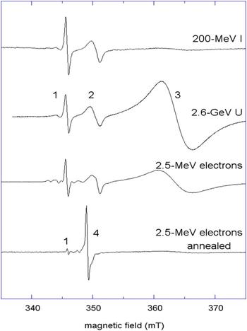
In insulators and semiconductors, charged defects are produced in the band gap. This effect is at the origin of the term color center that designates neutral or charged vacancies or interstitials in ionic crystals. In particular, paramagnetic F centers (neutral anion vacancies with one trapped electron) were thoroughly investigated in alkali halides and to a lesser degree F and F+ centers in oxides like MgO and Al2O3 Reference Henderson and Wertz102 with EPR and OA. More recently, an extensive study was carried out on F+-type centers in YSZ produced by elastic collisions induced by electron and ion irradiations as for MgO. Reference Shen, Feng, Tang, Valdez, Wang and Sickafus103 Hole centers produced by electronic excitations with charged particle or photon irradiations can also be detected by these techniques with great sensitivity, like the V− centers produced in MgO. Reference Henderson and Wertz102 Figure 4 displays an example of EPR spectra collected on irradiated YSZ single crystals.
RT-EPR spectra for B//<100> of YSZ (100) single crystals irradiated under 200-MeV I ions at 1 × 1013 cm−2, 2.6-GeV U ions at 1 × 1013 cm−2, and 2.5-MeV electrons at 2.9 × 1018 cm−2, annealed at 973 K: lines 1–2 and 1–4 were assigned to F+-type centers, whereas line 3 was associated with T-centers (Zr3+ ions in trigonal point symmetry). Reference Costantini, Beuneu, Morrison Smith, Devanathan and Weber47

EPR spectra allow detecting a small amount of electronic spins ∼1016 irrespective of sample volume. Moreover, the different charge states of paramagnetic species can be sorted out on the basis of the specific g-tensor principal values and hyperfine splitting. Angular dependence of the g-factor and hyperfine coupling constant can also provide valuable information about point-defect anisotropy. However, interference with EPR signals of paramagnetic impurities is always possible. For wide band gap materials, combining the EPR signal with OA and photoluminescence spectra may lift uncertainties. A great deal of work has been devoted to tracking point defects induced by irradiation in ceramics using OA as in MgAl2O4. Reference Summers, White, Lee and Crawford44,Reference Ibarra, Mariani and Jimenez de Castro104 Although defects can be more directly studied in iono-covalent materials than in metals, the presence of both cation and anion sublattices and the multiplicity of possible charge states make the number of basic defects much more important.
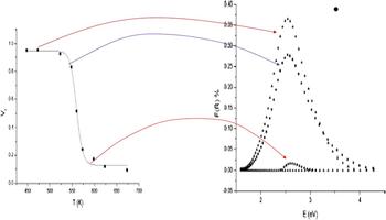
These spectroscopic techniques are extensively used to characterize defects produced by irradiation in silicates but do not give any indication about the local distortion they are liable to induce and accompanying structural modifications. To illustrate this point, Fig. 5 shows a correlation between the volume fraction of the tetragonal phase produced by irradiation in monoclinic zirconia Reference Simeone, Baldinozzi, Gosset, Mazerolles and LeCaër105 and the amount of F+ centers measured by OA in the material. Reference Simeone, Baldinozzi, Gosset, LeCaër and Mazerolles106
Correlation between the tetragonal volume fractions of m-ZrO2 sample irradiated at room temperature under a flux of 1010 cm−2 s−1 by 800 keV Xe ions determined from XRD measurements (left graph) and the amount of oxygen vacancies (F+ centers). Comparison between these two graphs clearly points out that the tetragonal volume fraction is associated with the existence of these charged defects in irradiated pure monoclinic zirconia. Copyright 2004, The American Physical Society. Reproduced with permission from Ref. 105.

Because these spectroscopic techniques are bulk sensitive techniques, they do not provide any spatial resolution. Raman and infrared spectroscopic techniques, sensitive to phonon vibrations, can give some clues on relaxations associated with the creation of defects. The study of peaks above 1000 cm−1 has shown the existence of Si–Si and C–C bonds in irradiated SiC. Reference Sorieul, Allard, Morin, Boizot and Calas107,Reference Zhang, Bae, Sun, Wang, Ishimaru, Zhu, Jiang and Weber108 Low-energy peaks (below 1000 cm−1) also give accurate information in relation to structural modifications of crystal structures induced by low-energy ions or in the wake of swift heavy ions. Reference Velisa, Thome, Trocellier, Serruys, Debelle, Garrido and Miro109 This technique was applied to demonstrate that pure monoclinic zirconia is transformed into tetragonal zirconia under ion-beam irradiation Reference Simeone, Dodane-Thiriet, Gosset, Beauvy and Daniel110 and track defects in UO2 irradiated with low-energy particles. Reference Guimbretière, Desgranges, Canizarès, Carlot and Caraballo111
B. Diffraction techniques
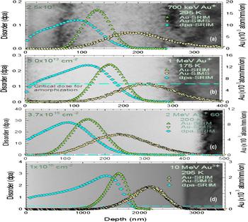
Different diffraction techniques are used to track modifications in the long-range order of ceramics subjected to radiation damage. TEM is commonly used in studies devoted to the analysis of the microstructure of ceramics under irradiation. This technique can provide a clear insight into mechanisms involving radiation damage and snapshots of the microstructure in real space. Figure 6 shows the impact of the implantation profile on the c-SiC microstructure obtained from a cross-sectional TEM micrograph of SiC irradiated with Au ion beams of different energies.
SRIM predictions and SIMS measurement are superimposed upon TEM images for (a) 700 keV Au+ irradiation in 6H–SiC at 295 K under a flux of 1010 cm−2 s−1 to an ion fluence of 2.5 × 1013 cm−2, (b) 1.0 MeV Au+ irradiation at 175 K to an ion fluence of 5 × 1013 cm−2, (c) 2.0 MeV Au+ irradiation with 60° incidence angle at 295 K to an ion fluence of 3.7 × 1015 cm−2, and (d) 10 MeV Au3+ irradiation at 295 K to an ion fluence of 1015 cm−2. Diamond-shaped boxes indicate the SRIM calculated damage profile. The triangle-shaped boxes indicate the Au profile in units of atoms per nanometer and per ion. The dashed line indicates the critical dose value for amorphization. Reference Zhang, Bae, Sun, Wang, Ishimaru, Zhu, Jiang and Weber108 Discrepancies between SRIM simulations and SIMS measurements are discussed in detail in the original paper. Copyright 2005, AIP Publishing LLC. Reproduced with permission from Ref. 108.

The contrast arising from local changes in diffracting conditions enables measuring strain fields. However, the main feature of this technique is a direct determination of the space group from the collected diffraction patterns. Combining diffraction and direct observation of the microstructure, it is possible to point out specific domains. This bright field analysis was applied to study the zirconia transformation induced by irradiation. Reference Simeone, Baldinozzi, Gosset, Mazerolles and LeCaër105 Nowadays, high-resolution transmission electron microscopy (HRTEM) offers the unique opportunity to extract information over a typical size of a few nanometers. The detailed structure of radiation-damaged areas is generally blurred though due to the high local strains induced by the presence of defects. In conventional TEM, this makes it difficult and impossible to find appropriate diffraction conditions to select equivalent atomic rows with precision. HRTEM images, showing rows of atoms, enable the study of stacking faults and dislocation loops in ceramics by selecting very small areas Reference Yasunaga, Yasuda, Matsumura and Sonoda46 and the method was applied to discuss phase transformations in spinel. Reference Ishimaru, AfanasyevCharkin and Sickafus112,Reference Baldinozzi, Simeone, Gosset, Surble Mazerolles and Thomé113 Because the energy momentum transfer vector q = k scatt − k inc Reference Cowley114 ( k scatt and k inc are the wave vectors associated with the scattering and incident wave, respectively), is large in electron diffraction experiments (few hundreds of nm−1 for 400 kV microscope) compared with x-ray radiations (few tens of nm−1 for Cu Kα radiations), energy filtering TEM in combination with an imaging technique offers the opportunity to collect diffraction patterns over a large q range. For polycrystalline samples with substantial mosaicity, it is then possible to apply the kinematic theory of diffraction neglecting multiple scattering and then compute a pair distribution function (PDF). PDFs are of prime importance for the study of amorphization in irradiated ceramics and the method has been used to discuss amorphization in SiC. Reference Hirotsu, Afanasyev Charkin, Siockafus and Ishimaru115 HRTEM also offers the opportunity to apply quantitative spectroscopic techniques like electron energy loss spectroscopy, Reference Nipoti116 high angular resolution electron channeling, Reference Soeda and Kinoshita Zaluzec117 or electron dispersive x-ray spectroscopy to quantify the concentration of species in ceramics over few nanometers. Reference Luneville118
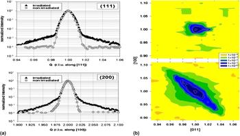
The main limitation of TEM is due to the fact that observations must be performed on thin films. Because the foils used in TEM are only a few hundreds of nanometers thick, the observed microstructure is not necessarily representative of the bulk. Free surfaces are powerful sinks, Reference Nichols119 which are able to substantially modify point defect concentrations as they annihilate there. The effect of this artifact on the microstructure is difficult to assess and obviously depends on the material studied. As ceramics studied in the nuclear industry possess high melting points, diffusion of point defects in the thin foils is limited at cryogenic or room temperature. It must also be noticed that the electron beam impinging the surface of the sample is also liable to enhance the surface diffusion of light elements (Li, Mg, Al). Because TEM is a diffraction technique, Reference Cowley114 it possesses a resolution window which is small (below 1 nm) and large clusters cannot be observed. Moreover, unit cell parameters obtained from TEM diffraction patterns are not accurate. Unit cell determination, atomic positions, and composition changes in irradiated ceramics can be obtained from the analysis of x-ray and neutron diffraction patterns collected on polycrystalline samples for which the kinematic approximation of diffraction applies. For polycrystalline samples, microstructural changes like strain fields and/or characteristic size of coherent diffracting domains can also be obtained. Moreover, diffuse scattering has long been applied to track point defect induced strain fields in irradiated single crystals. However, the analysis of diffuse scattering maps induced by defects is not straightforward. Figure 7 displays the elastic strain induced by the formation of lithium nanocrystals in Li2O single crystals irradiated with electrons. Because of the large penetration depth of x-rays and neutrons, such analyses must be performed on single crystals or polycrystalline samples irradiated over a large volume (few micrometers). Reference Krexner, Prem, Beneu and Vajda120 Thus, this technique does not allow determining the effect of damage induced by defects in solids irradiated with low-energy ions because the damaged volume remains always smaller than the x-ray penetration depth (or the extinction length for single crystals).
(a) Comparison of the scattering intensity (on a logarithmic scale) observed in irradiated Li2O at room temperature by 1 MeV electron beam under a flux of 1011 cm−2 s−1 and nonirradiated Li2O along radial scans through the (111) and (200) peaks. In the reference sample, the peak shape is a Gaussian upon a flat background whereas scattering due to distortion is visible following the electron irradiation 123. (b) Normalized iso-intensity contours of elastic diffuse scattering around the (111) peak of a nonirradiated (top) and an irradiated (bottom) crystal of Li2O. The large ellipsoid in blue is the signature of a large strain field induced by Li nanoclusters. Copyright 2003, The American Physical Society. Reproduced with permission from Ref. 120.

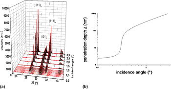
To track the structural and microstructural evolution of ceramics irradiated with low-energy ions, the glancing incidence x-ray diffraction (GIXRD) technique may be used. Reference Simeone, Gosset, Bechade and Chevarier121 By adjusting the incidence angle of the x-ray beam, it is possible to characterize radiation damage over a length scale of a few hundreds of nanometers, which is the typical depth over which damage is induced with low-energy ions. Specific features and limitations of this technique have been discussed. Reference Simeone, Baldinozzi, Gosset and Berar122 The analysis of Bragg peaks collected in GIXRD provides information on the structure and the microstructure (strain field, coherent diffracting domains, etc.). Figure 8 displays the variation of diffraction patterns collected as a function of depth on irradiated m-ZrO2 samples. The analysis of these diffraction patterns is a means of quantifying the radiation-induced monoclinic-to-tetragonal phase transformation in pure zirconia. Reference Simeone, Gosset, Bechade and Chevarier121
Variation of the diffraction patterns (a) of m-ZrO2 irradiated with 800 keV Ar ions at room temperature under a flux of 1010 cm−2 s−1 and a fluence of 1014 cm−2 s−1 collected at different incidence angles associated with different penetration depth of the x-ray beam (b). From the analysis of these diffraction patterns, the volume fraction of the tetragonal phase produced under irradiation can be extracted and compared with the damage profile extracted from simulations. Reference Simeone, Gosset, Bechade and Chevarier121

Spinel transformations Reference Simeone, Gosset, Bechade and Chevarier121 and the evolution of metallic carbides Reference Gosset, Dollé, Baldinozzi, Simeone and Thomé48 have also been studied this way. In this approach, the Rietveld method is used to refine diffraction patterns. Moreover, it also offers the opportunity to probe the impact of radiation damage over large space scales (few nanometers) due to its large coherent volume. Reference Simeone, Baldinozzi, Gosset and Berar122 However, this method then fails to track radiation-induced amorphization. The development of new synchrotrons may offer the opportunity to determine accurate PDFs from total scattering measurements.
From this rapid discussion, it appears that many powerful experimental tools are now emerging to probe the nature and amount of defects induced by irradiation as well as structural and microstructural changes associated with the presence of excess defects.
IV. LONG-TERM MICROSTRUCTURAL EVOLUTION OF CERAMICS
As in metallic materials, irradiation induces drastic modifications of the microstructure, point defect production and clustering, phase transformations and amorphization. However, due to the complexity of chemical bonds and crystalline structures, the predictions of long-term radiation-induced microstructural changes are scarce in ceramics compared with metals and alloys. The number of systems and initial microstructures, the range of irradiation conditions (characteristics of incident charged particles, temperature, and atmosphere) make this topic difficult to review comprehensively although this has been attempted in the past. Reference Limoge and Barbu123 Our aim here rather than a comprehensive review is to show, through various examples taken from a range of different materials (MgO, α-Al2O3, UO2, CeO2, ZrO2), some of the aspects that make these metal oxide systems very different from alloys.
A. Irradiation induced microstructural evolutions in “simple” oxides and radiation induced diffusion
The combination of high-energy electron irradiations and subsequent or simultaneous observation in a high-voltage transmission electron microscopy Reference Pells and Shikama25,Reference Shikama and Pells124 provides a unique insight into basic radiation effects since electrons are liable to produce point defects only. Using electrons for inducing damage greatly simplifies the system in comparison with situations where cascades are produced, as can be the case with ions or neutrons. This type of experiment bears the added attraction of enabling a selective displacement of atoms lying on one or the other sublattice, in cases where displacement energies of the metallic element are substantially different from that of oxygen. Reference Stathopoulos and Pells8,Reference Soullard27 In their study of α-Al2O3, Stathopoulos and Pells Reference Stathopoulos and Pells8 irradiated thin foils at temperatures ranging between 300 and 1100 K with 300 keV electrons, which is above the electron energy required to displace aluminum ions (175 keV) but significantly below that required to displace oxygen ions (390 keV). They observed the formation and growth (above 800 K) of interstitial dislocation loops of stoichiometric composition lying on basal or prismatic planes. Because only cation defects are produced upon irradiation, the formation of a stoichiometric dislocation loop requires the movement of oxygen ions to occur in which electrostatic interactions must play a key role. The authors propose a scenario to explain their observations involving first an aggregation of Al ions between either basal or prismatic planes subsequent to which oxygen ions diffuse into the defect structure to form the stable loops. This first example illustrates the close relationship between primary defect production and charged defect diffusion properties. It is reasonable to assume here that electrostatic interactions may induce cooperative effects between the oxygen and metal sublattices.
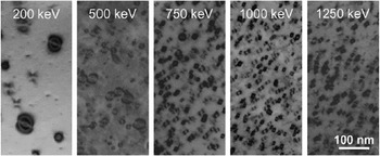
More recently, basic studies have been carried out on the CeO2 fluorite structure considered to be a surrogate for UO2. Reference Yasunaga, Yasuda, Matsumura and Sonoda9,Reference Yasunaga, Yasuda, Matsumura and Sonoda46 The afore-mentioned authors irradiated thin CeO2 foils with electrons in the 200 keV to 3 MeV energy range and at temperatures between 300 and 1073 K. In the lower energy range (200–500 keV), presumably lower than that required to displace Ce atoms from their sites, they observed the formation of larger dislocation loops characterized by a very strong strain contrast. The loops shown in Fig. 9 are lying on {1 1 1} planes with a Burgers vector parallel to the <1 1 1> direction. These extended defects are assumed by the authors to result from the insertion of a layer of oxygen atoms based on two arguments: the electron energy lies well below that required to displace cerium ions and HRTEM observations indicate a stacking fault but the absence of an additional layer of cerium atoms. One may also add that the large strain fields observed in the lattice are an indication that the dislocation loops are charged.
BF-TEM images of nonstoichiometric interstitial type loops in CeO2 irradiated with different energy electron beams, indicating for the left-hand micrograph in particular, unusually marked strain contrast. Copyright 2008, Elsevier. Reproduced with permission from Ref. 46.

This latter point has been extensively reported with regard to YSZ also in the work of Yasuda et al. Reference Yasuda, Kinoshita, Matsumura and Ryazanov125 They irradiated YSZ samples with low-energy ions such as 300 keV O+ ions and subsequently re-irradiated them with low-energy (200 keV) electrons, i.e., again below the threshold energy for displacing zirconium ions. In doing so, they observed the very rapid growth of large defect clusters (roughly 1 nm/s), which induced a considerable strain field as would be expected from a charged defect cluster. The defect reaches a threshold size (roughly 1 µm) and then breaks down into a dislocation tangle. The process of nucleation, growth, and breakdown of defects repeats itself under continued electron irradiation. These results are interpreted by the authors as resulting from the formation of charged oxygen platelets possibly involving electronic excitation effects inferred from the unexpectedly high mobility of oxygen point defects.
Ryazanov et al. Reference Ryazanov, Yasuda, Kinoshita and Klaptsov126 have attempted to interpret observations by Yasuda et al. Reference Yasuda, Kinoshita, Matsumura and Ryazanov125 based on rate equations which take charge effects into account explicitly. In this work, the authors show how an internal electric field may be created in the vicinity of the charged oxygen dislocation, electric field which in turn gives rise to an additional stress field around the dislocation. The authors consider a diffusion equation for free electrons, coupled with the electrical potential created around the charged defect cluster. The theoretical loop growth rate is compared to experiment. Charge accumulation is assumed to take place on the loops which trap free electrons that are created as a result of the interaction of the sample with the impinging electron beam. As a result of the concomitant accumulation of oxygen atoms and electrons, the induced strain around the defect rises until the yield stress is reached and plastic deformation occurs through dislocation multiplication.
We have seen how a selective displacement of a given lattice may be a means of producing charged extended defects that require taking into account Coulomb interactions over a longer range than when point or extended defects are neutral. However, it is interesting to note that in many cases, when the energy of impinging electrons increases or when ion beams or fission fragment recoils are used either producing displacements on both sublattices or full damage cascades, interstitial dislocation loops appear to be stoichiometric in character. This is reported to be the case in α-Al2O3, Reference Howitt and Mitchell127 MgO, Reference Youngman, Hobbs and Mitchell128 CeO2 Reference Yasunaga, Yasuda, Matsumura and Sonoda46 for electron energies above 1250 keV, UO2 Reference Whapham and Sheldon7,Reference Soullard27 for fission fragment induced damage and for high-energy electrons above 1.6 MeV. Reference Soullard129
In the case of MgO, Kinoshita et al. Reference Kinoshita, Hayashi and Kitajima130 have attempted to model the microstructural evolutions in electron-irradiated foils based on chemical rate theory equations. These were adapted from models developed for metals by taking into account point defect formation and diffusion specific to both sublattices. The authors derive from confrontation of theory with TEM observations migration energies of interstitials and vacancies. Stoichiometric loops are assumed.
In MgO Reference Youngman, Hobbs and Mitchell128 and in fluorite UO2 Reference Whapham and Sheldon7,Reference Soullard27,Reference Van-Sambeek, Averback, Flynn, Yang and Jäger131 and CeO2, Reference Yasunaga, Yasuda, Matsumura and Sonoda46 perfect interstitial dislocation loops are reported which have a (1/2) <1 1 0> Burgers vector and lie in neutral {1 1 0} planes. One of the major differences observed between the work of Yasunaga et al. on CeO2 and the work of Soullard on UO2 is that in the latter case there is no sign of oxygen loops even below the electron energy required to displace uranium atoms (1.6 MeV). However, it is worth mentioning that the studies were carried out 30 years apart. The fact that the interstitial loops formed are stoichiometric, as is reported in these studies, requires that charged interstitials be trapped in stoichiometric proportions. This is difficult to conceive since in the fluorite structure materials UO2 and CeO2, cations have much lower diffusion coefficients than anions, and much greater migration energies. This was recently confirmed from DFT calculations. Reference Dorado132 So, if growth is controlled by the slower of the two species, then charge accumulation must occur on the loops. In addition, in Soullard's work, Reference Soullard27 the high-energy electron irradiation experiments were carried out at room temperature and interstitial loops are observed to multiply and grow as the electron fluence increases demonstrating that uranium interstitials migrate. In fact, Soullard proposes an estimate for the migration energy of the uranium interstitial of roughly 0.3 eV, albeit based on a large number of hypotheses. The lowest interstitial migration energy found for uranium by Dorado et al. Reference Dorado132 was 4.1 eV and involved an indirect mechanism. Such widely differing migration energy values may suggest either a charge transfer effect or cooperative effects between the oxygen and uranium sublattices. In the former case, the charge transfer would drastically reduce the migration barrier of the defect, as suggested for the migration of neutral oxygen interstitials in ZrO2. Reference Kotomin and Popov52 Cooperative effects between the oxygen and uranium sublattices have been shown to decrease the uranium vacancy migration barriers in UO2. Reference Dorado132
A word must be said about radiation induced or enhanced diffusion. In metals, simplified rate theory approaches have adequately explained results from experiments seeking to characterize this phenomenon. Reference Sizmann133 Under simplifying assumptions of homogeneously distributed point defect sinks, rate equations for both vacancies and interstitial atoms are set up and solved assuming that a steady state situation is reached. The defect production rate is balanced off by the rates at which defects recombine (mutual recombination) or are eliminated at sinks of prescribed concentration (elimination of point defects at fixed sinks).
Two limiting cases were derived. At lower temperatures, under steady state conditions controlled by mutual recombination of Frenkel pairs, the enhanced diffusion coefficient is proportional to the square root of the defect production rate. The activation energy is given by half the migration enthalpy of the slowest moving point defect. At high defect sink concentrations however, where concentrations are controlled by the rate at which defects are eliminated at internal sinks, there exists a range of temperatures in which enhanced diffusion is independent of temperature and proportional to the defect production rate. To what extent does this theory also apply to cations or anions in oxide systems?
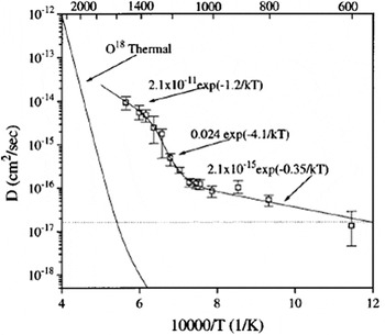
Van Sambeek et al. Reference Van-Sambeek, Averback, Flynn, Yang and Jäger131 carried out mainly oxygen radiation enhanced self-diffusion experiments in MgO in a wide range of temperature and fluxes and analyzed their results in the light of this theory. Figure 10 illustrates some of the most relevant results. Only in the higher temperature range is it possible to relate convincingly the temperature and ion flux dependence of the diffusion coefficient to one of the two limiting cases above: square root dependence of diffusion coefficient upon ion flux with an activation energy approximately half that of the vacancy migration energy. In this case, recombination-limited diffusion is apparent. In an intermediate-temperature range, the higher than expected activation energy was interpreted by the authors as the dissociation energy of small vacancy clusters. At lower temperatures, the diffusion coefficients nearly exhibit a linear dependence upon flux and low activation energy are compatible with sink limited recombination. Why the two rate limiting cases should occur in reverse order with respect to metals is unclear but it appears from this work that the adaptation of rate models for metals to oxides requires further effort. The different range of interactions between mobile defects and fixed, possibly charged dislocations must constitute a major difference with metals that models based on rate theory should take into account.
Arrhenius representation of radiation-enhanced oxygen self-diffusion coefficients subjected to a 2 MeV Kr ion irradiation in MgO. Dashed lines represent ballistic mixing and continuous lines represent thermally activated diffusion. Copyright 1998, AIP Publishing LLC. Reproduced with permission from Ref. 131.

To conclude this section, we have seen that one of the major differences between oxides and metals lies in the existence of both charged point and extended defects. The formal charge of point defects may vary depending upon temperature and irradiation conditions and migration barriers can be very sensitive to that. Frenkel pair recombination volumes are also generally much greater than in metals due to the long-range Coulomb interactions. Under certain conditions also, dislocation loops appear to be charged, inducing uncommonly high strain fields around them and creating an additional length scale determined by electrostatic interactions. Any predictive approach to these microstructural effects seems to require additional equations describing the conservation of defects on both sublattices along with the conservation of electrons and holes taking into account the dielectric material properties. Such approaches, which should be supported by first principles and MD, are far from common and only limited contributions may be found in the literature.
From an experimental point of view, more basic data on different oxide systems are definitely required to help sort through the incredibly complex picture and pick out general trends and important factors. A good starting point has to be radiation-induced diffusion studies in which microstructural evolutions and atom movement are intimately connected and have the power to provide a comprehensive picture of the material behavior.
B. Phase transformation and amorphization induced by irradiation
Under irradiation, ceramics are constantly subjected to damage accumulation and annealing as a result of the creation of defects and their elimination as they diffuse to sinks or annihilate. Their response to these opposing forces (damage accumulation and restoration) is varied: some ceramics retain their crystal structure or even recrystallize (UO2), others undergo phase transformations (such as pure zirconia) or amorphize (such as certain pyrochlores). We are firstly concerned here with providing the reader not only with a feel for the incredible wide variety of responses of ceramics to irradiation but also with ascertaining whether a rationale may be drawn from this complex picture. The follow-up to this is the identification of areas that require more attention if we are to arrive at a reasonably predictive approach to these important long-term radiation-induced consequences.
A great deal of work has focused on oxides which have representatives in all the four classes of response (damage accumulation with conservation of structure, recrystallization, phase transformation, and amorphization).
Symmetry and compacity are attributes which are extremely favorable to radiation resistance. As seen in the previous section, rocksalt (MgO), fluorite (YSZ, CeO2, UO2, PuO2) or hexagonal oxides (α-Al2O3), i.e., oxides in which anions and cations occupy all Wyckoff positions, appear to have no alternative under irradiation but to establish smaller scale patterns of themselves: they form stoichiometric, interstitial dislocation loops that grow into dislocation networks. These materials are possibly good candidates for testing traditional rate theory approaches but the essentially unexplained fact remains that radiation enables atomic movement that would otherwise be completely insignificant (see Sec. IV. A).
Another type of response to radiation observed is the structural transformation from a lower to a higher symmetry phase and an interesting example is pure zirconia which has three different phases: monoclinic, tetragonal, and cubic (YSZ). This oxide goes through a displacive phase transition as the temperature rises from a monoclinic to a more symmetric tetragonal phase. Similarly, under irradiation and irrespective of the nature of the energy deposition, pure zirconia shows the same transition which has been ascribed to a strain field induced by the accumulation of F+ centers. These defects are formed in all the three types of zirconia, but if irradiation of the tetragonal phase proceeds further, the material does not change to the cubic phase. Reference Simeone, Baldinozzi, Gosset, Mazerolles and LeCaër105 In fact, there are reports that nanograined pure tetragonal zirconia is subject to amorphization probably because of large local strain fields Reference Meldrum, Boatner and Ewing134 produced by the increasing F+ centers. The reason for this is no doubt that unlike the monoclinic phase, the tetragonal phase is not sensitive to the strain field.
Radiation tolerant MgAl2O4 responds yet differently. Under certain high-energy irradiation conditions, dislocation loops are indeed observed, Reference Soeda and Kinoshita Zaluzec117 but they are not under irradiation conditions (mainly low-energy ions) similar to those alluded to the above for zirconia, single phase MgO, or α-Al203 (of which MgAl2O4 spinel may be considered a binary compound). This is thought to be due to the fact that antisite defects Reference Simeone, Dodane-Thiriet, Gosset, Beauvy and Daniel110 (Mg on Al sites and vice versa) can form in spinel and experimental evidence for this has been well supported by MD simulations. Reference Bacorisen61 Similar results Reference Simeone, Dodane-Thiriet, Gosset, Beauvy and Daniel110 were observed on MgCr2O4 and ZnAl2O4 which may themselves be considered as MgO–Cr2O3 and ZnO–Al2O3 binary oxide compounds. The spinel example is an illustration that the crystal structure, and more specifically, the number and nature of sublattices that make up the ceramic can play an important role in controlling its long-term microstructure. Note that antisite defect or dislocation loop formation does not exempt these oxides from amorphization. MgAl2O4 single crystals irradiated with 400 keV Xe ions lose their long-range order at low temperature (100 K) and high doses (25 dpa). A similar loss of long-range order is observed in α-Al2O3 irradiated with 1.5 MeV Xe ions at 20 K and 4 dpa, illustrating that strain fields resulting from irradiation can be responsible for amorphization, irrespective of what actually induces the strain field in the first place (antisites, dislocation loops, or F+ centers in the case of tetragonal ZrO2). Increasing the irradiation temperature is expected to decrease the steady state defect concentration under irradiation, with obvious consequences on the threshold dose required for amorphization.
Pyrochlores, composition A2B2O7 where A and B are cations, have a distinct tendency to amorphize when they are not able to disorder to a more symmetric and stable fluorite phase, such as occurs with Gd2Zr2O7 or Am2Zr2O7. Reference Ewing135,Reference Belin, Martin, Valenza and Scheinost136 The irradiation conditions that lead to amorphization are very much dependent upon the nature of A and B cations and an interpretation was given based on the accumulation antisite defects. Reference Sickafus56 Antisite defects indeed produce very large local strains which the Li and Johnson formalism, Reference Li and Johnson137 used to interpret the crystal to glass transition, predicts may lead to amorphization. Based on a wide range of TEM observations Reference Li and Johnson137 of ion-irradiated pyrochlores of different compositions and supported by MD calculations, the authors claimed that the key parameter was the ratio of the two cation atomic radii; quantity which determines the level of strain induced by antisite defects, Reference Sickafus56,Reference Sickafus138 hence the propensity of a pyrochlore to amorphize. In this approach, the authors Reference Sickafus56,Reference Sickafus138 do not account for the relaxation of the crystal structure when calculating antisite defect energies.
Several alternative interpretations of the amorphization process have emerged more recently. One is based on MD Reference Lefèvre, Costantini, Esnouf and Petite42 calculations which appear to indicate that it is driven by the formation of B–B dumbbells in displacement cascades; covalent bonding being conducive to amorphization. The explanation for the amorphization of pyrochlores is similar to the structural freedom approach developed by Gupta Reference Gupta139 and applied by Hobbs Reference Hobbs, Sreeram, Jesurum and Berger140 to the stability of glasses under irradiation. The other involves the oxygen sublattice. In the previous interpretations, amorphization of spinels or pyrochlores appears to result from the complete destabilization of the more disordered cation sublattice. However, theories are now emerging in which the authors claim that amorphization of titanium dioxide and pyrochlores Reference Whittle, Rios, Smith, Zaluzec and Lumpkin141 is the result of two antagonistic processes: the first involves the removal of oxygen atoms from their original lattice sites by elastic or inelastic collisions and the second the reconstruction of the anion sublattice through diffusion processes. According to these authors, the amount of cation antisites is not the key parameter. Recent ab initio MD calculations Reference Xiao, Weber, Zhang, Zu and Li142 seem to confirm this assumption and provide an atomistic picture for pyrochlore amorphization. During the structural transition, O2-like molecules are formed, and this anion disorder further drives cation disorder eventually leading to an amorphous state.
These studies highlight the complex interplay between both sublattices and its impact on the radiation tolerance of oxides.
For materials exhibiting a large time scale for recombination of point defects resulting from radiation damage, the amount of local distortion will obviously depend upon whether displacement cascades overlap. The multiple impact models firstly derived by Gibbons, Reference Gibbon143 were applied by Weber Reference Weber144 with some degree of success to explain observed amorphization in zircon and SiC. Moreover, some authors also discussed synergetic effect of inelastic and elastic stopping power on the amorphization of SiC Reference Zhang, Varga, Ishimaru, Edmondson, Xue, Liu, Moll, Namavar, Hardiman, Shannon and Weber145,Reference Zheng, Shannon, Allen, Morgan, Szlufarska and Jamison146 to discuss fine variations of the amorphization threshold as a function of the incident ion energy.
V. CONCLUSION
The complex nature of bonds between atoms in ceramics leads to a broad range of crystal structures characterized by multiple sublattices. In this report, we provide an overview of the exceptional variety of radiation effects that this induces. The main difference with defects encountered in metals is that they are charged so in addition to basic Frenkel disorder, vacancies, interstitials, and dumbbells, one is also faced with Schottky disorder and a whole gamut of variably charged structures. Because predicting the number of stable defects left over from a damage cascade is an important elementary step toward modeling a microstructure, we first review and discuss experimental data related to this and the propensity of numerical methods to reproduce the data. It transpires that the discrepancies between TDEs measured and those determined from numerical methods probably stem from the fact that MD models usually involve fixed charges and are very sensitive to interatomic potentials. Ab initio MD models provide an encouraging option for capturing some of the charge transfer phenomena and eventually predicting defect concentrations left over from damage cascades. From a computational stand point, the existence of a band gap makes calculating defect energies an extremely complex task from first principles, but experimentally, it enables spectroscopic techniques such as EPR, Raman, or OA to be implemented. The second part of this document is devoted to spectroscopic techniques in particular. Abundant examples of their usefulness in tracking charged defects are given. However, tracking individual defects is insufficient for providing larger scale information of microstructural changes. This is why they should be associated with TEM or specific structural analyses based on x-ray or neutron diffraction. In recent years, with the expansion of synchrotron radiation in particular, a very wide range of techniques has become available and we discuss some of the issues experimentalist are confronted with when attempting to choose the most appropriate option to characterize usually small damaged volumes. The last part is devoted to giving the reader a feel for the incredible range of microstructural evolutions encountered in ceramics. Different types of response to irradiation (loops, antisites, colored centers, etc.) are outlined with various examples of charged structures that require Coulomb interactions to be incorporated in rate theory descriptions. Although no comprehensive theory is yet available for describing the range of higher scale microstructural changes observed in these materials, it is possible to attempt to rationalize matters. Radiation defects induce strain fields that can either lead to a phase transformation to a higher symmetry structure if it is available and sensitive to strain fields, to recrystallization or to full blown amorphization. In oxides, it also appears that several schools of thought exist on what the underlying characteristic is, if any, that determines this behavior. In the last ten years, focus has been put on the open cation sublattice that is susceptible to disorder and its contribution to strain-induced amorphization. More recently, approaches have emerged that highlight the importance of damage annealing on the oxygen sublattice. Whatever the explanation, one cannot help thinking that diffusion processes and cooperative effects between the cation and anion sublattices play a key role at several time and length scales. How else could one interpret the formation of neutral dislocation loops in fluorite structures at temperatures that should preclude cation movement?
Until now, the clustering and recombination of defects has only really been tackled qualitatively. This is both because of the difficulties in identifying the basic mechanisms experimentally and because we are still missing microstructural models, based on atomic scale modeling capable of describing damage cascades predictively, that account for the key role of Coulomb interactions.