Introduction
Anisakidosis is a parasitic disease of humans that develops following the ingestion of raw or undercooked marine fish infected with larvae of the Anisakidae family (Audicana & Kennedy, Reference Audicana and Kennedy2008). Recent studies have reported the allergenicity of Anisakis spp. in humans and the possible diagnosis thereof (Daschner et al., Reference Daschner, Cuellar and Rodero2012; Mattiucci et al., Reference Mattiucci, Fazii, De Rosa, Meucci, Megna, Glielmo, De Angelis, Costa, Meucci, Calvaruso, Sorrentini, Palma, Bruschi and Nascetti2013).
Based on data described by Huizinga (1965), Ansary et al. (Reference Ansary, Moghaddar and Esmaeili2009) demonstrated that fish inhabiting the Mehran River in Iran were infected with third-stage (L3) Contracaecum osculatum and suggested that infections in humans might cause gastrointestinal ulcers and bleeding, triggering serious illness and death in severe infections. Additional reports have confirmed that humans can be infected with either Contracaecum spp. (Schaum & Müller, Reference Schaum and Müller1967) and/or Pseudoterranova sp. (Skirnisson, Reference Skirnisson2006), underscoring the need to screen fish for these pathogens before human consumption.
Despite the possibility of these parasites causing harm to humans, few reports describe serologic responses against Contracaecum spp. following infection. In addition, most models have examined pathology associated with Anisakis spp. infections following the intraperitoneal introduction of antigens. It has been suggested that the presence of only one live larva in the gastric cavity is sufficient to elicit an immune response and produce gastrointestinal pathology in humans. To date, no infection models have used live Contracaecum spp. larvae for antigen stimulation in mice.
The aim of this study was to investigate serum reactivity and to assess gastrointestinal damage in mice infected with live Anisakis spp. or Contracaecum spp. larvae via orogastric inoculation.
Materials and methods
Study design
This study represents a pilot experiment designed to characterize the immune responses and pathology associated with the presence of L3 Anisakis spp. or Contracaecum spp. larvae in the gastric cavity of mice. A total of 18 Balb/c mice, aged 2 months, were inoculated via gavage with a single live Anisakis spp. or Contracaecum spp. L3 larva and five control mice received 0.9% NaCl solution via the same route. Mice were anaesthetized by subcutaneous injection of xylazine, 10 mg/kg body weight (Rompun®, Bayer, São Paulo, Brazil) and ketamine (Francotar®, Virbac SA, Brazil), 10 mg/kg body weight.
Collection and examination of larvae
Larvae of both nematodes were obtained from three fish species: pink cusk-eel (Genypterus brasiliensis, Regan, 1903), cutlassfish (Trichiurus lepturus, Linnaeus, 1758) and red porgy (Pagrus pagrus, Linnaeus, 1758). Fish were purchased from a local fish market in the municipality of Niterói, state of Rio de Janeiro, Brazil, and were transported to the UFF School of Veterinary Medicine for collection of nematodes from their viscera. The harvested nematodes were then transported to the UFF Laboratory of Immunobiology at the Institute of Biology. The number of larvae harvested from each fish ranged from five to eight and they were primarily located in the mesentery, stomach and intestinal serosa. Larvae were identified by optical microscopy, taking into account the established differences among the ventricles, intestinal caecum, number of rectal glands and presence of the mucron, which is absent in Contracaecum spp. (Saad et al., Reference Saad, Vieira and Luque2012). Larvae were stored in 0.9% NaCl solution until the start of the experiment. Immediately before placement in the inoculation tube, larvae were transferred to a Petri dish containing 5 ml of 50 mm HCl to encourage movement.
Gastric intubation and pathology
A non-toxic, commercially available size 4 paediatric silicon tube was used to inoculate mice via gavage with Anisakis spp. or Contracaecum spp. L3 larva (Figueiredo et al., Reference Figueiredo, Cardoso, Teixeira, Lopes, Sao Clemente and Vericimo2012). Introduction of the larvae into the gastric cavity was successful. After inoculation, mice were monitored for breathing difficulties related to the procedure and for the development of any allergic symptoms (Li et al., Reference Li, Schofield, Huang, Kleiner and Sampson1999) and/or gastrointestinal manifestations associated with systemic disease. After 42 days, the mice were euthanized and gastrointestinal tract specimens were collected and analysed using ImagePro 4.5® (Media Cybernetics, Silver Spring, USA). Histologically, the specimens were investigated for the presence of oedema, neutrophils, eosinophils and other inflammatory cells, and eosinophilic granuloma.
Antigens
Once the respective parasites had been identified, somatic antigens were obtained using a Potter homogenizer. The resulting homogenate was then centrifuged and supernatants collected and stored. To obtain excretory/secretory antigens, live larvae were incubated in 0.9% NaCl solution containing 50 mm HCl at 37°C for 18 h and the supernatants then collected (Caballero et al., Reference Caballero, Moneo, Gomez-Aguado, Corcuera, Casado and Rodriguez-Perez2008), protein concentrations determined (Lowry et al., Reference Lowry, Rosebrough, Farr and Randll1951) and antigens stored at − 70°C until use.
Enzyme-linked immunosorbent assay
The presence of serum immunoglobulin (Ig)G and IgE responses specific to Anisakis antigens was determined by enzyme-linked immunosorbent assay (ELISA). Briefly, 50 μl of the Anisakis antigen solution containing 20 μg protein/ml was added to each well of a 96-well microplate (MaxiSorp™ Surface, Nunc, Roskilde, Denmark), which was incubated overnight at 4oC. After washing with PBS-T (phosphate-buffered saline, 0.05% Tween 20), the plates were blocked with PBS-1% gelatin (PBS-G) for 2 h at room temperature. For the determination of IgG and IgE, 50 μl of duplicate serum samples were analysed from serial dilutions of 1:10 and 1:50 v/v, respectively. PBS-G was added to each well. After a 2 h incubation at 37°C, the plates were again washed with PBS-T and 50 μl of horseradish peroxidase (HRP) goat anti-mouse IgG and IgE (Sigma-Aldrich, St. Louis, Missouri, USA) was added to each well for 1 h at 37°C according to the manufacturer's instructions. Reactivity was visualized following the addition of o-phenylenediamine and H2O2 (diluted in citrate phosphate buffer, pH 5.0) and the reaction stopped after 20 min with 2 n sulphuric acid. Optical density (OD) was determined using an automated plate reader (Anthos 2010, Krefeld, Germany) at 490 nm. Results are expressed as the mean OD of duplicate wells.
Data analysis
Serological data were plotted according to the best view using the general linear model. Tested parameters were expressed as mean and standard deviation. P values < 0.05 were considered statistically significant, and determined using analysis of variance (ANOVA) with Tukey's post-hoc test.
Results and discussion
No complications were noted during the infection procedure, particularly respiratory distress, suggesting that the gavage procedure was successful and caused no adverse effects. Furthermore, in the days and weeks following the procedure, no behavioural or systemic changes were observed in the animals in the experimental or control groups. No animals in either group presented any signs of acute intestinal disturbances or systemic allergic conditions, and at no time was there evidence of L3 larva elimination in the faeces. No histopathological changes in the gastrointestinal tract were observed.
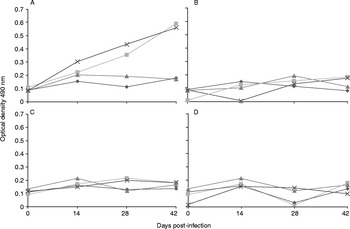
Significant serum IgG somatic and anti-excretory/secretory responses were elicited following gastric inoculation of Anisakis (P< 0.001) (fig. 1A). When the IgE anti-excretory/secretory Anisakis response was analysed by the Tukey post-hoc test, no significantly different responses to excretory/secretory antigens were observed (P= 0.0978); however, the IgE anti-Anisakis somatic antigen response was positive in consecutive samples examined between 0 and 42 days (P< 0.001) compared to controls. This mild reaction was not considered significant (fig. 1C). No somatic or excretory/secretory IgG or IgE antibody responses specific to Contracaecum spp. were observed (fig. 1B and D).
Optical density values up to 42 days post-infection in mice to show (A) anti-Anisakis IgG (immunoglobulin G) responses specific for excretory/secretory antigens (ES) and somatic (S) antigens; (B) no anti-Contracaecum IgG responses; (C) no anti-Anisakis IgE (immunoglobulin E) responses to ES nor S antigens; and (D) no anti-Contracaecum IgE responses to ES nor S antigens; ( ) IgG and IgE ES larval antigen, (
 ) IgG and IgE S larval antigen and (
 ) (
 ) controls.

Mouse models have been developed as a means of gaining a better understanding of the pathology associated with A. simplex L3 infections (Cho & Lee, Reference Cho and Lee2006). Previous studies have reported that the administration of Anisakis L3 larvae via the gastric route (Perteguer & Cuellar, Reference Perteguer and Cuellar1998; Perteguer et al., Reference Perteguer, Cuellar, Guillen, Aguila, Fenoy, Chivato and Laguna2003) elicited nematode-specific antibodies; however, these studies did not quantify the number of larvae used for respective inoculations. The experiment described herein examined pathological and immune responses to inoculation with a single L3 larva, although it is possible that human infections are the result of more than one larva.
Different inoculation routes, such as intraperitoneal (Rodriguez et al., Reference Rodriguez, Romaris, Lorenzo, Moreno, Bonay, Ubeira and Garate2006), intramuscular, intravenous and intranasal (Kirstein et al., Reference Kirstein, Horsnell, Nieuwenhuizen, Ryffel, Lopata and Brombacher2010) have been utilized to study immune responses elicited by Anisakis in mice. However, few analyses examining allergic responses in the gastrointestinal mucosa, or eosinophilic granuloma development, associated with Anisakis infections or L3 larva migration have been carried out. Jones et al. (Reference Jones, Deardorff and Kayes1990) described several histopathological findings in an animal model in which L3 Anisakis larvae were surgically introduced into the abdominal cavity. Based on these observations, it is likely that T-helper (Th)1 and Th2 type responses would be elicited following orogastric inoculation, suggesting that a model of intestinal allergic responses can be developed as a means of examining disease presentation, which is poorly defined in humans.
Although this topic is still a matter of controversy, it is not known whether live worms must be present to elicit allergic symptoms (Audicana et al., Reference Audicana, Ansotegui, De Corres and Kennedy2002; Alonso-Gomez et al., Reference Alonso-Gomez, Moreno-Ancillo, Lopez-Serrano, Suarez-De-Parga, Daschner, Caballero, Barranco and Cabanas2004). Somatic antigens are the most abundant and cross-reactive Anisakis antigens, making it difficult to diagnose infections caused by related species (Weiler, Reference Weiler2007). These antigens are synthesized in body structures and can be obtained by homogenizing whole larvae. However, excretory/secretory antigens help parasites penetrate the gastric and intestinal mucosa (Valls et al., Reference Valls and Pascual2003), and, as shown in this report, the presence of L3 Anasakis larvae in the gastrointestinal tract elicited serum IgG production with specificity for both types of antigens. It is currently accepted that Anisakis sensitization is only produced by living larvae and that Ani s 7 is the most important excretory/secretory A. simplex allergen (Anadon et al., Reference Anadon, Romaris, Escalante, Rodriguez, Garate, Cuellar and Ubeira2009).
Responses elicited against Anisakis spp. were significantly different from reactions elicited against Contracaecum spp. L3 larva of Contracaecum spp. are structurally similar to Anisakis spp. and range from 3 to 6 cm in length, with an average thickness of 0.16 cm. Both species possess a flat tooth with a ventrally located excretory pore. The excretory channel is located at the basal and lateral strands in a classic butterfly form (Laffon-Leal et al., Reference Laffon-Leal, Vidal-Martinez and Arjona-Torres2000). These same authors demonstrated that Contracaecum spp. larvae migrated into fish muscles in a manner similar to that of Anisakis larvae, making it difficult for health authorities to ignore this infection. Contracaecum spp. has only rarely been implicated as a causative agent of disease (Schaum & Müller, Reference Schaum and Müller1967; Shamsi & Butcher, Reference Shamsi and Butcher2011). Although Contracaecum spp. is described as being found infrequently in this region of the planet (Mattiucci & Nascetti, Reference Mattiucci and Nascetti2008), recent evidence shows the presence of larvae of Anisakis and Contracaecum in fish for human consumption (Dias et al., Reference Dias, Sao Clemente, Pinto and Knoff2011; Fontenelle et al., Reference Fontenelle, Knoff, Felizardo, Lopes and Clemente2013).
The absence of serological conversion in mice inoculated with Contracaecum spp. is difficult to explain, since this nematode shares similar basic structures and similar somatic and excretory/secretory antigens with Anisakis spp. Perhaps genetic analyses will identify differences between these species that may explain the different disease manifestations observed, and may explain further why there was an absence of sensitization following inoculation with a live Contracaecum spp. larva.
In conclusion, the data presented in this report demonstrate that introduction of a live Contracaecum spp. larva into the gastrointestinal tract failed to elicit sensitization in mice.
Financial support
This research was partially supported by Proppi – Universidade Federal Fluminense (Fopesq-2013).
Conflict of interest
None.
Ethical standards
This study was approved by the Animal Research Ethics Committee of the Universidade Federal Fluminense (UFF) Centre for Laboratory Animals (038/2009).