

Sir Henry Dale is regarded by many as the founder of pharmacology. He was awarded the Nobel Prize in 1936 together with Otto Loewi for his discovery of chemical neurotransmission (hotly debated at that time between chemical and electrical communication between cells). He identified acetylcholine (ACh) and a substance related to adrenaline as the two neurotransmitters in the autonomic nervous system. John Eccles, one of the major figures in the neurotransmission field and also a Nobel Prize winner in 1963, formulated ‘Dale's Principle’ that came to be known as ‘one nerve utilises one transmitter’.Reference Eccles1 This was, however, misleading because what Dale actually claimed was that the same transmitter was released from both central and peripheral endings of sensory nerves,Reference Dale2 which did not exclude the possibility of more than one transmitter in nerve terminals.
In my laboratory in Melbourne in the early 1960s, we discovered from experiments using an autonomic nerve-smooth muscle preparation – the guinea-pig taenia coli – that neurotransmission could not be accounted for by the two classical autonomic transmitters (by then Von Euler had been awarded a Nobel Prize for discovering that ‘the substance related to adrenaline’ was noradrenaline (NA)) and evidence was presented for non-adrenergic, non-cholinergic (NANC) neurotransmission in the gutReference Burnstock, Campbell, Bennett and Holman3 and later in other organs.Reference Burnstock4 We then addressed the question of what the NANC neurotransmitter might be and examined many substances, including monoamines, neuropeptides and amino acids, to see if they satisfied the criteria for establishing a neurotransmitter set out by Eccles.Reference Eccles5
These were:
(1) synthesis and storage in nerve terminals;
(2) release by a Ca2+-dependent mechanism;
(3) mimicry of the neurogenic response by exogenous application;
(4) inactivation by ectoenzymes and/or neuronal uptake
(5) modification of response by drugs in a comparable manner to that of the neurogenic response.
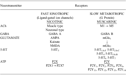
None of these substances were satisfactory and I was influenced by two important papers, one by Drury and Szent-Györgyi,Reference Drury and Szent-Györgyi6 which showed that purines had extracellular signalling actions on heart and blood vessels and another by HoltonReference Holton7 that claimed that ATP was released during antidromic stimulation of sensory nerves supplying the ear artery, to investigate ATP as a possible contender. The results were strikingly clear (see Figure 1) and we claimed in 1970 that ATP or a related nucleotide may be the neurotransmitter in NANC nerves supplying the gut,Reference Burnstock, Campbell, Satchell and Smythe8 and in a Pharmacological Reviews article entitled ‘Purinergic nerves’ soon after, the word ‘purinergic’ was introduced to represent ATP, a purine nucleotide (Figure 2).Reference Burnstock9 The hypothesis was rejected by many scientists in the following 20 years, probably because ATP was well established as an intracellular energy source and it seemed unlikely that such a ubiquitous molecule would also be an extracellular signalling molecule. It was only when the receptors for ATP and its breakdown product adenosine were cloned and characterised in the early 1990s that the concept of purinergic signalling became widely accepted (Table 1).Reference Abbracchio and Burnstock10–Reference Burnstock12 Recent cloning of ATP receptors in primitive invertebrates and green algae suggests that ATP might have been utilised as an extracellular signalling molecule very early in evolution.Reference Burnstock and Verkhratsky13
(a) Excitatory junction potentials (ejps) in response to repetitive stimulation of sympathetic nerves at 1 Hz in the guinea-pig vas deferens. The upper trace records the tension, the lower trace the electrical activity of the muscle recorded extracellularly by the sucrose gap method. Note both summation and facilitation of successive junction potentials. At a critical depolarisation threshold an action potential is initiated which results in contraction. (Reproduced from Ref. Reference Burnstock and Costa52 with permission of Chapman and Hall.) (b) The effect of various concentrations of α,β-methylene ATP on ejps recorded from the guinea-pig vas deferens (intracellular recording). Top panel: The control responses to stimulation of the motor nerves at 0.5 Hz are shown on the left. After at least 10 trains in the continuous presence of the indicated concentration of α,β-methylene ATP, ejps were recorded using the same stimulation parameters. The ejps are clearly reduced in magnitude in the presence of α,β-methylene ATP. Notice also that in control cells several large spontaneous ejps were seen, whereas after α,β-methylene ATP no spontaneous ejps were recorded. (Reproduced from ref. Reference Sneddon and Burnstock21 with permission from Elsevier). (c) Spritzed ATP, but not NA, mimicked the ejp recorded in the vas deferens. (Reproduced from ref. Reference Burnstock and Verkhratsky53 with permission from Springer.)

(a) Release of endogenous ATP from control (n = 32), 6-hydroxydopamine (6-OHDA)-pretreated (n = 7), tetrodotoxin (TTX)- (16 μM, n = 7) and guanethidine (GUAN)-exposed (5 μg/ml, n = 7) guinea-pig vas deferens during field stimulation at 8 Hz (0.5 ms, 20 V). Upper panel: mean ± SEM nmol of ATP released/min per g of vas deferens. Lower panel: contractile responses to field stimulation. The vas deferens were stimulated for 1 min as denoted by the upward bracket. ***P < 0.001. (b) Upper panel: release of endogenous ATP from control (n = 32) and reserpine-pretreated (n = 12) guinea-pig vas deferens during field stimulation at 8 Hz (pulse width 0.5 ms, 20 V), mean ± SEM nmol of ATP released/min per g of vas deferens. Lower panel: contractile responses to field stimulation. The vas deferens were stimulated for 1 min as denoted by the upward bracket. Note that the second slow phase of the mechanical response has gone in the reserpine-pretreated tissue. ((a) and (b) reproduced from Ref. Reference Kirkpatrick and Burnstock24 with permission from Elsevier.) (c) Contractions produced in the isolated saphenous artery of the rabbit following neurogenic transmural stimulation (0.08–0.10 ms; supramaximal voltage) for 1 s at the frequencies indicated (triangles). Nerve stimulations were repeated in the presence of 10 μM prazosin (a1-adrenoceptor antagonist) added before (Continued) desensitisation of the P2 purinoceptors with 10 μM α,β-methylene ATP as indicated by the arrows. (Reproduced from ref. Reference Burnstock and Warland54 with permission from Elsevier.) (d) Intracellular recording of the electrical responses of single smooth muscle cells to field stimulation of the sympathetic motor nerves (the pulse width was 0.1 ms at 0.5 Hz, indicated by •). Top panel (i): control response of the muscle. Note that to each individual stimulus there was a rapid depolarisation, and as the train of pulses progressed, a slow depolarisation developed. Similar responses were obtained in bottom panel (i), which are also control responses in Krebs solution. Top panels (ii) and (iii) show the effect of phentolamine (2 × 10⁻⁶ M, added to the bathing solution). The fast depolarisations produced by each stimulus were not reduced, but there was a progressive reduction in the size of the slow depolarisation, which was almost abolished after 6 min. Bottom panel (ii) shows the effect of a higher concentration of α,β-methylene ATP. Here the fast depolarisation was totally abolished, whilst the slow depolarisation persisted, although reduced to some extent. Subsequent addition of phentolamine (2 × 10⁻⁶ M), together with α,β-methylene ATP, abolished the neurogenic response completely (iii). (Reproduced from Ref. Reference Sneddon and Burnstock25 with permission from Elsevier.)
Comparison of fast ionotropic and slow metabotropic receptors for acetylcholine (ACh), γ-aminobutyric acid (GABA), glutamate, and 5-hydroxytryptamine (5-HT) with those proposed for ATP. (Updated and reproduced from Ref. Reference Burnstock59 with permission from John Wiley and Sons)
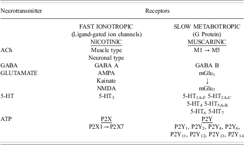
AMPA, 2-(aminomethyl)phenylacetic acid; NMDA, N-methyl-D-aspartate
In the early 1970s, through experiments carried out with Che Su and John Bevan during a sabbatical leave in Los Angeles, an important new finding emerged, namely that ATP was released from sympathetic nerves supplying the taenia coli as well as from intrinsic NANC inhibitory nerves.Reference Su, Bevan and Burnstock14 This was suggestive that ATP may be a cotransmitter with NA in sympathetic nerves and, together with a number of other hints in the literature (e.g. Refs Reference Stjärne and Lishajko15–Reference Langer and Pinto17), I questioned Dale's Principle and formally proposed the concept of cotransmission and reviewed the evidence in general, but in particular that ATP and NA were cotransmitters in sympathetic nerves.Reference Burnstock18 Early electrophysiological experiments concerned with sympathetic neurotransmission in the vas deferens showed excitatory junction potentials (ejps) that summed and facilitated until, at a critical threshold depolarisation, a spike was initiated associated with contraction (Figure 1(a)).Reference Burnstock and Holman19 We were puzzled at that time, when NA was considered to be the sole transmitter in sympathetic nerves, that adrenoceptor antagonists did not block the ejps. However, over 20 years later, we showed that α,β-methylene ATP (α,β-meATP), which desensitises the ATP receptors,Reference Kasakov and Burnstock20 blocked the ejpsReference Sneddon and Burnstock21 (Figure 1(b)) and the responses to ATP mimicked the ejps (Figure 1(c)), that it became clear that ATP and NA were cotransmitters in the sympathetic nerves, a claim already made earlier on the basis of ATP mimicry of non-adrenergic contractile responses by Westfall and colleagues.Reference Westfall, Stitzel and Rowe22 The release of ATP during stimulation of sympathetic nerves to the vas deferens was shown to be blocked by tetrodotoxin (showing that it was released from nerves), by guanethidine, an antagonist known to act by preventing release of sympathetic neurotransmitters and by 6-hydroxydopamine, known to destroy sympathetic nerves (see Figure 2(a)). An alternative hypothesis was proposed at the time that NA was the sole transmitter in sympathetic nerves and that the ejps were the result of activation of γ-receptors.Reference Neild and Hirst23 However, a key experiment was carried out, which showed that reserpine, which depletes NA, while blocking the slower second phase of the nerve-mediated contraction, did not abolish the fast first phase of the contraction or the release of ATP, which negated this hypothesis (Figure 2(b)).Reference Kirkpatrick and Burnstock24 Sympathetic cotransmission to blood vessels was also establishedReference Sneddon and Burnstock25–Reference Katsuragi and Su28 (see for example Figure 2(c) and (d)). Figure 3 shows the currently accepted model of sympathetic cotransmission. While neuropeptide Y is also contained and released by most sympathetic nerves, it rarely acts as a cotransmitter, but rather as a prejunctional neuromodulator inhibiting the release of the cotransmitters and/or as a postjunctional neuromodulator enhancing the actions of NA and ATP.Reference Cheung29, Reference Ellis and Burnstock30 The proportions of ATP and NA in sympathetic nerves varies considerably in different organs and species and during development and ageing (see Ref Reference Burnstock27). For example, while NA is the dominant neurotransmitter in renal arteries, the sole neurotransmitter in sympathetic nerves supplying the rabbit jejunal artery and intestinal arterioles is ATP, with NA acting as a neuromodulator of ATP release.Reference Ramme, Regenold, Starke, Busse and Illes31, Reference Evans and Surprenant32 Later, the concept of ‘chemical coding’ was formulated,Reference Furness, Morris, Gibbins and Costa33 and it was shown that ATP was often accompanied by neuropeptides as well as classical transmitters in nerve terminals (see Ref. Reference Burnstock34).
Schematic of sympathetic cotransmission. ATP and NA released from small granular vesicles (SGV) act on P2X and α1 receptors on smooth muscle, respectively. ATP acting on inotropic P2X receptors evokes excitatory junction potentials (EJPs), an increase in intracellular calcium ([Ca2+]i) and fast contraction; while occupation of metabotropic α1 adrenoceptors leads to production of inositol triphosphate (InsP3), an increase in [Ca2+]i and slow contraction. Neuropeptide Y (NPY) stored in large granular vesicles (LGV) acts after release both as a prejunctional inhibitory modulator of release of ATP and NA and as a postjunctional modulatory potentiator of the actions of ATP and NA. Soluble nucleotidases are released from nerve varicosities, and are also present as ectonucleotidases. (Reproduced from Ref. Reference Burnstock and Verkhratsky55 with permission from Elsevier.)
ATP and ACh were identified as cotransmitters in some parasympathetic nerves (e.g urinary bladder,Reference Burnstock35 carotid bodyReference Zhang, Zhong, Vollmer and Nurse36 and presynaptic terminals in the enteric plexusReference Burnstock37). Resistance to atropine, an antagonist to muscarinic ACh receptors, to parasympathetic nerve stimulation to the urinary bladder was recognised at the end of the nineteenth century.Reference Langley and Anderson38 However, for many years this was assumed to be due to the inaccessibility of the neurotransmitter ACh to its receptors on smooth muscle. However, in 1972 it was shown that the atropine-resistant component of the responses of the bladder to nerve stimulation satisfied the criteria for ATP as a cotransmitter.Reference Burnstock, Dumsday and Smythe39, Reference Burnstock, Cocks, Crowe and Kasakov40 ATP mimicked the non-cholinergic responses (Figure 4(a)), ATP was shown to be released in a Ca2+-dependent manner during parasympathetic nerve stimulation (Figure 4(b)) and α,β-meATP to block both the responses to nerve stimulation and ATP (Figure 4(c)). P2X1 receptors were shown to be localised with immunohistochemistry on bladder smooth muscle.
(Continued)
(a) Left-hand side: mechanical responses of the guinea-pig taenia coli to intramural nerve stimulation (NS, 1 Hz, 0.5 ms pulse duration, for 10 s at supramaximal voltage) and ATP (2 × 10⁻⁶ M). The responses consist of a relaxation followed by a ‘rebound contraction’. Atropine (1.5 × 10⁻⁷ M) and guanethidine (5 × 10⁻⁶ M) were present. (Reproduced from Ref. Reference Burnstock and Wong56 with kind permission of the Nature Publishing Group.) Right-hand side: a comparison of the contractile responses of the guinea-pig bladder strip to intramural nerve stimulation (NS: 5 Hz, 0.2 ms pulse duration and supramaximal voltage) and exogenous ATP (8.5 μM). Atropine (1.4 μM) and guanethidine (3.4 μM) were present throughout. (b) Effect of changing the calcium ion (Ca2+) concentration on the release of ATP from the guinea pig isolated bladder strip during stimulation of intramural nerves. Upper trace: mechanical recording of changes in tension (g) during intramural nerve stimulation (NS: 20 Hz, 0.2 ms pulse duration, supramaximal voltage for 20 s). Lower trace: concentration of ATP in consecutive 20 s fractions of the superfusate. The Ca2+ concentration in the superfusate varied as follows: (i) 2.5 mM (normal Krebs); (ii) 0.5 mM; (iii) 0.25 mM; (iv) 2.5 mM. The successive contractions were separated by 60 min intervals as indicated by the breaks in the mechanical trace. Atropine (1.4 μM) and guanethidine (3.4 μM) were present throughout. The temperature of the perfusate was between 22°C and 23°C. ((a) right-hand side and (b) Reproduced from Ref. Reference Burnstock, Cocks, Crowe and Kasakov40 with permission of the Nature Publishing Group). (c) The effect of α,β-methylene ATP (α,β-meATP) on the response of isolated guinea-pig bladder strips to nerve stimulation (NS), ATP (Δ) and histamine (Hist). Upper trace: Control responses; lower trace, desensitisation attained by five successive applications of α,β-meATP (50 μM, ▴), at 4-min intervals, completely abolished nerve-mediated and ATP induced contractions, although histamine-induced contraction is only slightly reduced. (Reproduced from Ref. Reference Kasakov and Burnstock20 with permission from Elsevier.)
Our earlier suggestion that ATP was the transmitter on NANC enteric nerves was later modified to show that ATP, nitric oxide and vasoactive intestinal polypeptide were cotransmitters in these enteric inhibitory nerves (Figure 5), again the proportions varying considerably in different regions of the gut an in different species.Reference Burnstock37, Reference Belai and Burnstock41
Schematic representation of non-adrenergic non-cholinergic (NANC) inhibitory nerves in the gut. Neurotransmitters and/or agonists: VIP, vasoactive intestinal polypeptide; NO, nitric oxide; ATP, adenosine 5′-triphosphate; AD, adenosine; PG, prostaglandins. Antagonists or inhibitors: L-NAME, N G-nitro-L-arginine methyl ester; RB2, Reactive blue 2; 8-PT, 8-phenyltheophylline. Responses: +, excitatory; −, inhibitory. (Reproduced from Ref. Reference Burnstock57 with permission from Springer.)
Evidence has been presented that many sensory nerves release ATP as a cotransmitter with calcitonin gene-related peptide and substance P, for example in the skin (Figure 6) and with glutamate in terminals of primary afferent sensory nerves in the dorsal horn of the spinal cord.Reference Burnstock34, Reference Burnstock42
(a) Schematic representation of the basis of the ‘axon reflex’ in the skin leading to vasodilation and inflammation. It is suggested that calcitonin gene-related peptide (CGRP), substance P (Subst. P) and/or ATP are released during antidromic activation of sensory collaterals. (Modified from Ref. Reference Burnstock58 with permission from Williams and Wilkins, Co.) (b) The figure on the left shows skeletal neuromuscular transmission in the adult, with ACh acting via nicotinic receptors on the post-synaptic membrane, while the ATP released during transmission acts as a prejunctional neuromodulator via P1 (A1) receptors of ACh release after breakdown to adenosine (AD). The figure on the right shows that in the developing myotube motor nerves release ACh and ATP as cotransmitters acting on nicotinic and P2X ion channel receptors on the post-synaptic membrane, respectively. ATP, after breakdown to adenosine, acts on prejunctional P1 (A1) receptors to inhibit release of ACh.
While it is well established that ACh and ATP are coreleased from motor nerves, ACh is the principal neurotransmitter in the mature skeletal neuromuscular junction, while the ATP released is broken down by ecto-nucleotidases to adenosine where it acts as a prejunctional modulator of ACh release and ATP as a postjunctional potentiator of the action of ACh (Figure 6(a)). However, in the developing myotube, both ATP and ACh act as cotransmitters via P2X and nicotinic ion channel receptors (Figure 6(b)).Reference Kolb and Wakelam43
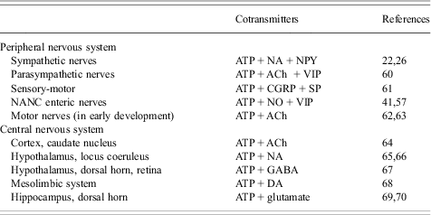
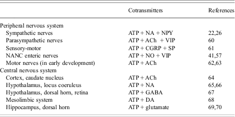
It is now widely accepted that ATP is a cotransmitter not only in sympathetic and parasympathetic nerves, but in most, if not all, nerves in both the peripheral and central nervous systems (see Table 2).Reference Burnstock44 The physiological significance of cotransmission has been explored (see Figure 7),Reference Burnstock45 suggesting that cotransmission offers a number of ways of modulating local activity at peripheral sites, rather than the direct inflexible control by the central nervous system that was emphasised earlier.
Table showing cotransmitters in the peripheral and central nervous systems. (Compiled from Ref. Reference Burnstock12.)
(Continued)
Spectrum of signalling variations offered by cotransmission (solid line arrows = neurotransmission; broken line arrows = pre- or post-junctional neuromodulation). (a) Fast transmission is usually produced by small molecules (C1) released at low frequency nerve stimulation acting on ionotropic receptors (R1), whereas slow transmission is usually produced by release of peptides (C2) or other molecules at high frequency stimulation acting on G-protein-coupled receptors (R2). (b) Cotransmitters C1 and C2 can both be fast messengers acting via ionotropic receptors (R1 and R2). (c) Cotransmitters C1 and C2 act on receptors (R1 and R2) localised on different postjunctional cells. (d) Cotransmitters C1 and C2 not only act postjunctionally via R1 and R2 receptors but can also act as prejunctional modulators to either inhibit (−) or enhance (+) the release of C1 and/or C2. (e) Cotransmitters C1 and C2 act synergistically to enhance the combined responses produced via R1 and R2 receptors. (f) Cotransmitters C1 and C2 act to inhibit the responses evoked via R1 and/or R2 receptors. (g) Cotransmitter C1 evokes neurotransmission via R1 receptors, while C2 evokes long-term (trophic) responses of postjunctional cells via R2 receptors. (h) Cotransmitter C1 produces excitation via R1 receptors when the postjunctional smooth muscle target has a low tone, with C2 having little influence; however, when the smooth muscle tone is high, the dominant response might be relaxation produced by C2 via R2 receptors. (i) Substance C3 is taken up by nerve terminals, rather than being synthesised and stored as is true for the cotransmitters C1 and C2. C3 can then be released on nerve stimulation to act on postjunctional R3 receptors. In these circumstances, C3 would be known as a ‘false transmitter’. (j) A coexisting substance C3 (often a peptide) can be synthesised and stored in a nerve, but not act directly via a postjunctional receptor to produce changes in postjunctional cell activity. It could, however, act as a prejunctional inhibitor (+) of the release of the cotransmitters C1 and C2, or as a postjunctional enhancer (þ) of the responses mediated by R1 and R2. (Modified from Ref. Reference Burnstock45 with permission from Elsevier.)
Plasticity of expression of purinergic cotransmitters has been described in several disease situations, as well as in development and aging in the nerves that remain after trauma or surgery (see Refs Reference Burnstock34 and Reference Bao, Eriksson and Stjärne46). For example, in the healthy human urinary bladder less than 3% of the response to parasympathetic nerve stimulation is purinergic, although purinoceptors are expressed in the smooth muscle (in contrast to the 40–60% component of parasympathetic cotransmission in rodent bladders). However, in pathological conditions, such as interstitial cystitis, obstructed and neurogenic human bladder, up to 40% of the response is purinergic (see Refs Reference Burnstock35, Reference Palea, Artibani, Ostardo, Trist and Pietra47 and Reference Smith and Chapple48). There are also reports that the purinergic component of sympathetic cotransmission is much increased in spontaneous hypertensive rats.Reference Vidal, Hicks and Langer49, Reference Bulloch and McGrath50 Sympathetic pain, which involves an increase in sympathetic nerve activity, whether causalgia with sympathetic hyperinnervation or reflex sympathetic dystrophy with increased sympathetic activity, was not attenuated by adrenoceptor antagonists (although guanethidine, which prevents the release of the cotransmitter ATP as well as NA, had some success) (see Ref. Reference Burnstock51), suggesting that ATP may be the neurotransmitter involved in sympathetic pain (see Figure 8).
Sympathetic pain that occurs during both causalgia and reflex sympathetic dystrophy, where there is increased release of sympathetic neurotransmitters. It is suggested that pain is mediated largely via ATP acting on P2X2/3 receptors on nociceptive sensory nerve fibres. It is further proposed that the cotransmitters noradrenaline (NA) and neuropeptide Y (NPY) act as postjunctional enhancers of the activation of P2X2/3 receptors as well as adenosine after breakdown of ATP.
Geoffrey Burnstock, PhD DSc FAA FRCS(Hon) FRCP(Hon) FMedSci FRS, is currently President of the Autonomic Neuroscience Centre, UCL. He gained his PhD from UCL in 1957), DSC (Melbourne 1971), was Professor of Zoology, Melbourne University (1964), HOD Anatomy and Developmental Biology UCL (1975). He had been elected to the Australian Academy of Sciences (1971), Royal Society (1986), Academy of Medical Sciences (1998). He is a Honorary Fellow of the Royal College of Surgeons (1999), Physicians (2000). He has been awarded the Royal Society Gold Medal (2000), Copernicus Gold Medal (Ferrara, 2009), Gaddum Memorial Award (2010), Academia Europaea Erasmus Medal (2012). He has Honorary Degrees from the Universities of Antwerp, Frankfurt and Leipzig. He has been Editor-in-Chief of Autonomic Neuroscience (Elsevier), Purinergic Signalling (Springer), Open Neuroscience & Open Pharmacology Journals (Bentham). He is best known for his seminal discovery of purinergic signalling. He has supervised over 100 PhD and MD students, and has over 1500 publications, cited >80,000 times.