INTRODUCTION
Records of Gastrotricha from deep sea sites such as abyssal plains and special habitats like hydrothermal vents are scarce (Radziejewska, Reference Radziejewska2002; Gutzmann et al., Reference Gutzmann, Martínez Arbizu, Rose and Veit-Köhler2004; Kieneke & Zekely, Reference Kieneke and Zekely2007). At the same time they expand the range of marine benthic ecosystems in which new taxa of that phylum can potentially be discovered. A valuable number of samples and specimens from different deep sea expeditions show a high dispersal of Gastrotricha (unpublished preliminary data) and, amongst others, yielded a specimen of an undescribed species of the taxon Thaumastoderma. Like recently described species of this taxon (see e.g. Hummon, Reference Hummon2008), members of Thaumastoderma live in medium to coarse sediments of the eulittoral zone while few species are reported from deeper sites (Clausen, Reference Clausen2004). The occurrence of a species of Thaumastoderma in silty deep sea sediment is somehow surprising and possibly underlines the discreteness of this new species.
Until recently, the genus Thaumastoderma comprised sixteen described species. Many species are very similar, so in some cases a successful identification is dependent on a unique combination of characters, many of which are shared by other species (see table 5 in Clausen, Reference Clausen2004). Instead of arguing in a traditional taxonomic fashion, I here describe the new species on the basis of a phylogenetic analysis of the genus. Hence, the species discrimination and taxonomic affiliations shall be evidenced by the use of potential autapomorphies and synapomorphies, respectively. One aim of the present investigation is to test whether it is possible to reconstruct a reasonable phylogenetic scenario for these very similar and character-poor species.
MATERIALS AND METHODS
Sampling and specimen preparations
The sample containing the single specimen of an undescribed Thaumastoderma species was collected during the Andeep-1 cruise which was carried out in Antarctic waters of the Drake Passage (ANT XIX/3; RV ‘Polarstern’; January–February 2002). As sampling gear, a multiple corer (MUC) has been used at seven different stations (see Martínez Arbizu, Reference Martínez Arbizu2003; Gutzmann et al., Reference Gutzmann, Martínez Arbizu, Rose and Veit-Köhler2004). The study area comprised two transects/depth gradients (eastern and western of the Shackleton Fracture Zone) with depths from 2274 to 5194 m. For meiofauna investigation the upper 10 cm of sediment of each core (128 cores in total, see Gutzmann et al., Reference Gutzmann, Martínez Arbizu, Rose and Veit-Köhler2004), together with the filtrate of the supernatant water, were fixed with buffered formalin at a final concentration of about 4%. The specimen of Thaumastoderma was found in core number 5 of MUC-deployment 99-5 at a ground depth of 5191 m (latitude: 61°7.09′S; longitude: 59°16.50′W). Several other specimens of the taxon Gastrotricha were extracted from the cores of the Andeep-1 cruise that will be subject of another study.
The meiofauna was extracted from the sediment samples by differential flotation and centrifugation using Levasil®. Metazoan specimens were filtered using a sieve with a mesh size of 40 µm. Subsequently, the meiofauna was stained with an aqueous solution of rose Bengal to ease identification and counting under a stereo microscope. From single specimens, glycerol wholemounts were prepared using a mixture of paraffin and beeswax (ratio 2:1). The specimen was observed and documented with a Leica DMLB compound microscope equipped with differential interference contrast and a digital camera Olympus Color View I. Drawings were made using a camera lucida mounted on the Leica microscope. Positions and ranges of internal and external structures of the new species are provided in percentage body units (U), where the value of 100 refers to the total body length (the pair of elongated cirrata tubes not considered). Percentage body units and given length are taken from the holotype.
Phylogenetic analysis
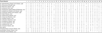
Data matrix: for the cladistic analysis of the genus Thaumastoderma, a data matrix has been coded including all sixteen described species of the genus plus the new species (Table 1). The matrix was built in the nexus format (Maddison et al., Reference Maddison, Swofford and Maddison1997) using the Nexus Data Editor 0.5.0 (Page, Reference Page2001a). For testing monophyly of the genus and for character polarization, three outgroup representatives have also been included: one member of the taxon Turbanellidae (Turbanella cornuta) and two species of the taxon Thaumastodermatidae (Tetranchyroderma arcticum and Acanthodasys lineatus). In total, 37 unordered and equally weighted characters (approximately one half binary (absent/present) characters and the rest multistate characters, see Appendix) have been sampled. The original species descriptions served as the data source (see Table 1).
Species-character matrix of sixteen described species of Thaumastoderma, three outgroup representatives and the new described species. A question mark (?) refers to unknown data, a dash (–) indicates inapplicable character states. For character state descriptions see Appendix.

Some of the characters depend on the existence of a superordinate structure (see Appendix). If this is absent in a certain species, the subordinate character(s) has to be coded as inapplicable (–). Although some systematists claim for character independence, the inapplicable coding is regarded as a good strategy for extracting phylogenetically relevant information from hierarchically structured data (e.g. existence, different numbers and shapes of a character). Otherwise, information could be lost when transforming a set of dependent characters into a single multistate one (Lee & Bryant, Reference Lee and Bryant1999). However, since the algorithms of many computer programs treat inapplicable character states as ‘?’ (no information), one has to be careful when optimizing characters onto a phylogenetic tree.
Tree search: for finding an optimal tree, the species-character matrix was calculated with the computer program PAUP* v4.0b10 (Swofford, Reference Swofford2002). A heuristic search with 1000 replicates was run with a set rearrangement limit of 10,000,000 for each replicate; Turbanella cornuta was fixed as outgroup. Other parameters for the heuristic-search were as follows: starting trees via random stepwise addition, branch swapping by TBR, number of maximum trees to be saved automatically increased and elimination of ‘zero branches’. The strict consensus tree was evaluated in order to find possible monophyletic subgroups and evolutionary novelties (apomorphies) within Thaumastoderma. For this purpose, the program MacClade 4.0 (Maddison & Maddison, Reference Maddison and Maddison1989, Reference Maddison and Maddison2000) has been used (ACCTRAN-optimization).
Bremer or decay indices (Bremer, Reference Bremer1988, Reference Bremer1994) were calculated with PAUP* using the parsimony ratchet approach (Müller, Reference Müller2004). Readable commands for PAUP* were generated with the program PRAP (Müller, Reference Müller2004) using the original data matrix and the strict consensus tree of the heuristic-search. The number of ratchet iterations used for evaluating each node was set to a value of 10. Bootstrap values (Felsenstein, Reference Felsenstein1985) were obtained by a bootstrap search calculated in PAUP* with 250 bootstrap replicates. The search settings were equal to the heuristic-search but with replicate number per bootstrap replicate reduced to five. For assessing the results from the calculation of decay indices and bootstrap values and for exporting the tree files, the program TREEVIEW 1.6.5 (Page, Reference Page2001b) was used.
RESULTS
Thaumastoderma antarctica sp. nov.
TYPE MATERIAL
The single specimen of Thaumastoderma antarctica sp. nov. mounted on a microscopic slide (holotype) is deposited in the collection of the Forschungsinstitut und Naturmuseum Senckenberg (inventory number SMF 4); coordinates: 61°7.09′s 59°16.50′W; water depth: 5191 m.
DIAGNOSIS
Very small Thaumastoderma with one pair of simple spatulate tentacles, one pair of long, rod-shaped lateral and one pair of shorter anterior tentacles. Cephalic cirri present, eye spots lacking. Cuticular armature of small-sized tetrancres. Five pairs of dorsal cirrata, last pair strongly elongated and thickened, all others of equal length. Four pairs of dorsal epidermal glands with conspicuous slit-like pores. Adhesive tubes: two pairs of anterior adhesive tubes, six ventro-lateral adhesive tubes per side, ventral tubes lacking, up to four posterior adhesive tubes per side; two or three positioned on each of the paired posterior pedicles, the remaining two tubes lateral to the pedicles.
ETYMOLOGY
The species name antarctica refers to the position of the type locality that lies in Antarctic waters of the South Atlantic Ocean.
DESCRIPTION
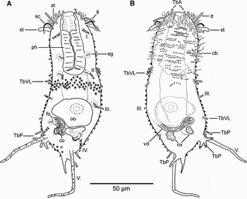
Habitus: Thaumastoderma antarctica is a compact, tongue-shaped gastrotrich with no noticeably separated head and with slightly convex frontal and blunt caudal ends (Figures 1 & 2A, B). In the region of the mature oocyte, the trunk is slightly wider than in the anterior half. At the angles of the caudal end, a pair of pedicles is situated bearing some of the posterior adhesive tubes (see below). Thaumastoderma antarctica has a body length of 115 µm and widths of 38 µm and 45 µm at U30 and U70, respectively.
Schematic drawings of Thaumastoderma antarctica sp. nov. (A) Dorsal view. Tetrancres only partially indicated; (B) ventral view. Abbreviations: at, anterior tentacle; cb, ciliary bases; co, caudal organ; eg, epidermal glands; fo, frontal organ; lt, lateral tentacle; oo, mature oocyte; ov, putative ovaries; ph, pharynx; sc, sensory cirri; st, spatulate tentacle; TbA, anterior adhesive tubes; TbP, posterior adhesive tubes; TbVL, ventro-lateral adhesive tubes; vd, vas deferens; I.–V., dorsal cirrata pairs 1–5.

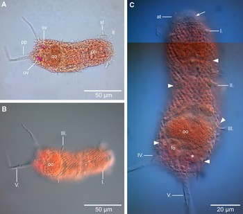
Light microscopic images of Thaumastoderma antarctica sp. nov. (A & B) Captures of the habitus showing some features of the internal and external morphology; (A) ventral view; (B) dorsal view; (C) high-magnification close-up of the trunk, dorsal view. Notice the cuticularized, slit-like openings of the epidermal glands (white triangles) and the area that is not covered with tetrancres (arrow). Abbreviations: at, anterior tentacle; fo, frontal organ; lt, lateral tentacle; oo, mature egg; ov, putative ovaries; ph, pharynx; pp, posterior pedicle with adhesive tubes; st, spatulate tentacle; I.–V., dorsal cirrata pairs 1–5; asterisk, premature oocyte; A, bright field illumination; B and C, differential interference contrast.

Sensory structures: like most members of the genus, Th. antarctica possesses a characteristic set of paired sensory organs at the head region (at, lt, st; Figures 1, 2A). These organs, position at approximately U10, insert laterally as 10 µm long spatulate tentacles (st) of 3 µm width. Directly anterior to the spatulate tentacles and inserting a little more dorso-laterally, there is a pair of 15 µm long cirri (sc) that probably consist of compound cilia. The cirri seem to be flexible and are bent caudally. Around U5, a pair of 12 µm long and 1 µm wide straight tentacles inserts laterally on the head, i.e. the lateral tentacles (lt). At the same level (U5), but more dorsally, there is a fourth pair of shorter (7–10 µm long, 1 µm wide) tentacles, i.e. the anterior tentacles (at). In addition to the four sets of solid sensory organs, there are numerous cilia at the sides and ventral surface of the head region and around the mouth opening, which are probably sensory cilia. I have not observed any trace of pigmented eye spots in Th. antarctica.
Adhesive tubes: as in the congeners, one can distinguish cirrata-type tubes from ‘ordinary’ adhesive tubes in Th. antarctica. Therefore, I treat them separately in the following description. The new species has five pairs of cirrata tubes situated at U15, 45, 65, 90 and 95 (Figure 1A). All five pairs insert dorsally but the first two pairs insert a little more medially than do pairs III–V. All cirrata tubes show a characteristic content of vesicles or secretion droplets. Pairs I–IV are approximately 10 µm long and 1.5 µm wide. However, pair V is conspicuously elongated and thickened compared to all other cirrata tubes. Each cirrata tube V is about 45 µm long and 3 µm wide at the base tapering to 1.5 µm at the tip. The tips of all five pairs of cirrata tubes are more or less directed dorso-caudo-laterally.
Thaumastoderma antarctica has two pairs of frontally directed anterior adhesive tubes (TbA) situated along the caudal margin of the mouth opening (U2). Six pairs of ventro-lateral adhesive tubes (TbVL) are arranged as follows: one pair at U35, an inconspicuous group of four pairs between U40 and U55 and one pair at U80 (Figure 1B). There are four posterior adhesive tubes (TbP) at the left but only three tubes at the right side of the animal. Three or two tubes, respectively, are arranged at the caudal pedicles. The additional pair of posterior adhesive tubes inserts lateral to the caudal pedicles around U90. There are no further tubes between both caudal pedicles. All adhesive tubes are 5 to 7 µm long and 1 µm wide, only the tubes of the caudal pedicles are slightly longer.
Cuticular armature: the dorsal and dorsolateral surface of Th. antarctica is densely covered with minute tetrancres except for a narrow anterior zone at the dorsal side of the head region that lacks cuticular differentiations (Figure 2C). There are seventeen longitudinal rows of dorsal plus dorso-lateral ancres that alter more or less regularly. However, the number of ventro-lateral rows could not be defined. Tetrancres slightly increase in size from frontal to the mid trunk before decreasing again toward the caudal end. Ancres of the mid trunk region measure 2×2 µm only. Hence, they are the smallest ancres reported from any Thaumastoderma species.
Epidermal glands: there are at least three pairs of dorso-lateral epidermal glands and an additional unilateral gland indicates a fourth pair. The four pairs are located at U35, U45, U70 and U85. Except for the first pair, the epidermal glands are closely associated with the dorsal cirrata tubes II to IV. Some of the globular to pear-shaped glands contain a kind of rod-shaped content. A striking feature are the external pores. Each gland opens via a cuticularized, slit-like pore similar to the stomata of a leaf epidermis (Figures 1A, 2C).
Internal anatomy: the subterminal mouth opening is followed by a spacious, 10 µm wide buccal cavity (U0–U15). Thaumastoderma antarctica has a 16 µm wide pharynx reaching to U45 where it passes into the intestine. Distinct pharyngeal pores could not be observed but, focused to the ventral side, two pharyngeal bulges are barely visible at U40 that possibly lead to the ventro-lateral pores. The intestine has a width of 20 µm and tapers toward the caudal end. I could not detect an anus.
The holotype contains a large mature egg in a dorso-median position (U65–U85) that has a dimension of 31 µm to 25 µm (Figure 2A–C). A second smaller, premature oocyte lies caudally on the right side (Figure 2C). In that area (U85–U90) on the left side, there is a set of two accessory reproductive organs, an anterior frontal organ and slightly caudo-ventral to that organ, a globular caudal organ. Both organs contain curled spermatozoa visible as filamentous structures. Focused to the ventral side, one can see a vas deferens running from the right side of the trunk toward the accessory reproductive organs where it is apparently connected to one of them. However, I failed to detect the related testis, but, the vas deferens bends frontally alongside the mature oocyte where I suspect the singular male gonad is present. Further ventral to the accessory organs and the premature oocyte (around U90), there is a pair of red structures (obviously disproportionately stained by the rose Bengal), around 5 µm in diameter each. Possibly, these structures display the paired ovary of Th. antarctica.
Ciliation: locomotory cilia cover the ventral surface of Th. antarctica and are arranged in several transverse bands (Figure 1B). It seems that up to three consecutive rows of cilia form each band. However, density of cilia decreases toward the caudal end.
TAXONOMIC AFFINITIES
The erection of a new species and the taxonomic placement of this species are performed in a strict phylogenetic sense. Hence, a cladistic analysis of all known species of the taxon Thaumastoderma has been carried out. More general results of this analysis are given below. Here, I focus on the closest relatives and the related apomorphies of the new species Thaumastoderma antarctica. The new species is characterized by at least three apomorphic features that are unique within the whole genus: the pattern of only the last pair of cirrata tubes being elongated and the possession of very small tetrancres (a maximum diameter/length of 2 µm). The conspicuous slit-like pores of the epidermal glands of Th. antarctica are another unique character within this genus. Moreover, the last pair of dorsal cirrata tubes is not only elongated but also more than four times longer than the remaining cirrata. Another unambiguous character transformation concerns the number of three posterior adhesive tubes per caudal pedicle. However, this possible apomorphic character has to be treated carefully since three tubes per pedicle also occur in Thaumastoderma renaudae (see Table 1) and because the single specimen of the new species shows an asymmetric pattern: it possesses three tubes on one and only two tubes on the other pedicle.
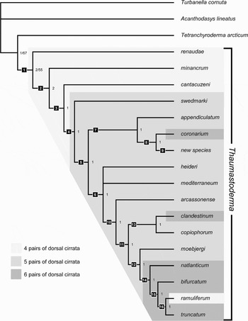
According to the analysis, two species are closely related to Thaumastoderma antarctica: Th. coronarium as the sister species and Th. appendiculatum as the sister species of the latter two (Figure 3). The sister group relationship of Th. antarctica sp. nov. and Th. coronarium is characterized by the shared possession of more than fourteen dorsal and dorso-lateral longitudinal rows of tetrancres and by the shared reduction to a maximum of five pairs of epidermal glands. The relationship of Th. antarctica sp. nov., Th. coronarium and Th. appendiculatum depends on the loss of ventral adhesive tubes within their common stem lineage. However, the complete reduction of these structures is homoplastic and does also occur in the stem lineages of the species Th. bifurcatum and Th. copiophorum (see Table 1).
Strict consensus tree calculated from three equally parsimonious trees found by the heuristic search. Tree length (included characters only): 90 steps, CI: 0.49, RI: 0.55. Numbers in the right of the nodes indicate decay values plus bootstrap percentages if higher than 50%. Numbered black squares indicate possible autapomorphies (unambiguous character transformations). (1) Presence of one pair of cephalic spatulate tentacles and one pair of lateral tentacles; (2) presence of one pair of cephalic anterior tentacles, reduction of anterior adhesive tubes from four or more pairs to a maximum of three pairs; (3) presence of cephalic sensory cirri (compound cilia); (4) number of dorsal cirrata tubes increased from four to five pairs; (5) cuticular armature enlarged to large-sized tetrancres; (6) number of anterior adhesive tubes increased from maximum three pairs to four or more pairs; (7) loss of ventral adhesive tubes; (8) number of dorsal plus dorso-lateral tetrancres increased to fourteen or more rows, number of epidermal glands reduced to five pairs; (9) cuticular armature transformed to small-sized tetrancres, number of posterior adhesive tubes per caudal pedicle increased to three, most caudal pair of dorsal cirrata tubes strongly enlarged and thickened, slit-like openings of epidermal glands; (10) loss of cephalic sensory cirri; (11) first and last pair of cirrata tubes elongated; (12) lateral-most posterior adhesive tube twice as long as the remaining tubes; (13) presence of accessory dorsal cirrata tubes; (14) number of dorsal cirrata tubes enlarged from five to six pairs; (15) presence of cephalic sensory cirri; (16) number of ventro-lateral adhesive tubes increased to twenty or more pairs. The areas in the tree highlighted by different shades of grey indicate the evolutionary increment of dorsal cirrata tubes (compare with autapomorphies 4 and 14).

ECOLOGICAL REMARKS
Station 99-5, the one from which Thaumastoderma antarctica was obtained, is the deepest station of the Andeep-1 cruise. As in the other stations of both, a western and eastern transect, the sediment mainly consists of silt with a small fraction of clay and fine sand. Although a general trend of decreasing amount of organic content with increasing depth was observed, Station 99-5 shows an elevated value of nutritive substances (chloroplastic pigment equivalents: 4.3 µg/ml sediment; total organic matter: 6.3%; see Gutzmann et al., Reference Gutzmann, Martínez Arbizu, Rose and Veit-Köhler2004).
Phylogeny of Thaumastoderma Remane, 1926
The heuristic search yielded three equally parsimonious trees (EPTs) with a tree length of 90 steps varying in the position of two species only. Hence, the strict consensus tree shows almost complete resolution and is used for the following description and evaluation of intra-generic relationships inferred by the cladistic analysis (Figure 3). All three EPTs come from a common tree island that was hit 472 times during the 1000 replicates. However, bootstrap values above 50% could only be found for the common node of Thaumastoderma and that of Thaumastoderma plus Tetranchyroderma arcticum (Figure 3).
The consensus tree suggests a phylogeny of Thaumastoderma which does not have an initial split leading to two separated and equally species-rich clades. Instead, there is one direct stem lineage as a ‘backbone’ from which single species or, in one case, a group of three species successively split off. The character optimization yielded several evolutionary transformations along the direct stem lineage that can be interpreted as apomorphies for the resolved monophyletic groups (see Figure 3). According to this, the genus Thaumastoderma is characterized by the existence of one pair of spatulate tentacles plus one pair of lateral tentacles. Within Thaumastoderma, a more derived monophyletic fraction of species (Th. heideri to Th. truncatum) is characterized by the possession of four or more pairs of anterior adhesive tubes. Regarding the dorsal cirrata tubes, there is an interesting evolutionary trend within Thaumastoderma: the number of cirrata pairs successively increases from four pairs in most basal species (Th. renaudae to Th. cantacuzeni) over five pairs in the stem lineage of Th. swedmarki plus the remaining species up to six pairs in the stem lineage of Th. natlanticum to Th. truncatum. However, there are as well a few cases where the number of cirrata tubes was reduced secondarily (see Figure 3). Given the limited outgroup species used in this analysis, the taxon Thaumastoderma is most closely related to Tetranchyroderma.
DISCUSSION
Gastrotrichs are already reported to inhabit deep sea habitats (Radziejewska, Reference Radziejewska2002; Gutzmann et al., Reference Gutzmann, Martínez Arbizu, Rose and Veit-Köhler2004). However, there is only a single record of gastrotrichs with resolution at the species level: the recently described macrodasyid Desmodasys abyssalis is reported from a hydrothermal vent of the east Pacific Ocean (Kieneke & Zekely, 2007). Therefore, the description of Thaumastoderma antarctica represents the second species-level record of Gastrotricha inhabiting the abyss. Furthermore, specimens of the paucitubulatan genus Musellifer are known from deep sea samples (see notes in Kieneke & Zekely, 2007). These initial records possibly indicate an underestimated abyssal diversity of the taxon Gastrotricha. For instance, Antarctic deep sea samples yielded an unexpected high level of new biodiversity in other taxa such as isopod crustaceans (Brandt et al., Reference Brandt, Gooday, Brandão, Brix, Brökeland, Cedhagen, Choudhury, Cornelius, Danis, De Mesel, Diaz, Gillan, Ebbe, Howe, Janussen, Kaiser, Linse, Malyutina, Pawlowski, Raupach and Vanreusel2007). Even though one single record of Th. antarctica does not allow any conclusions on distribution or abundance, the occurrence of a Thaumastoderma species in silty sediment is remarkable since all known species are reported only from medium to coarse sands, coral fragments or shell gravel of the eulittoral or shallow sublittoral zone (a few species occur at deeper sites, see e.g. Clausen, Reference Clausen2004). The outstanding content of organic matter at Station 99-5 may hint at a certain specialization of the new species and explain the restriction to this locality.
Using a single specimen for the description of a new species might of course provide reason for criticism. However, scientists working on a much more abundant and diverse group inhabiting the deep sea floor, the harpacticoid copepods, regularly encounter similar problems (e.g. George, Reference George2006; Wandeness et al., Reference Wandeness, George and Santos2009; George & Plum, Reference George and Plum2009). George & Plum (Reference George and Plum2009) argue that every species description is a testable hypothesis (even when based on a single individual) and that it is favourable to document morphological diversity rather than lose it. The unique features of Thaumastoderma antarctica, i.e. minute tetrancres, strongly elongated last cirrata pair and possibly a number of three posterior adhesive tubes per caudal pedicle which are supported by the results of the phylogenetic analysis of the genus Thaumastoderma legitimize the establishment of a new morphospecies. Information on morphological variation, however, could not be obtained and has to be covered when, for instance, future cruises may recover additional specimens of Th. antarctica.
Although recently there has been noticeable research activity to resolve the internal relationships (i.e. the relationships among the genera) of the phylum Gastrotricha (e.g. Marotta et al., Reference Marotta, Guidi, Pierboni, Ferraguti, Todaro and Balsamo2005; Todaro et al., Reference Todaro, Telford, Lockyer and Littlewood2006; Kieneke et al., Reference Kieneke, Riemann and Ahlrichs2008), analyses of delimited ingroups, for instance single families or even genera, are scarce. One example is the phylogenetic discussion dealing with the internal relationships of the exclusive freshwater family Dasydytidae (Kisielewski, Reference Kisielewski1991). A possible problem one would face when dealing with intra-family phylogeny is that monophyly is not proven for all gastrotrich families (e.g. Hochberg & Litvaitis, Reference Hochberg and Litvaitis2000; Kieneke et al., Reference Kieneke, Riemann and Ahlrichs2008). On the other hand, monophyly of most gastrotrich genera is not doubted. However, this has not been proven by means of phylogenetic analyses in many cases. The results of the present phylogenetic analysis do not contradict monophyly of the taxon Thaumastoderma. Furthermore, it reveals possible apomorphic features that support its monophyly. With regard to some members of the putative sister taxon Tetranchyroderma, a result that is congruent with other phylogenetic analyses (e.g. Hochberg & Litvaitis, Reference Hochberg and Litvaitis2000), which also possess rod-like or spatula-like head appendages like Tetranchyroderma bunti or T. sardum, an apomorphy ‘existence of spatulate tentacles and lateral tentacles’ might be diminished. However, the fact that all species of Thaumastoderma bear two, possibly three pairs of anterior sensory tentacles (this depends on the situation in Th. renaudae that possibly lost the anterior tentacles) is unique and a solid autapomorphy of that taxon. Furthermore, a homology of tentacles in Thaumastoderma and other species could only be proven or dismissed by means of ultrastructural studies of such organs. To date, only a single study of such tentacles exists, e.g. Tetranchyroderma papii (Gagné, Reference Gagné1980).
The topology of internal relationships of Thaumastoderma does not justify the erection of subgenera since the analysis suggests a series of successive speciations rather than a basal split into two equal subgroups (i.e. putative subgenera). However, the character optimization yields a scenario for character evolution within Thaumastoderma; some of the inferred transformations possibly represent innovations which enabled the coverage of new niches. One obvious trend concerns the dorsal cirrata tubes, the number of which is successively enlarged along the direct stem lineage. Given the suggested scenario, we also have to accept that there could have been events of reverse evolution as in the case of sensory cirri (compound cilia): they evolved within the stem lineage of Thaumastoderma exclusive of Th. renaudae, then lost and later evolved again in the stem lineage of Th. bifurcatum+Th. ramuliferum+Th. truncatum (Figure 3). However, such a result can be due to an insufficient documentation of some species or to an event of convergent evolution (i.e. that the cirri detected in several species are non-homologous). At this point it has to be stressed that the different species of Thaumastoderma—and that of many gastrotrich genera in general—are morphologically very uniform. This led to species discriminations based on unique combinations of selected characters rather than on single autapomorphic characters (see table 5 in Clausen, Reference Clausen2004). Under such circumstances, it is difficult to find characters or character states that can be identified as potential synapomorphies used for reconstructing phylogenetic relationships. An analogous phenomenon has been observed within Encentrum, a species-rich genus of the rotifer family Dicranophoridae. Here, most species possess individual apomorphies but putative synapomorphies between species or groups of species are lacking (Riemann et al., Reference Riemann, Kieneke and Ahlrichs2009). In such highly radiated and possibly evolutionary young taxa, the use of other character systems such as DNA sequence data may be obligatory. Nevertheless, it was possible to obtain a testable scenario for the relationships within Thaumastoderma although the number of characters is limited.
ACKNOWLEDGEMENTS
The crew of RV ‘Polarstern’ and the scientific team is greatly appreciated for taking the sediment cores during the Andeep-1 cruise. Many thanks go to the technical staff of the DZMB for sorting the raw samples. Comments provided by Rick Hochberg and two anonymous referees significantly improved this manuscript.
APPENDIX
In the following, characters and character state descriptions are listed. Where it was necessary, supplementary comments are provided. Most of the character states are linked to a distinct species (morphological types, see Kieneke et al., Reference Kieneke, Riemann and Ahlrichs2008) to ease comparability.
1. Anterior conical papillae: existence: (0) absent; (1) present—type Th. coronarium.
2. Spatulate tentacles: existence: (0) absent; (1) present—type Th. heideri.
3. Spatulate tentacles: number: (0) one simple pair—type Th. moebjergi; (1) two simple pairs (bases adjoined)—type Th. swedmarki; (2) compound pair (proximal branch)—type Th. ramuliferum; (3) compound pair (distal branch)—type Th. cantacuzeni.
4. Knobbed tentacles: existence: (0) absent; (1) present—type Th. minancrum.
5. Anterior (rod-shaped) tentacle: existence: (0) absent; (1) present—type Th. moebjergi.
6. Lateral (rod-shaped) tentacle: existence: (0) absent; (1) present—type Th. moebjergi.
7. Sensory ciliary cirri: existence: (0) absent; (1) present—type Th. mediterraneum.
8. (Pigmented) eye spots: existence: (0) absent; (1) present—type Th. heideri.
9. Anterior adhesive tubes: number: (0) two AATs per side—type new species; (1) three AATs per side—type Th. minancrum; (2) four AATs per side—type Th. clandestinum; (3) five AATs per side—type Th. moebjergi; (4) six AATs per side—type Th. arcassonense; (5) ten AATs per side—type Tetranchyroderma arcticum. This character was excluded from the heuristic search. Instead, the following character 10 has been applied in which distinct numbers are grouped to few ranges.
10. Anterior adhesive tubes: number (grouped): (0) up to three AATs per side; (1) four or more AATs per side.
11. Ventrolateral adhesive tubes: existence: (0) absent—type Turbanella cornuta; (1) present—type Acanthodasys lineatus.
12. Ventrolateral adhesive tubes: number (maximum values): (0) five per side—type Th. minancrum (the single pair of lateral tubes in Th. minancrum is here treated as the first pair of ventro-lateral tubes!); (1) seven per side—type new species; (2) eight per side—type Th. natlanticum; (3) twelve per side—type Th. moebjergi; (4) thirteen per side—type Th. appendiculatum; (5) fifteen per side—type Th. renaudae; (6) sixteen per side—type Th. heideri; (7) seventeen per side—type Th. copiophorum; (8) twenty per side—type Th. ramuliferum; (9) more than 20 per side—type Th. truncatum/Tetranchyroderma arcticum. This character was excluded from the heuristic search. Instead, the following character 13 has been applied in which distinct numbers are grouped to few ranges.
13. Ventrolateral adhesive tubes: number (grouped): (0) absent; (1) up to ten tubes per side; (2) eleven to nineteen tubes per side; (3) twenty and more tubes per side.
14. Ventral adhesive tubes: existence: (0) absent—type Acanthodasys lineatus; (1) present—type Tetranchyroderma arcticum.
15. Ventral adhesive tubes: number: (0) one tube per side—type Th. moebjergi; (1) two tubes per side—type Th. renaudae; (2) three to five tubes per side—type Tetranchyroderma arcticum; (3) six per side—type Th. natlanticum.
16. Lateral adhesive tubes: number (maximum values): (0) absent; (1) more than 22 per side—type Turbanella cornuta.
17. Dorsolateral adhesive tubes: number (maximum values): (0) absent; (1) fifteen per side—type Acanthodasys lineatus; (2) more than 20 per side—type Turbanella cornuta.
18. Dorsal cirrata tubes: number of pairs (maximum values): (0) absent; (1) one pair—type Tetranchyroderma arcticum; (2) four pairs—type Th. minancrum; (3) five pairs—type Th. heideri; (4) six pairs—type Th. natlanticum.
19. Dorsal cirrata tubes: length allocation: (0) all cirrata of equal length—type Th. natlanticum; (1) first and last pair of cirrata elongated—type Th. moebjergi; (2) only last pair of cirrata elongated—type new species; (3) penultimate pair shortened—type Th. cantacuzeni; (4) every second pair of cirrata shortened—type Th. swedmarki.
20. Dorsal cirrata tubes: median pair strongly elongated: (0) absent; (1) present—type Th. truncatum.
21. Accessory dorsal cirrata: existence: (0) absent; (1) present—type Th. copiophorum.
22. Accessory dorsal cirrata: number (maximum values): (0) one pair—type Th. clandestinum; (1) three pairs—type Th. appendiculatum.
23. Lateral cirrata: existence: (0) absent; (1) present—type Tetranchyroderma arcticum.
24. Ventrolateral cirrata: existence: (0) absent; (1) present—type Th. appendiculatum.
25. Ventrolateral cirrata: number (maximum values): (0) two pairs—type Th. clandestinum; (1) four pairs—type Th. appendiculatum.
26. Posterior adhesive tubes: number: (0) four tubes per side—type Th. minancrum; (1) five tubes per side—type Th. arcassonense; (2) six tubes per side—type Th. heideri; (3) seven tubes per side—type Th. clandestinum; (4) eight tubes per side—type Th. moebjergi; (5) nine tubes per side—type Th. natlanticum; (6) ten tubes per side—type Tetranchyroderma arcticum (the cirrata tubes at the posterior pedicles are excluded, but the ‘lateral tubes’ are included and treated as posterior adhesive tubes in Tetranchyroderma arcticum). This character was excluded from the heuristic search. Instead, the following character 27 has been applied in which distinct numbers are grouped to few ranges.
27. Posterior adhesive tubes: number (grouped): (0) up to six PATs per side; (1) seven to nine PATs per side; (2) ten or more PATs per side.
28. Posterior adhesive tubes: length of lateralmost tube: (0) as long as other PATs; (1) twice or even longer as other PATs—type Th. minancrum; (2) shorter than other PATs—type Th. cantacuzeni.
29. Posterior pedicles: existence: (0) absent; (1) present—type Th. moebjergi.
30. Posterior pedicles: number of adhesive tubes: (0) two PATs per pedicle—type Th. moebjergi; (1) three PATs per pedicle—type Th. renaudae; (2) four PATs per pedicle—type Acanthodasys lineatus; (3) all PATs on pedicles—type Turbanella cornuta.
31. Cuticular armature: (0) cuticular differentiations absent; (1) uniancres—type Acanthodasys lineatus; (2) tetrancres—type Th. moebjergi.
32. Ancres: number of longitudinal rows (dorsal plus dorsolateral rows, maximum values): (0) eight—type Th. moebjergi; (1) ten—type Th. bifurcatum; (2) eleven—type Th. minancrum; (3) twelve—type Th. heideri; (4) thirteen—type Th. copiophorum; (5) fourteen—type Th. coronarium; (6) fifteen—type Acanthodasys lineatus; (7) seventeen—type new species. This character was excluded from the heuristic search. Instead, the following character 33 has been applied in which distinct numbers are grouped to few ranges.
33. Ancres: number of longitudinal rows (grouped): (0) up to thirteen rows; (1) fourteen and more rows.
34. Ancres: width (maximum values): (0) large ancres (>5 µm)—type Th. moebjergi; (1) medium-size ancres (2–5 µm)—type Th. minancrum; (2) small ancres (<2 µm)—type new species; (3) very large ancres (>10 µm)—type Th. copiophorum.
35. Ancres: separation: (0) ancres close to each other—type Th. minancrum; (1) ancres widely separated—type Th. cantacuzeni.
36. Epidermal glands: number of pairs (maximum values): (0) four—type new species; (1) five—type Th. coronarium; (2) six—type Th. minancrum; (3) seven—type Th. copiophorum; (4) eight—type Th. moebjergi; (5) nine—type Th. arcassonense; (6) ten—type Th. natlanticum; (7) more than ten—type Turbanella cornuta. This character was excluded from the heuristic search. Instead, the following character 37 has been applied in which distinct numbers are grouped to few ranges.
37. Epidermal glands: number of pairs (grouped): (0) up to five pairs; (1) more than five pairs; (2) more than ten pairs.