Sudden, unexpected, non-traumatic death is uncommon in young people, with a reported incidence rate of 6.2/100,000 annually in a cohort aged 20–40 years,Reference Shen, Edwards, Hammill, Bailey, Ballard and Gersh 1 and within a cohort of 6.3 million military recruits aged 18–35 yearsReference Eckart, Scoville and Campbell 2 the incidence was 13/100,000 recruit-years. When it does occur, cardiac causes predominate.Reference Shen, Edwards, Hammill, Bailey, Ballard and Gersh 1 , Reference Corrado, Basso, Poletti, Angelini, Valente and Thiene 3 – Reference Corrado, Thiene and Pennelli 5 The reported rates of sudden cardiac death are higher for males than for females.Reference Corrado, Thiene and Pennelli 5 – Reference Virmani, Robinowitz and McAllister 7 The overall rates (male and female) vary in the literature from 1.8/100,000 per yearReference Papadakis, Sharma, Cox, Sheppard, Panoulas and Behr 8 to 2.85/100,000 person-years.Reference Margey, Roy and Tobin 9 The incidence for males alone is as high as 6.68/100,000 person-years.Reference Eckart, Shry and Burke 6 When considering cardiac death, Coronary heart disease (CHD) is often thought of as a disease of older adults, but it is not limited to those over 65, being the leading cause of death in the United Kingdom among men aged 35–64 years.Reference Finegold, Asaria and Francis 10 Furthermore, it is reported as either the most frequentReference Corrado, Basso, Poletti, Angelini, Valente and Thiene 3 , Reference Papadakis, Sharma, Cox, Sheppard, Panoulas and Behr 8 or the second most frequent, after sudden arrhythmic death syndrome,Reference Eckart, Shry and Burke 6 , Reference Margey, Roy and Tobin 9 cause of sudden cardiac death in young people (⩽35 years). These variations in the predominate cause of sudden cardiac death in the young reflect study methodology and populations studied, as well as the reporting of sudden arrhythmic death syndrome in hearts lacking structural abnormalities. Even in those ⩽35 years of age, the rates of coronary artery disease vary, with a significant increase in those aged over 30 years. In those aged 20–29 years, it has been reported that 13–24% of sudden deaths are caused by atherosclerotic coronary artery disease, rising to 42–58% in those aged 30–40 years.Reference Shen, Edwards, Hammill, Bailey, Ballard and Gersh 1 , Reference Drory, Turetz and Hiss 4
It is apparent that premature atheroma is often associated with a lack of preceding symptoms, with sudden cardiac death being the first clinical manifestation of the disease.Reference Corrado, Basso, Poletti, Angelini, Valente and Thiene 3 , Reference Corrado, Thiene and Pennelli 5 In contrast to atherosclerosis in older adults, several studies have demonstrated that there is a much higher rate and often predominance of single-vessel disease in premature atheroma, with a high proportion of cases involving only the left anterior descending coronary artery.Reference Corrado, Basso, Poletti, Angelini, Valente and Thiene 3 – Reference Corrado, Thiene and Pennelli 5 , Reference Virmani, Robinowitz and McAllister 7 The plaques themselves are more frequently fibrous lacking a lipid core,Reference Corrado, Basso, Poletti, Angelini, Valente and Thiene 3 , Reference Corrado, Thiene and Pennelli 5 , Reference Henriques, Van der Wal, Van der Loos and Becker 11 and “non-atheromatous” atherosclerosis has been shown to be associated with younger age.Reference Tavora, Cresswell, Li, Ripple, Fowler and Burke 12 The presence of thrombosis associated with plaque rupture has been shown to be predicted by the lipid core area of the plaque,Reference Schmermund, Schwartz and Adamzik 13 and there is evidence that fibrous plaques are infrequently complicated by thrombosis.Reference Corrado, Basso, Poletti, Angelini, Valente and Thiene 3 In addition, it has been shown that rupture and subsequent thrombotic occlusion are associated with thin cap atheroma;Reference Tavora, Cresswell, Li, Fowler and Burke 14 , Reference Falk, Nakano, Bentzon, Finn and Virmani 15 however, within the literature, the contribution of thrombosis to death in young people varies, accounting for only 22% of cases in one studyReference Corrado, Basso, Poletti, Angelini, Valente and Thiene 3 as opposed to 56% and 68% in other publications.Reference Virmani, Robinowitz and McAllister 7 , Reference Tavora, Li, Ripple, Fowler and Burke 16
To our knowledge, this is the first autopsy study of premature atheroma in the United Kingdom. The aims of this study were to review the cases of premature atherosclerosis referred to the Cardiac Risk in the Young Cardiovascular Pathology Unit, Imperial College, London, and Royal Brompton Hospital, London, to compare the demographics, clinical details, and macroscopic and microscopic findings with those already published, and to add to the body of data regarding sudden death due to atherosclerosis in young adults.
Materials and methods
An observational retrospective study of cases of sudden cardiac death in the young (age ⩽35 years) associated with premature atheroma, referred between 1995 and 2013, was carried out. These cases had been referred to the Cardiac Risk in the Young Cardiovascular Pathology Unit, Imperial College, London, and Royal Brompton Hospital, London, for a specialist opinion. In 37/46 cases, the complete heart was referred for dissection at the Cardiac Risk in the Young Unit; in 9/46 cases, the heart was examined by the referring pathologist, and only slides were sent for review.
The demographic information recorded included the person’s age at death, sex, and body mass index. The body mass index was categorised according to the World Health Organisation classification into underweight (body mass index <18.5), normal (body mass index 18.5–24.99), overweight (body mass index 25–29.99), and obese (body mass index ⩾30). The history considered relevant was the circumstances of death, any previous cardiac history or other relevant medical history, any family history of cardiac disease, any medications the person was taking, and any history of recreational drug or alcohol use and a smoking history. The circumstances of death were categorised as follows: bed, deceased found in bed; at rest; exertion; drugs/alcohol, deceased reported to have consumed alcohol or taken non-prescription drugs around the time of death; or no details provided. Death was considered to be a sudden cardiac death when all other causes of death were excluded (including toxicology) and death occurred within 1 hour of the acute onset of symptoms, or the person was last seen alive, fit, and well 24 hours before death occurred.Reference Basso, Burke and Fornes 17
With regard to the macroscopic examination of cardiac tissues, the presence or absence of acute and/or chronic infarction (or fibrosis) and the details of vessels involved due to atherosclerosis – the number of vessels, the percentage stenosis, and the presence of thrombus – were documented. Premature atheroma was considered significant, and the cause of death – where all other causes were excluded – if it resulted in ⩾75% occlusion of a coronary artery, resulting in a pinpoint lumen of <1 mm diameter.Reference Basso, Burke and Fornes 17 , Reference Davies 18
Microscopic features that were considered included the presence or absence of contraction band necrosis, acute infarction and chronic infarction, the composition of atherosclerotic plaques, and when a thrombus was present whether this was associated with plaque erosion or rupture. In addition, the presence of any dual pathology was noted.
This study falls under Research Ethics Committee reference number 10/H0724/38 of the National Research Ethics Service.
Results
Demographics
A total of 46 referral cases were received between 1995 and 2013, in whom the cause of death was due to premature atheroma; 33 cases were male (72%). The median age at death was 30 years (IQR 27, 32). The median age for females was 31 years (IQR 29, 31), and the median age for males was 30 years (IQR 25, 32). The age range was 11–35 years. Data regarding body mass index were available for 24 cases, and in two cases the deceased were described in the post-mortem report as being obese, but no body mass index was provided. The mean body mass index was 30 kg/m2 (SD±5.65). Overall, three patients had a normal body mass index, eight patients were overweight, and 15 (62.5%) were obese. In a cohort of 46 persons (33 male, 13 female, median age 24.5 years (IQR 19, 31)) who died as a result of sudden arrhythmic death syndrome, the mean body mass index was 26 kg/m2 (SD±6), which was significantly lower than that in the cohort with premature atheroma (p=0.01). In this sudden arrhythmic death syndrome comparison cohort, 17% were obese.
History
According to the clinical details available, the circumstances of death were as follows: at rest (n=21), exertion (n=7), in bed (n=7), related to drugs/alcohol (n=4), and unknown (n=7). Symptoms were reported before death in only five cases (shortness of breath n=1, chest pain n=1, palpitations n=1, and feeling unwell n=1).
Previous cardiac history was available for 10 cases (arrhythmia n=8, chest pain n=1, and other n=1). In two cases, there was a history of hypercholesterolaemia, one with confirmed familial hypercholesterolaemia; two cases had a history of hypertension, one of whom also had diabetes.
Among all, one patient had a family history of sudden cardiac death, but this was related to cardiomyopathy; nine patients had a family history of heart disease, and in three cases there was death of a first-degree relative under the age of 50, with two of them confirmed as heart attacks.
With regard to drugs, alcohol, and smoking, information was limited. In seven cases, there was a documented history of use of one or more Class A drugs such as amphetamine, cocaine, and heroin, and in one case there was a history of cannabis use. No toxic levels of drug were reported in patients. A smoking history was available only in five cases, detailing four patients as smokers and one as non-smoker.
Macroscopic and microscopic examination of the heart
The mean heart weight in males was 413 g (SD±66) and in females was 327 g (SD±59).
In 28 cases, only one coronary artery was involved by significant atheroma. In 21 out of these 28 cases, the vessel involved was the left anterior descending coronary artery. Overall, the left anterior descending coronary artery was involved in 39/46 cases. The type of plaque was clearly documented in 28 cases. Fibrous rich plaques were most frequent (12/28), followed by mixed fibrous-lipid plaques (9/28). Predominantly, lipid plaques accounted for only 7/28 cases.
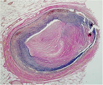
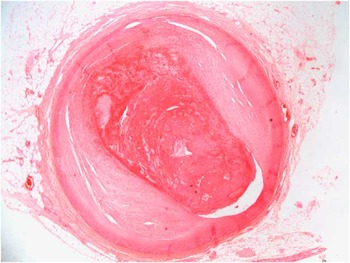
The cause of vessel occlusion was significant atheroma (⩾75%) alone in 27/46 cases (Fig 1). Thrombosis was reported in 16/46 cases (Fig 2). In two cases, a section of the involved coronary artery was not referred; therefore, the cause of occlusion could not be confirmed. With respect to the cases of thrombosis, 10/16 were associated with erosions (6/11 males and 4/5 females with thrombosis), but only 2/16 were associated with rupture (both in males). In four cases, the presence or absence of erosion or rupture associated with the thrombus was not documented.
Fibrous atherosclerotic plaque (trichrome stain).

Coronary artery occlusion due to thrombus.

On macroscopic examination, evidence of acute infarction was reported in 3/46 cases, the presence or absence of acute or chronic infarction was not reported on macroscopic examination in four cases. Microscopically, the three acute infarcts were confirmed. In addition, 10 cases showed regional contraction band necrosis alone, and eight cases showed acute infarction with neutrophilic infiltration.
Evidence of chronic infarction or fibrosis was described macroscopically in 7/46 cases, five of the cases showing replacement fibrosis were confirmed microscopically; however, on microscopic examination, 2/7 of these cases showed acute infarction only with no fibrosis. On microscopic examination, fibrosis was identified in a further six cases. Overall, 32/46 cases had evidence of acute or chronic damage.
In one case, there were features of arrhythmogenic right ventricular cardiomyopathy involving the right ventricle with epicardial replacement of the myocardium by fat and fibrous tissue, in addition to significant atheroma in the left anterior descending coronary artery. Finally, in one case, there was left ventricular hypertrophy (>20 mm thickness), in addition to 60% occlusion of the left anterior descending coronary artery, which, although non-significant, was thought to have acted synergistically with the left ventricular hypertrophy to result in fatal cardiac arrhythmia and sudden death.
Discussion
This study highlights several important aspects of premature atheroma in young people and its identification at autopsy. The male:female ratio was ~2.5:1, reflecting the predominance of coronary artery disease in men over women in the pre-menopausal age groups; 15 of the 24 cases with body mass index data available were obese (62.5%), which is high compared with that in the United Kingdom overall, where 26.1% of adults are categorised as obese.Reference Zaninotto, Head, Stamatakis, Wardle and Mindell 19 In addition, the mean body mass index of this cohort dying as a result of premature atheroma was significantly higher compared with, although slightly younger, a cohort of persons dying as a result of sudden arrhythmic death syndrome. Thus, the usual risk factors for ischaemic heart disease, such as obesity, smoking, hypercholesterolaemia, hypertension, and diabetes, also apply to younger patients. Obesity is increasing in young people and is set to increase to 33% in the next decade;Reference Zaninotto, Head, Stamatakis, Wardle and Mindell 19 thus, premature atheroma is expected to increase as well. Smoking is also linked to surface erosions, which feature in this study.Reference Barua and Ambrose 20
The circumstances of death vary in the literature. The most frequent circumstance of death was at rest, consistent with the findings of Corrado et al,Reference Corrado, Basso, Poletti, Angelini, Valente and Thiene 3 , Reference Corrado, Thiene and Pennelli 5 who reported 16/19 deaths occurred while the person was sedentary; however, other studies report that the majority of deaths occurred during exertion, with Virmani et alReference Virmani, Robinowitz and McAllister 7 reporting that 36/48 deaths occurred during or soon after exercise.
In concordance with the literature,Reference Corrado, Basso, Poletti, Angelini, Valente and Thiene 3 , Reference Corrado, Thiene and Pennelli 5 , Reference Virmani, Robinowitz and McAllister 7 single-vessel disease predominates (28/46 cases). Unlike coronary artery disease in older people, fibrous rich or mixed fibrous-lipid plaques were more common (80%), as has been previously reported,Reference Corrado, Basso, Poletti, Angelini, Valente and Thiene 3 , Reference Corrado, Thiene and Pennelli 5 , Reference Henriques, Van der Wal, Van der Loos and Becker 11 , Reference Tavora, Li, Ripple, Fowler and Burke 16 and it has been postulated that this reflects the stages of development of atheromatous plaques.Reference Corrado, Basso, Poletti, Angelini, Valente and Thiene 3 These plaques occur in younger patients and also in pre-menopausal women.
In this study, there was more non-thrombotic occlusion compared with thrombotic occlusion. The predominance of non-thrombotic occlusion has been previously reported.Reference Corrado, Thiene and Pennelli 5 Virmani et alReference Virmani, Robinowitz and McAllister 7 reported thrombi in 27/48 cases of coronary artery disease in patients <30 years, but of the 21 who died suddenly only eight (39%) had thrombosis. The low proportion of cases with thrombosis is consistent with the predominance of fibrous and mixed fibrous-lipid plaques, as rupture and thrombosis are associated with lipid-rich plaquesReference Corrado, Basso, Poletti, Angelini, Valente and Thiene 3 and thin cap atheromas.Reference Tavora, Cresswell, Li, Fowler and Burke 14 , Reference Falk, Nakano, Bentzon, Finn and Virmani 15 Coronary artery erosion is more common in younger people.Reference Tavora, Li, Ripple, Fowler and Burke 16 It has been proposed that the pathophysiology of fatal arrhythmia in these non-thrombotic cases may be due to coronary vasospasm.Reference Corrado, Thiene and Pennelli 5
As evidenced by the data, acute or chronic infarction is not always identified macroscopically by the pathologist, emphasising the importance of histological examination. In 10 cases, there was evidence of regional contraction band necrosis without further evidence of acute infarction. This should be interpreted with caution, particularly if cardiopulmonary resuscitation has been performed, as it may be a reflection of this process, rather than true evidence of myocardial ischaemia occurring before death. The distribution of the contraction band necrosis should be noted, as ischaemia restricted to the territory of the occluded coronary artery is more likely to relate to ischaemia than widespread contraction band necrosis in the context of cardiopulmonary resuscitation.
In the absence of evidence of infarction, the role of coronary artery disease in death must be considered, especially if there is no thrombosis occluding a vessel. In 14/46 cases in this series, there was no identifiable substrate – that is, evidence of contraction band necrosis or acute or chronic infarction – for arrhythmia. In this situation, it is important to raise the possibility of an electrical abnormality such as a channelopathy also being present. Coronary artery disease may not always act alone to cause death, and should be interpreted in the context of other findings, as a recent paper emphasises.Reference Papadakis, Raju and Behr 21 If there is significant hypertrophy with even a moderate degree of atherosclerosis, this may be sufficient to cause a fatal arrhythmia. In addition, once coronary artery disease is identified, the pathologist must be sure not to miss other potentially inherited pathologies, which could have contributed to or directly caused death, such as arrythmogenic right ventricular cardiomyopathy, for which family members should be screened.
As with all autopsy cases, clinicopathological correlation is fundamental. In this study, there was a lack of clinical information, particularly relating to risk factors and lifestyle. Smoking and drug histories should be obtained where possible, in order to further investigate factors contributing to the presence of premature atheroma. Use of cocaine and amphetamine – and its derivativesReference Lucena, Blanco and Jurado 22 – Reference Kaye, Darke, Duflou and McKetin 24 – are known to cause a wide spectrum of cardiovascular pathology, including accelerated atherosclerosis, and therefore their use, as well as toxicological investigation, should always be considered.
Careful examination of the coronary arteries is essential, even in young people with no preceding history. Autopsy examination not only provides an accurate cause of death but is also important for detecting inheritable disease and preventing further deaths. In a high proportion of cases,Reference Corrado, Thiene and Pennelli 5 sudden death is the first manifestation of coronary artery disease, and its identification should prompt investigation for dyslipidaemias in family members.
Conclusions
Precocious atheroma can cause sudden death in young people. Careful examination of the coronary arteries is essential in all cases even in young people with no preceding history. Histological examination of the myocardium is also essential to detect infarction. Autopsy examination not only provides an accurate cause of death but is also important for detecting inheritable disease and preventing further deaths, and the presence of premature atheroma should prompt investigation for dyslipidaemias in family members.
Acknowledgement
The authors thank the charity Cardiac Risk in the Young for funding for this project (cry@c-r-y.org.uk, www.c-r-y.org.uk).
Financial Support
This work was funded by the charity Cardiac Risk in the Young.
Conflicts of Interest
None.
Ethical Standards
The authors assert that all procedures contributing to this work comply with the ethical standards of the relevant national guidelines on human experimentation and with the Helsinki Declaration of 1975, as revised in 2008, and has been approved by the institutional committees. This study falls under Research Ethics Committee reference number 10/H0724/38 of the National Research Ethics Service.