Congenital adrenal hyperplasia (CAH) is a group of autosomal recessive disorders of adrenal steroidogenesis characterised by a complete or partial deficiency of one of the adrenal enzymes necessary for the synthesis of cortisol from cholesterol. The resulting decrease in cortisol or in cortisol and aldosterone production is associated with changes in the levels of sex steroid hormones, which interfere with primary and secondary sexual development from fetal to adult life. Cortisol synthesis is positively regulated by adrenocorticotropic hormone (ACTH) produced by the anterior pituitary, which in turn is controlled by corticotrophin-releasing hormone (CRH) secreted by the hypothalamus. Low levels of cortisol synthesis lead to an overproduction of ACTH; this continuously stimulates the adrenal gland (in order to produce more cortisol), giving rise to hyperplasia of the adrenal tissue (Refs Reference Itoi, Seasholtz and Watson1, Reference Waterman and Bischof2).
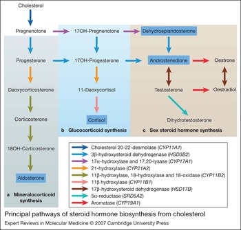
The three main categories of steroid hormones (mineralocorticoids, glucocorticoids and adrenal androgens) are synthesised in the adrenal cortex from cholesterol. The enzymes (and corresponding genes) necessary for the successive steps of steroid hormone biosynthesis are depicted in Figure 1. The different clinical phenotypes of CAH are best understood by a comprehensive analysis of the enzymatic deficiency and the effects resulting from: (1) low levels of hormones with decreased production; (2) accumulation of intermediary hormones upstream of the enzymatic defect; and (3) the physiological actions of the three major steroid hormones.
Principal pathways of steroid hormone biosynthesis from cholesterol. Major products of adrenal steroidogenesis are indicated with a blue box. Enzymatic activities catalysing each bioconversion (coloured arrows) are shown in the key below, where genes encoding each enzyme are given in italics (CYP gene symbols show that the corresponding enzyme is a member of the cytochrome P450 superfamily). The steroidogenic acute regulatory protein (STAR) promotes cholesterol transport to the inner mitochondrial membrane (not shown) (Ref. Reference Miller140). The first step of steroidogenesis is the conversion of cholesterol to pregnenolone; this requires three reactions (20α-hydroxylation, 22-hydroxylation, and cleavage of cholesterol side chain), which are all catalysed by cholesterol 20-22-desmolase (Ref. Reference Miller88). (a) Mineralocorticoid synthesis. The biosynthesis of mineralocorticoids (in the zona glomerulosa), requires the action of 3β-hydroxysteroid dehydrogenase, which converts pregnenolone to progesterone. This is subsequently converted to deoxycorticosterone by 21-hydroxylase. The last enzymatic steps that culminate in aldosterone biosynthesis are performed by a single enzyme – aldosterone synthase (encoded by CYP11B2 gene) – which has three enzymatic activities: 11β-hydroxylation of deoxycorticosterone, 18-hydroxylation and 18-oxidation of corticosterone. (b) Glucocorticoid synthesis. To produce the glucocorticoid cortisol (in the zona fasciculata), the CYP17A1-encoded enzyme catalyses the bioconversion of pregnenolone to 17-hydroxypregnenolone. 3β-hydroxysteroid dehydrogenase uses 17-hydroxypregnenolone as a substrate, converting it to 17-hydroxyprogesterone, which is also produced by the 17α-hydroxylation of progesterone. 21-hydroxylase mediates the bioconversion of 17-hydroxyprogesterone to 11-deoxycortisol, which is converted into cortisol by the action of 11β-hydroxylase. (c) Sex steroid hormone synthesis. The production of adrenal androgens results from the conversion of 17-hydroxypregnenolone to dehydroepiandrosterone by the CYP17A1 enzyme as a result of its 17,20-lyase activity, and dehydroepiandrosterone is subsequently converted by 3β-hydroxysteroid dehydrogenase to androstenedione. The latter is also converted by the CYP17A1 enzyme from 17-hydroxyprogesterone (Ref. Reference Donohoue, Parker, Migeon and Scriver7). There are several isozymes with 17β-hydroxysteroid dehydrogenase activity. While type 5 isozyme HSD17B5 (encoded by AKR1C3) converts androstenedione into testosterone in the adrenal gland during early fetal life (Ref. Reference Goto5), other isozymes, like HSD17B3 and HSD17B1, are responsible for testosterone biosynthesis in the testes and ovaries. During puberty, aromatase converts androstenedione and testosterone to oestrone and oestradiol, respectively. Testosterone is metabolised to dihydrotestosterone by 5α-reductase, predominantly in genital skin and in other androgen target tissues.

Aldosterone, the major mineralocorticoid synthesised by the zona glomerulosa of the adrenal cortex, regulates the excretion of electrolytes in the kidney and maintains intravascular volume and blood pressure: it enhances the reabsorption of sodium, accompanied by water and chloride retention, and excretion of potassium ions into the urine (Ref. Reference Williams and Williams3). Aldosterone biosynthesis is regulated mainly by potassium levels and by the renin–angiotensin system. The renin hormone – produced by the kidney in response to hypotension, lowered intravascular volume, or hyperkalaemia – cleaves angiotensinogen (produced by the liver) to angiotensin I. This peptide, under the action of angiotensin-converting enzyme (mainly present in the pulmonary endothelium), is converted to angiotensin II. Angiotensin II stimulates the zona glomerolosa to increase the biosynthesis of aldosterone (Ref. Reference Migeon, Donohoue, Kappy, Blizzard and Migeon4).
Cortisol, the principal glucocorticoid, is produced by the adrenal zona fasciculata. Cortisol has multiple and important functions in most of the metabolic pathways: it plays a key role in blood sugar maintenance, promoting gluconeogenesis during periods of physical or psychological stress, regulates blood pressure and homeostasis, and supports cardiovascular and immunological functions. In addition, it was recently demonstrated for the first time that during early fetal life, between 8 and 10 weeks post-conception, cortisol synthesised by human adrenal cortex is able to down-regulate androgen biosynthesis by suppression of the fetal hypothalamic–pituitary–adrenal axis. This negative feedback mechanism, occurring during a critical period of primary sexual differentiation, provides transient physiological conditions that promote the normal differentiation of female external genitalia (Ref. Reference Goto5).
Dehydroepiandrosterone and androstenedione are the adrenal androgens produced mainly by the adrenal zona reticularis. In normal physiological conditions, these two sex steroids (which are weak androgens) have no significant function in primary sexual differentiation during early male and female fetal life. However, during this period of life, some of the enzymatic defects that impair cortisol biosynthesis lead to accumulation of sex hormone precursors, which are converted to high doses of dehydroepiandrosterone and androstenedione and subsequently metabolised to testosterone and dihydrotestosterone, mainly in peripheral tissues. These two more potent androgens induce virilisation of the external genitalia in females, causing, at birth, various degrees of sexual ambiguity, as discussed below (Refs Reference White6, Reference Donohoue, Parker, Migeon and Scriver7, Reference Moore and Persaud8).
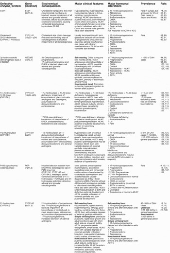
The enzymatic deficiencies that most frequently cause CAH are defects in 21-hydroxylase, 11β-hydroxylase, 3β-hydroxysteroid dehydrogenase, and less frequently 17α-hydroxylase/17,20-lyase, cholesterol 20-22-desmolase and the transporter protein StAR (steroidogenic acute regulatory protein). In addition, a new form of CAH was recently described, characterised by deficiencies in both 21-hydroxylase and 17α-hydroxylase/17,20-lyase activities as a result of mutations in the electron donor enzyme P450 oxidoreductase (POR) (Refs Reference Fluck9, Reference Fluck and Miller10). Most patients with POR deficiency have skeletal malformations, genital anomalies in both sexes and abnormal steroid profiles (Refs Reference Fluck and Miller10, Reference Fukami11, Reference Huang12). The clinical and molecular features of all these enzymatic defects are summarised in Table 1. For deficiency in 21-hydroxylase, the main subject of this paper, a more detailed review is presented in the next sections.
Summary of the various enzymatic defects that cause congenital adrenal hyperplasia

Abbreviations: ACTH, adrenocorticotropic hormone; CAH, congenital adrenal hyperplasia; DHEA, dehydroepiandrosterone; FSH, follicle-stimulating hormone; hCG, human chorionic gonadotropin; LH, luteinising hormone.
Clinical features of 21-hydroxylase deficiency
21-hydroxylase deficiency (21-OHD) is the most common cause of CAH, accounting for 90–95% of all cases. Patients with 21-OHD have a deficiency in cortisol biosynthesis, which in most of the severe cases is also accompanied by aldosterone deficiency. The decreased adrenal secretion of cortisol gives rise, because of the absence of negative feedback to the hypothalamus and pituitary, to an increased secretion of CRH and ACTH. The steroid precursors prior to this enzymatic deficiency (progesterone and 17-hydroxyprogesterone) are accumulated and shunted through the adrenal androgen biosynthetic pathway. The increased secretion of adrenal androgens from the eighth week of gestation, and the resulting production of high levels of testosterone and dihydrotestosterone, particularly affects sexual differentiation in females, and causes advanced somatic development in both sexes during childhood (Ref. Reference White and Speiser13). Clinically, although there is a continuous spectrum of phenotypic manifestations associated with this disease, it has been useful to divide 21-OHD into three forms: salt-wasting classical 21-OHD, simple virilising classical 21-OHD, and nonclassical 21-OHD.
Classical 21-OHD
Classical 21-OHD occurs in two major forms: salt-wasting and simple virilising. Patients with renal salt-wasting account for three-quarters of classical 21-OHD cases. In addition to having a severe cortisol deficiency, they do not synthesise enough aldosterone and therefore are not able to maintain sodium homeostasis. Severely affected patients usually present between the first and fourth weeks of age with hyponatraemia, hyperkalaemia, hyperreninaemia, hypovolaemic shock and hypoglycaemia. Other symptoms are also common, such as lethargy, poor feeding, vomiting and failure to thrive. In the female fetus, androgen excess causes variable degrees of external genitalia virilisation, and consequently newborn females have genital ambiguity or male-resembling external genitalia with bilateral cryptorchidism, which may result in errors of sex assignment at birth. In the male fetus, the testicular androgens normally produced at high levels induce the normal masculinisation of the external genitalia, and the excess of adrenal androgens usually has no effect until childhood, although sometimes a macrogenitossomia at birth is observed. By contrast to the situation in affected newborn females, where the presence of genital ambiguity contributes to the diagnosis of severe 21-OHD, the normal external genitalia in affected newborn males do not alert paediatricians to the diagnosis of a severe form of CAH before the onset of a salt-wasting crisis. This critical health condition can lead to death if appropriate medical assistance is not available (Refs Reference White and Speiser13, Reference Kovacs14).
The simple virilising form of 21-OHD occurs in around a quarter of the cases of classical 21-OHD. In this form of CAH, as 21-hydroxylase activity is residual but not complete, and because the adrenal gland is stimulated by the renin–angiotensin system, adequate levels of aldosterone are still produced, allowing normal sodium homeostasis. Adrenal glands under continuous stimulation by increased levels of ACTH may synthesise approximately normal levels of cortisol, although cortisol precursors are still accumulated and consequently directed into the biosynthetic pathway of adrenocortical androgens. As mentioned above, the peripheral conversion of high levels of dehydroepiandrosterone and androstenedione into testosterone and dihydrotestosterone results in external genitalia masculinisation in the 46,XX fetus, and females are consequently born with variable degrees of external genitalia virilisation, ranging from mild hypertrophy of the clitoris and partial fusion of the labioscrotal folds to a phallic urethra and a complete fusion of the labioscrotal folds. It is important to emphasise that for children with external genitalia ambiguity or with normal male genitalia without palpable testes, the possibility of 21-OHD should be considered (among the different causes of genital ambiguity, 21-OHD is the most frequent). In the 46,XY fetus, the adrenal excess of androgen precursors has no effect on the external genitalia masculinisation. Later in life, untreated children of both sexes with simple virilising 21-OHD develop signs of androgen excess that usually include premature appearance of pubic hair, followed by development of axillary hair, acne, facial hair, rapid linear growth and advanced bone age, although, due to premature epiphyseal fusion, patients usually have a short adult stature. After childhood, untreated females do not undergo normal puberty and in most cases present clitoral enlargement, hirsutism, male-pattern baldness, cystic ovaries, and amenorrhoea or irregular menses, and consequently primary infertility or reduced fertility. Prepubertal males usually have progressive penile enlargement, small testes and later in life invariably show seriously impaired fertility (Ref. Reference White and Speiser13).
The phenotypic variability in degree of external genitalia ambiguity in females with classical 21-OHD might reflect differences in the efficiency of conversion of the adrenal androgens into the two more potent androgens, as well as the androgen receptor gene expression level, the affinity of the androgen receptor for its ligands (testosterone and dihydrotestosterone), and the efficiency of androgen receptor transactivation on target androgen-regulated genes (Ref. Reference Brinkmann15).
Nonclassical 21-OHD
The nonclassical form of 21-OHD is associated with less severe symptoms of hyperandrogenisation. This is due to a mild deficiency of 21-hydroxylase and to slightly elevated levels of 21-hydroxylase adrenal steroid precursors. Aldosterone biosynthesis is not disturbed in patients with nonclassical 21-OHD. The clinical manifestations of mild androgen excess are variable. Females are usually born with normal external genitalia (sometimes a mild clitoromegaly is observed), and both males and females may show signs of androgen excess at any time after postnatal development. Children may present with early pubic hair, precocious puberty, and accelerated growth associated with advanced bone age. Females may show one or more of the following symptoms: hirsutism, temporal baldness, delayed menarche, polycystic ovaries and menstrual irregularities. Adult males and females may have cystic acne, impaired fertility, and short stature due to premature epiphyseal fusion. In addition, asymptomatic patients may be identified and diagnosed during the familial screening of one affected individual (Ref. Reference New16).
Prevalence of CAH arising from 21-OHD
Neonatal screening for classical CAH arising from 21-OHD, established mainly in Caucasian populations, found an overall prevalence of approximately 1/15 000 live births, ranging from 1/10 000 to 1/16 000 in the USA, and from 1/10 000 to 1/25 000 in European countries (Refs Reference Pang17, Reference Therrell18, Reference van der Kamp and Wit19). The highest prevalence occurs in two geographically isolated populations: 1/280 among Yupik Eskimos (Alaska) and 1/2100 in the island of La Reunion (Indian Ocean) (Ref. Reference Pang17).
The milder, nonclassical form of 21-OHD is much more common, with a prevalence of 1/100 in the general population, although a higher ethnic-specific incidence is found among Ashkenazi Jews (1/27), Hispanics (1/40) and Yugoslavs (1/60) (Ref. Reference Speiser20).
Hormonal diagnosis of 21-OHD
The biochemical diagnosis of 21-OHD cannot be made through direct measurements of the enzymatic activity because 21-hydroxylase is essentially expressed only in the adrenal cortex. Instead, diagnosis of 21-OHD is based on elevated baseline and ACTH-stimulated levels of serum 17-hydroxyprogesterone, the main substrate of 21-hydroxylase, measured by immunoassay. In addition to elevated basal levels of 17-hydroxyprogesterone (usually higher than 10 000 ng/dl), patients with classical 21-OHD also secrete progesterone, dehydroepiandrosterone, androstenedione and testosterone in excess. Patients with the salt-wasting form show the highest 17-hydroxyprogesterone levels of >20 000 ng/dl, while patients with the simple virilising form usually have 17-hydroxyprogesterone concentrations of 10 000–20 000 ng/dl (Ref. Reference White and Speiser13); however, some patients with these two clinical forms present 17-hydroxyprogesterone levels that overlap, and a cut-off cannot be established. The nonclassical 21-OHD patients have lower 17-hydroxyprogesterone after ACTH stimulation (1000–10 000 ng/dl). Basal serum 17-hydroxyprogesterone levels in most of the latter patients are not distinguishable from heterozygotes for classical or nonclassical 21-OHD. The large data obtained from neonatal screening of CAH demonstrated that preterm and stressed newborns have higher 17-hydroxyprogesterone levels in serum than babies born at term, generating false-positive results. In order to avoid these false-positive results, cut-off values were established based on gestational age (in Japan and Europe) or on birth weight (in the USA). The neonatal screening is also not efficient in the identification of newborns with the nonclassical form of the disease. It is important to recognise that other steroidogenic deficiencies, such as 11β-hydroxylase deficiency and 3β-hydroxysteroid dehydrogenase deficiency type 2, may be misdiagnosed as 21-OHD. In these cases, it is important to perform a complete adrenocortical hormone profile and evaluate ratios of precursors versus products after ACTH stimulation (Refs Reference White and Speiser13, Reference van der Kamp and Wit19).
Molecular features of 21-OHD
The CYP21A2 locus
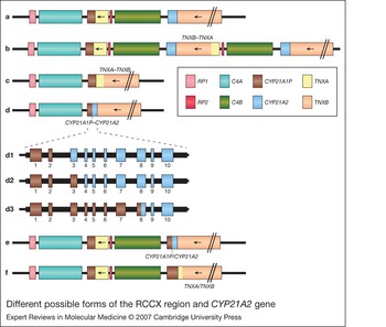
Human adrenal 21-hydroxylase is encoded by the CYP21A2 gene, also previously called CYP21 and CYP21B, which is located on 6p21.3 within class III HLA, ∼30 kb from a pseudogene (CYP21A1P; known also as CYP21P and CYP21A). These two genes share about 98% homology in their ten exons and about 96% in the introns, but CYP21A1P is inactive because of the presence of several deleterious mutations (Refs Reference White21, Reference Higashi22). The gene and the pseudogene are included in what is called the RCCX region (Fig. 2), which, in the most common alleles, has a bimodular form (RCCX–RCCX), composed of two sets of four genes arranged in tandem: RP1–C4A–CYP21A1P–TN XA–RP2–C4B–CYP21A2–TN XB (Ref. Reference Lee, Lee and Lin23). The C4A and C4B genes code for the fourth component of serum complement, RP1 encodes a putative nuclear protein similar to DNA helicase, and RP2 is a truncated, nonfunctional copy of RP1. TNXB encodes a putative extracellular matrix protein (tenascin X) and overlaps the CYP21A2 gene on the opposite DNA strand. Likewise, TNXA is a truncated copy of TNXB and overlaps CYP21A1P, also on the opposite DNA strand. Monomodular and trimodular alleles resulting from genetic recombination events have also been described (Ref. Reference Blanchong24).
The localisation and structure of the RCCX region.RP1, C4A, C4B, CYP21A2 and TNXB are functional genes; CYP21A1P, TNXA and RP2 are nonfunctional pseudogenes. TNXA and TNXB are encoded on the opposite DNA strand. The sizes (bp) of the CYP21A2 exons (blue boxes) are indicated above them; the sizes of the introns are indicated between the exons; the numbering of the exons is indicated beneath the boxes. The black solid line represents the untranslated region of the gene and the slashes indicate the gene or DNA sequence is longer than shown. Abbreviations: C2, complement component 2; C4A and C4AB, complement component 4A and 4B; CYP21A1P, cytochrome P450, family 21, subfamily A, polypeptide 1 pseudogene; CYP21A2, cytochrome P450, family 21, subfamily A, polypeptide 2; HLA, human leukocyte antigen; RD, also known as RDBP (RNA-binding protein); RP1 and RP2, also known as STK19 and STK19P, respectively (‘serine/threonine kinase 19 gene and pseudogene’); TNF, tumour necrosis factor; TNXA, tenascin XA pseudogene; TNXB, tenascin XB. The top half of the figure, showing the position of the RCCX region relative to neighbouring genes on chromosome 6, is adapted from Ref. 7: Donohoue, P.A., Parker, K. and Migeon, C.J. (1995) Congenital adrenal hyperplasia. In The Metabolic and Molecular Bases of Inherited Disease (7th edn) (Scriver, C.R. et al., eds), pp. 2929-2966, McGraw Hill, New York.

Mutations causing 21-OHD
In October 2006, the Human Gene Mutation Database (http://www.hgmd.cf.ac.uk/ac/index.php) had a total of 103 entries for CYP21A2 mutations: 87 point mutations or small deletions or insertions, seven splicing mutations, and nine gross deletions, duplications or gene conversions.
As a result of its structure of repeated genes arranged in tandem, the RCCX region is prone to a high frequency of recombination events, which can lead to (1) unequal crossing-overs during meiosis, resulting in a wide variety of arrangements depending on the breakpoints, such as gene duplications and gross gene deletions encompassing the C4 and CYP21A2 genes (Refs Reference White, New and Dupont25, Reference Werkmeister26), or to (2) large or short gene conversions in which the CYP21A1P mutations, which drastically impair its transcription and prevent the production of a functional enzyme, are transferred to CYP21A2 (Fig. 3). Gross deletions encompassing variable portions of C4A, CYP21A1P, TNXA, RP2, C4B, CYP21A2 and TNXB produce different types of chimaeras in the RCCX modules. The resulting alleles may have different extents of the CYP21A1P sequence in the 5′ portion of the 21-hydroxylase gene attached to the 3′ portion of CYP21A2 (CYP21A1P–CYP21A2 chimaeras), or a 5′ portion of the TNXA gene attached to the 3′ portion of TNXB (TNXA–TNXB chimaeras) (Ref. Reference Lee27). In both cases, the resulting allele does not have a functional CYP21A2 gene. The CYP21A1P–CYP21A2 chimaeras do not lead to the production of active 21-hydroxylase not only because of the deleterious mutations of the CYP21A1P in the 5′ portion, but also because the promoter sequence of the pseudogene has only 20% of the transcriptional activity of the CYP21A2 promoter (Ref. Reference Chang and Chung28). The TNXA–TNXB chimaeras are formed as a result of a gross deletion that includes the CYP21A2 gene (Ref. Reference Lee27).
Different possible forms of the RCCX region andCYP21A2gene. (a) Standard bimodular form of the RCCX region; (b) trimodular form of the RCCX region, with a duplication of RP2, C4B, CYP21A2 and TNXB, resulting in a chimaeric TNXB–TNXA gene; (c) monomodular form of the RCCX region arising from a deletion in the region TNXA–RP2–C4B–CYP21A2–TNXB, resulting in a chimaeric TNXA–TNXB gene; (d) monomodular form of the RCCX region arising from a deletion in the region CYP21A1P–TNXA–RP2–C4B–CYP21A2, resulting in a chimaeric CYP21A1P–CYP21A2 gene; (d1–3) three of the distinct CYP21A1P–CYP21A2 chimaeras described by Lee (Ref. Reference Lee27); (e) partial gene conversion of CYP21A2 to CYP21A1P; (f) complete CYP21A2 gene conversion to CYP21A1P and partial conversion of TNXB to TNXA. Abbreviations: see Figure 2 legend.

Some authors have reported alleles with duplications of the CYP21A2 gene (Refs Reference Friaes29, Reference Koppens, Hoogenboezem and Degenhart30, Reference Haglund-Stengler31, Reference Wedell, Stengler and Luthman32). This may complicate mutation analysis: one of the duplicated copies might have mutations that are detected by the traditional methods but the other one might be a functional copy, rendering this allele perfectly active. Different extents of gene conversions comprising several CYP21A2 exons have also been reported – namely gene conversions from exon 1 to 7, from exon 1 to 3, from exon 3 to 7, from exon 3 to 8, from exon 4 to 7, and from exon 5 to 8 (Refs Reference Friaes29, Reference Levo and Partanen33, Reference Dolzan34, Reference Kharrat35).
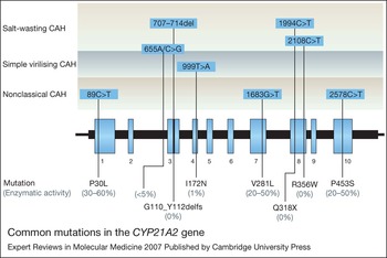
Apart from gene deletions and large gene conversions, there are eight mutations reported with a higher frequency in the CYP21A2 gene (Fig. 4): g.89C > T (p.P30L), g.655A/C > G (I2 splicing), g.707_714delGAGACTAC (p.G110_Y112delfs), g.999T > A (p.I172 N), g.1683G > T (p.V281L), g.1994C > T (p.Q318X), g.2108C > T (p.R356W) and g.2578C > T (p.P453S) (Ref. Reference White and Speiser13). Except for the last one, all the other seven mutations are present in CYP21A1P and are presumed to have been transferred to CYP21A2 by short gene conversions. The mutation g.2578C > T (p.P453S) has been suggested to be occasionally present in the pseudogene as a polymorphism, and transferred to CYP21A2 by gene conversion events just like the other most frequent point mutations (Ref. Reference Owerbach36). Three of these mutations are associated with the nonclassical form of 21-OHD, one is typically related to simple virilising classical 21-OHD, and three to salt-wasting classical 21-OHD. The mutation g.655A/C > G has been reported both in salt-wasting and in simple virilising cases. This mutation activates cryptic splicing receptor sequences causing the incorrect processing of almost all the mRNA. A small amount of normally spliced mRNA can be detected in cell cultures; therefore, in the absence of other mutations, small quantities of functional enzyme can be produced, thus preventing the salt-wasting crisis (Refs Reference Higashi37, Reference Lee and Chang38).
Common mutations in theCYP21A2gene. The schematic indicates the localisation of the eight most frequent CYP21A2 mutations in most populations, and the corresponding forms of congenital adrenal hyperplasia (CAH). The changes in amino acid sequence are given below the exons (except for 655A/C > G, which affects splicing), and enzymatic activity (based on in vitro studies; Ref. Reference White and Speiser13) resulting from each mutation compared with the normal 21-hydroxylase activity is indicated in parentheses. Figure adapted, with permission from Perrin White (University of Texas Southwestern Medical Center, Dallas, TX, USA) and The Endocrine Society, from Ref. Reference White and Speiser13: White, P.C. and Speiser, P.W. (2000) Congenital adrenal hyperplasia due to 21-hydroxylase deficiency. Endocr Rev 21(3), 245-291 (Copyright 2000, the Endocrine Society).

Molecular diagnosis of 21-OHD
As discussed above, 21-OHD is diagnosed biochemically by measuring levels of 17-hydroxyprogesterone after stimulation with ACTH, but this method does not differentiate clearly between heterozygous carriers and normal individuals (Ref. Reference Honour and Rumsby39). Therefore, the molecular diagnosis of 21-hydroxylase by detection of mutations in the CYP21A2 gene is of major importance to complement the biochemical diagnosis.
The direct screening of CYP21A2 mutations requires two steps. The first one is the isolation of the CYP21A2 gene from the pseudogene, since the latter is inactive and has most of the mutations of interest. Currently, this step is usually performed by PCR with primers specific for CYP21A2, which do not allow the concomitant amplification of CYP21A1P (Refs Reference Lee40, Reference Owerbach, Crawford and Draznin41). The product of this first step is used as template DNA for detection of mutations in the second step. The screening of the most common mutations is regularly performed by PCR-based and/or sequencing techniques. For example, allele-specific PCR using pairs of primers specific for the detection of each of the eight most frequent mutations has been widely used (Ref. Reference Wedell and Luthman42), but, more recently, multiplex mini-sequencing has allowed the concomitant screening of all the frequent mutations by single-base extension using a specific primer for each mutation and fluorophore-labelled ddNTPs, followed by separation of the fragments by capillary electrophoresis using an automated sequencing apparatus (Refs Reference Friaes29, Reference Krone43, Reference Keen-Kim44). When the mutations detected by these methods do not explain the phenotype exhibited by the patient, novel mutations can be searched for by screening the CYP21A2 exons by SSCP (single-strand conformation polymorphism) analysis, by DHPLC (denaturing high-performance liquid chromatography) analysis, or by direct sequencing of the several exons and noncoding regions of CYP21A2 (Refs Reference Orita45, Reference Abrams, Murdaugh and Lerman46, Reference Bobba47).
All the above-mentioned techniques fail, however, to detect gross gene deletions and duplications, as well as large gene conversions, which have a significant frequency in most populations (Ref. Reference White and Speiser13). These abnormalities are best characterised if family members are included, and by using laborious Southern-blotting methodology with the appropriate probes for CYP21 and for C4 genes. Traditionally, cDNA probes are used, but Southern blotting using genomic DNA CYP21A2 probes has also been described (Ref. Reference Morel and Miller48). The restriction enzymes most commonly used are TaqI and BglII, although different endonucleases have been utilised to characterise complex rearrangements and extra copies of C4 and CYP21A2 (Refs Reference Morel and Miller48, Reference Lee49). In addition, real-time quantitative PCR has been recently used to rapidly detect CYP21A2 deletions/conversions (Ref. Reference Olney50). However, this method is critically dependent on an accurate DNA quantification and showed some discrepancies when compared with Southern-blotting results, particularly when discriminating between two and three gene copies. Moreover, it does not allow the distinction between gene deletions and gene conversions or different combinations of alleles with distinct abnormalities. A semiquantitative strategy based on a two-step PCR and TaqI digestion has also been proposed (Ref. Reference Tukel51), and showed a very good concordance with Southern blotting, but the interpretation of the results might occasionally be difficult and misleading, since it relies on visual quantification (on agarose gels) of PCR/digestion products, which are dependent on PCR yields. For a rapid and easier detection of chimaeric genes and CYP21A2 deletions, Lee and co-workers have recently developed a simpler method based on extended PCR followed by TaqI restriction (Ref. Reference Lee, Lee and Lin23); however, this does not differentiate total gene conversions from CYP21A2 complete deletions, nor does it detect gene duplications. Thus, although these methods are very useful for diagnostic purposes because they provide a rapid, easy and economic means of detecting deleterious gene abnormalities resulting from recombination events, the results are often ambiguous and in those cases Southern-blotting analysis is still needed for confirmation and further characterisation of the RCCX modules.
Whenever a new genetic alteration is identified in either the coding sequence, intronic sequences or promoter region of the CYP21A2 gene, it is important to evaluate and predict its consequences for 21-hydroxylase activity at different levels: (1) the nature of the amino acid alteration, associated structural alterations and enzymatic or functional consequences; (2) possible alterations of the normal splicing sites (elimination or creation of new splicing sites); and (3) impairment of gene expression. It is of major importance to perform structural and functional studies in vitro, for a better prediction of the CAH phenotype associated with the mutation (Refs Reference Krone52, Reference Grischuk53, Reference Barbaro54).
Frequency of common CYP21A2 mutations
Rearrangements of the RCCX module resulting in CYP21A2 deletions or large gene conversions are one of the most common causes of 21-OHD worldwide, accounting for over 20% of defective CYP21A2 alleles in most populations (Refs Reference White and Speiser13, Reference Kotaska, Lisa and Prusa55). There are, however, some countries with frequencies under 10% for these types of mutations – namely Egypt, Mexico and Portugal (Refs Reference Friaes29, Reference Kotaska, Lisa and Prusa55, Reference Ordonez-Sanchez56). The mutation g.655A/C > G is by far the most common severe point mutation in the great majority of the published studies, with frequencies usually above 25%. However, there are some exceptions: in Austria, Finland, China and Iran (Refs Reference Levo and Partanen33, Reference Ko57, Reference Vakili58, Reference Dolzan59) g.999T > A (p.I172 N) was more common; in Portugal both these mutations were detected with the same frequency (Ref. Reference Friaes29); and in Tunisia, the most frequent mutation was g.1994C > T (p.Q318X) (Ref. Reference Kharrat35). Among nonclassical 21-OHD alleles, the most common mutation is usually either g.1683G > T (p.V281L) or g.89C > T (p.P30L) (Refs Reference Friaes29, Reference Kotaska, Lisa and Prusa55, Reference Dolzan59, Reference Asanuma60, Reference Mathur61). As Kotaska et al. (Ref. Reference Kotaska, Lisa and Prusa55) concluded, the overall frequency of mutation g.1683G > T (p.V281L) is higher in South Europe and North Africa, where the nonclassical form of the disease has a higher prevalence. However, the highest frequency of this mutation is registered among the Ashkenazi Jews (Jews of Eastern European origin), with a prevalence of more than 10% in the general population (Ref. Reference White and Speiser13).
Genotype–phenotype correlations
CYP21A2 mutations can be predicted to cause a certain phenotype – salt-wasting, simple virilising or nonclassical – on the basis of the reduction of the enzymatic activity they produce. However, most CAH patients are actually compound heterozygous for two or more mutations. Since 21-OHD is an autosomal recessive disease, the phenotype of the patient should reflect the mutation that is predicted to cause a less severe impairment of the enzymatic activity. This approach to predicting phenotype has been shown to be correct in 80% to ∼100% of cases for null mutations and patients with salt-wasting 21-OHD, and for the p.V281L mutation and patients with nonclassical 21-OHD (Refs Reference White and Speiser13, Reference Friaes29, Reference Stikkelbroeck62). Slight deviations to this correlation occur in the forms of the disease with intermediate severity and with mutations such as p.I172N, g.655A/C > G or p.P30L, which in vivo may produce variable 21-hydroxylase activity. Sometimes it is difficult to differentiate between the simple virilising and nonclassical forms in male patients, because their symptoms of androgen excess are usually not detected at birth, so they may be incorrectly assigned as nonclassical 21-OHD patients. Otherwise, some of the causes that contribute to discrepancies between the predicted and the patient's phenotype may be the association of mutations of different severities in the same or in distinct alleles, the existence of mutations that impair CYP21A2 gene expression, and the presence of alterations in other genes, which may disturb 21-hydroxylase function or expression. It has also been proposed that some alterations usually considered to be polymorphisms, such as p.K102R and p.S268T, might have a synergistic effect when present in the same allele, resulting in a decreased 21-hydroxylase activity (Ref. Reference Dolzan59). However, deviations to the predicted genotype–phenotype might also be caused by an incorrect genotyping. Studies based on the screening of known mutations fail to detect novel mutations, which might also affect significantly the enzymatic activity or expression. Moreover, the occurrence of allele drop-out, in which a certain allele is preferentially amplified by PCR, has been reported (Refs Reference Day63, Reference Day64, Reference Van de Velde65). Patients might then be incorrectly genotyped as homozygotes for a certain mutation, when they are in fact heterozygotes or hemizygotes.
Genetic counselling and prenatal diagnosis
Some of the above-mentioned genotyping limitations can be minimised by the study of the CYP21A2 gene of both parents of the patient, thus confirming if the detected mutations are in opposite alleles of the proband. Furthermore, this additional study provides data useful for genetic counselling of the patient and family, as well as for prenatal diagnosis in future pregnancies.
Pregnancies at risk for a child affected with classical CAH are subjected to prenatal therapy with dexamethasone, which must begin in the early first trimester in order to efficiently prevent female genital ambiguity. However, there is consensus that glucocorticoid therapy should be used with caution because of the associated risk of potential adverse effects both to the mother (such as weight gain, hyperglycaemia, cutaneous striae, irritability, gastrointestinal intolerance, and increased blood pressure) and the fetus (congenital malformations, intrauterine growth retardation, mood fluctuations and shyness) – although none of these risks has been proved to be directly associated with the treatment, with some studies reporting the same frequency of fetal malformations in pregnancies subjected to dexamethasone treatment and in the general population (Refs Reference White and Speiser13, Reference Lajic66). It is therefore desirable to perform prenatal diagnosis as early as possible, since only affected females will suffer from genitalia virilisation. If the fetus is male or does not carry CYP21A2 mutations in both alleles, dexamethasone therapy can be immediately suspended, saving both mother and fetus from unnecessary glucocorticoid exposure (Refs Reference White and Speiser13, Reference Lajic66).
In pregnancies at risk for a child affected with classical CAH, noninvasive early fetal sex determination from maternal plasma is now possible (Refs Reference Avent and Chitty67, Reference Bartha, Finning and Soothill68, Reference Hyett69), although conventional methodologies are still currently performed. Cells obtained by chorionic villus sampling (at 10–12 weeks of gestation) or by amniocentesis (at 14–16 weeks) can be cultured and used for sex determination by PCR amplification of the SRY (sex-determining region of the Y chromosome; no cell culture required in this case), fluorescence in situ hybridisation (FISH) or conventional karyotyping, as well as for searching for the CYP21A2 mutations previously identified in the parents. As a quality-control measure in prenatal diagnosis, fetal DNA should be retested using informative tandem repeats (VNTRs or STRs) to ensure lack of maternal contamination, while simultaneously allowing paternity confirmation. Preimplantation genetic diagnosis (Refs Reference Sermon70, Reference Renwick and Ogilvie71) has also been used for the diagnosis of 21-OHD (Ref. Reference Van de Velde65); however, despite the great advantage of the very early diagnosis, this methodology is currently carried out only in specialised centres (Ref. Reference Thornhill72).
It is advisable to confirm all prenatal diagnoses soon after birth because of the possibility of errors, including false-negative diagnosis. It is important to bear in mind that de novo mutations occur in the CYP21A2 gene with a considerable frequency of 1–1.5%, which can complicate molecular prenatal diagnosis of 21-OHD: the fetus might have a mutation that does not exist in the parents and that might not be screened on a first approach (Ref. Reference Mao73).
Research in progress and outstanding research questions
Intense research over the past three decades in human molecular biology associated with human endocrinology has produced a plethora of discoveries that encompass the identification of genes coding for enzymes, transcription factors, hormones and receptors, physiological functions of endocrine hormones and receptors, and the characterisation of the molecular pathology of numerous endocrinological diseases, including nearly all forms of CAH. In addition, with this knowledge it has been possible to offer prenatal diagnosis and genetic counselling to couples at risk of having an affected baby with the most severe forms of CAH. However, the prediction of phenotype based on genotype may fail in some situations, namely in the ones associated with mutations of intermediate severity, as occurs with some mutations identified in the deficiencies described above, especially those associated with variable expressivity. This absence of genotype–phenotype correlation is in part due to the incomplete understanding of gene expression regulation, which is dependent on multiple transcription factors (some of which are still unidentified) that interact with each other and with regulatory sequences. In addition, there are also rare forms of CAH and many cases of hyperandrogenic females for whom the molecular defect is unknown. For these issues, it is expected that in the near future new technologies like proteomics, tandem mass spectrometry, microchip arrays and ultra-rapid DNA genotyping and sequencing will contribute to the identification of new disease-associated genes and new biomarkers. After elucidating the physiological functions of these with the use of functional studies and animal models, it is expected that the data will help exclude the false-positive results frequently obtained with the conventional immunoassays used in neonatal screening programmes of CAH, quickly identify the molecular defects of all the CAH forms, and contribute to better genetic counselling and patient treatment.
In the future, with the new technologies of genome and proteome analysis, it will be possible to simultaneously evaluate multiple genes or gene products directly associated with CAH and involved in the same or in multiple physiological pathways. The integration of these data obtained from a large number of patients with pharmacogenomic studies and eventually with gene therapy will have a great impact in individual and global health care (Refs Reference Hsueh, Bouchard and Ben-Shlomo74, Reference Merke75).
At present, there is considerable expectation that gene therapy and autograft transplantation of genetically modified adrenal cortical stem cells or multipotent mesenchymal bone marrow cells may achieve a definitive cure for patients with CAH or with other endocrine pathologies (Ref. Reference Yanase76). Also currently under research are microRNA molecules that specifically regulate gene expression at the post-transcriptional level (Ref. Reference Cuellar and McManus77): these microRNAs might be applied in the treatment of hyperandrogenism by regulating specifically the expression of genes directly involved in adrenal androgen biosynthesis, such as CYP17A1.
All these subjects are currently under intensive research, so that future experts will have the opportunity to comprehensively, efficiently and simultaneously integrate the evaluation of multiple physiological systems of each patient and select innovative, individualised, curative therapeutics.
Acknowledgments and funding
We thank the anonymous peer reviewers for their critical reading of the manuscript and their constructive suggestions. J.G. thanks Centro de Investigação em Genética Molecular Humana da Universidade Nova de Lisboa for the support given to his research activities.