Introduction
Witherite is an important natural barium mineral, the main component of which is BaCO3 (Villiers, Reference Villiers1971; Holl et al., Reference Holl, Smyth, Laustsen, Jacobsen and Downs2000; Salvatori et al., Reference Salvatori, Muhr, Plasari and Bossoutrot2002). Because of its high specific gravity, low hardness, strong absorption of X-rays and gamma rays, witherite has many applications in oil and gas drilling, the chemical industry, light industry, metallurgy, building materials, medicine and other fields (Qi et al., Reference Qi, Ma, Cheng and Zhao1997; Gutmann and Chalup, Reference Gutmann and Chalup2000; Yu et al., Reference Yu, Cölfen, Xu and Dong2004). The formation of witherite originates from biochemical sedimentation of barium in sea water (Gao, Reference Gao1998; Tang et al., Reference Tang, Lin, Gao and Mu1998). Specifically, barium in the witherite successively undergoes the process of bioconcentration, deposition, dissolution, migration and redisposition. Biogenic organic matter plays a key role in enrichment and dissolution. The ore-forming barium ions are those combined with organic matter rather than free. These functional organic substances include proteins, polysaccharides and humic acids, as well as their decomposition products (Lü et al., Reference Lü, Liu, Liu and Wu2004). Because of the complexity of its metallogenic environment, witherite appears in many morphologies, including compact blocks, fibres and grape-like bunches (Tu, Reference Tu1999; Pi et al., Reference Pi, Jiang, Luo, Yang and Ling2014). Although many mineralogical researchers have investigated the genesis of witherite minerals in depth, the relationship between its diverse morphologies and its mineralisation environment is not well understood (Lü et al., Reference Lü, Liu, Liu and Wu2004). Nonetheless, there have been a few reports on the effects of different organic materials on the morphology of BaCO3. Yu and colleagues (Yu et al., Reference Yu, Cölfen, Tauer and Antonietti2005) prepared barium carbonate with a spiral configuration using a racemic block copolymer to induce crystal growth. Ma and colleagues obtained hollow barium carbonate graded by size in the presence of soluble starch (Ma et al., Reference Ma, Su, Yang, Li, Wang, Zhou and Yuan2012). Lv and colleagues synthesised different shapes of barium carbonate ranging in size from 130–380 nm that were induced by PVP (polyvinylpyrrolidone) and citric acid (Lv et al., Reference Lv, Sheng, Zhang and Sun2008). These studies have shown that organic materials can have a significant effect on the mineral's morphology. Many kinds of inorganic ions also influence the natural environment of witherite (Wang and Li, Reference Wang and Li1991). Analysis of witherite shows that it contains a number of impurities such as Ca, Sr and other inorganic metal elements (Baldasari and Speer, Reference Baldasari and Speer1979). How do these metal elements enter the mineral? Do they affect the mineralisation process and the mineral's morphology? Answering these questions has major practical significance for the genesis of witherite and its systematic exploitation and use as a mineral resource. In order to gain insight into the factors that influence the formation of witherite, this paper uses a simplified model involving Ca2+ (CaCO3), a common substitute for Ba2+ of witherite to simulate the inorganic environments of sea water, and mixed proteins (egg white) to simulate the organic environments of marine surface water. Their influence on the morphology of the mineral was studied, and the regulatory mechanisms were investigated in depth.
Experimental
Materials
All chemicals and reagents (except for the egg white and the ultra-pure water) were purchased from Kelong Chemical Company (Chengdu, China) and used without further purification. The water used in this experiment was ultra-pure water (resistivity 18.2 MΩ cm). The barium chloride (BaCl2⋅2H2O), sodium carbonate (Na2CO3) and calcium chloride (CaCl2) used were all analytical grade (Purity ≥ 99.7%). The hen eggs were purchased from Walmart (Mianyang, China) and egg white was extracted directly from them.
Mineralisation of witherite
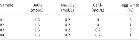
Mineralisation of witherite was carried out by direct precipitation. First, four solutions were prepared. Solutions #1 and #2 both consisted of a solution of 100 mL 1.6 mol/L BaCl2. Solutions #3 and #4 both consisted of 100 mL of a mixture of 1.6 mol/L BaCl2 and 0.2 mol/L CaCl2. Next, 1 mL of fresh egg white was added to solutions #2 and #4, respectively, and the resulting mixtures were then stirred magnetically for 10 mins. The composition of all four reaction solutions is displayed in Table 1. A 0.2 mol/L Na2CO3 solution also had to be prepared beforehand.
Experimental parameters of the witherite samples.

The mineralisation process was as follows: four copies of 100 mL Na2CO3 solution were added dropwise to reaction solutions #1–#4 under vigorous magnetic stirring, producing a white precipitate. After ageing for four hours, the suspension was centrifuged to extract a mineralised product. Next, this product was washed three times in ultra-pure water and absolute ethanol, and then freeze-dried. The resulting samples #1–#4 were collected for subsequent testing and characterisation.
Characterisation
The crystallisation of witherite was determined by powder X-ray diffraction (XRD) using an X-ray diffractometer (X'Pert PRO MPD, Netherlands Panalytical) at an operating voltage of 40 kV and an operating current of 40 mA. Cu radiation was used (λ = 1.54187 Å), the step interval was 0.03°, the scanning speed was set to 10°/min and the range was from 3° to 80° for the conventional diffraction angle.
The morphology of the samples was imaged by a field emission scanning electron microscope (FESEM, Ultra 55, German Zeiss); the surface of the samples was plated with Au, and the accelerated working voltage was 7 kV.
The samples were compressed using a KBr pellet press and their infrared absorption spectra were measured with a Fourier transform infrared (FT-IR) spectrometer (Spectrum One, American PE Instruments) over a wavelength range of 400–4000 cm–1.
High-resolution transmission electron microscopy (HRTEM) was undertaken using a Libra 200FE (Germany Zeiss) equipped with selected-area electron diffraction (SAED) and energy-dispersive spectroscopy (EDS). The accelerating voltage was 200 kV. Each sample was dispersed in anhydrous ethanol using ultrasound and tested on the copper net.
X-ray photoelectron spectroscopy (XPS) was performed on a Thermo Scientific CACALAB 250XI instrument to confirm the chemical state of each element in the pressed samples, with cut aluminium foil wrapped around them. Samples of 1 cm × 1 cm were prepared and tested on the sample table. The X-ray excitation source was monochrome AlKα (hν = 1486.6 eV) with a power setting of 150 W and a beam spot set to 500 μm.
The thermogravimetric test (TG) and differential thermal test (DSC) were performed on a NETZSCH STA 449F5 instrument with ambient air as the atmosphere and a heating rate of 10 K/min.
In the beginning, the molecular structure of the unit cell and super cell of witherite was constructed using Materials Studio version 8.0 (DS BIOVIA, Dassault Systèmes). The energy optimisation and molecular dynamics were accounted for by the Universal force field, which was implemented in the Forcite Calculation modules. Secondly, we calculated the surface energy of the corresponding crystal surface by the formula:
 $$E_{{\rm surf}} = \lpar {E_{{\rm slab}}-E_{{\rm bulk}}} \rpar /2A$$
Where, E slab is the total surface energy of the crystal surface, E bulk is the energy of the constructed crystal cell, and A represents the area.
Results and discussion
For witherite mineralisation, a simple direct precipitation was used. In addition to the sample synergistically regulated by Ca2+ and egg white, three other control samples were prepared in order to investigate the crystal growth mechanism: sample #1 is the blank control; sample #2 had only egg white added; sample #3 had only calcium added; and sample #4 contains both egg white and calcium. The egg white used in the experiment is composed of water (85%), protein (11.5%), carbohydrates (0.6%) and ash (0.5%). Table 1 gives the preparation parameters for all four samples. The solubility product constants (K sp) of BaCO3 and CaCO3 are 5.1×10⁻⁹ mol2/L2 and 3.36×10⁻⁹ mol2/L2, respectively (the K sp value of the insoluble precipitate is constant at room temperature and does not need to be calculated). Therefore, in theory, as long as [Ba2+]/[Ca2+] is greater than 1.52 in aqueous solution, Ba2+ will precipitate preferentially. In the experiments, however, even if the ratio of [Ba2+]/[Ca2+] is as high as 4:1, Ba(CaCO3) precipitates instead of pure BaCO3 (Supplemental Figs S1, S2). This may be due to local supersaturation of CO32– and heterogeneous nucleation of CaCO3 on the surface of BaCO3 particles formed in the beginning. In order to reduce the negative effects of these two phenomena, the amount of [CO32–] should be significantly lower than that of [Ba2+]. Indeed, in order to ensure precipitation of BaCO3 and inhibit precipitation of Ca2+, [Ba2+]/[Ca2+] and [Ba2+]/[CO32–] were both set at 8:1. In addition, the volume of the Na2CO3 solution must also be controlled strictly, with the result that the actual experimental ratio was much lower than the stoichiometric ratio. Control of concentration and volume is in fact the key factor for ideal mineralisation. For the sake of consistency, the same ratio was used for the three other control samples, and the specific parameter proportion is shown in Table 1 (where applicable).
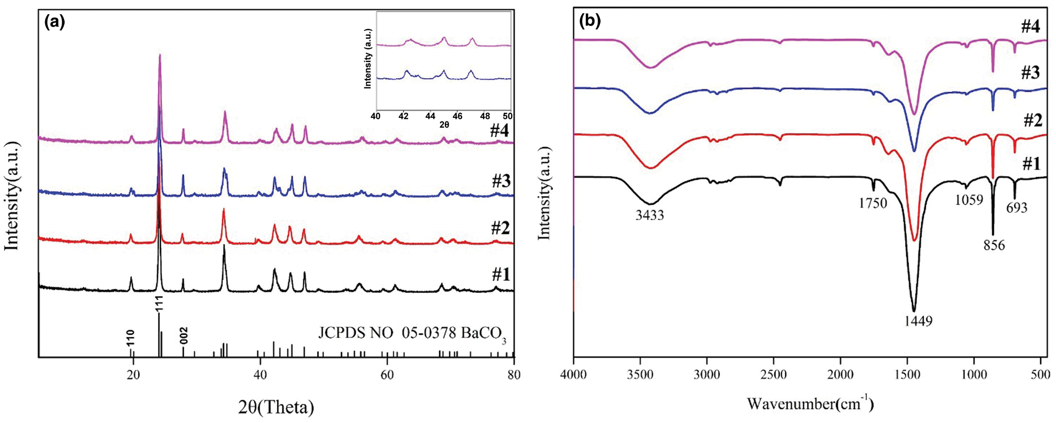
The XRD patterns of the samples are given in Fig. 1a. The lines at the bottom of the patterns correspond to the standard position and intensity of witherite (JCPDS No. 05-0378 – powder diffraction files from the International Centre for Diffraction Data, http://www.icdd.com/). It can be seen that the position and intensity of all the diffraction peaks of the samples are consistent with the standard data, and no other diffraction peaks appear. Therefore, the minerals prepared under the various conditions are all pure witherite crystals. As far as the crystalline phase is concerned, the addition of Ca2+ and egg-white proteins has no obvious effect on the mineralisation. Moreover, it also appears that weak diffraction peaks exist in some intervals in the diffraction patterns of samples #3 and #4, implying that Ca2+ may help improve the crystallinity of product. In addition, the peak intensity ratios between the (111) crystal face and the (002) crystal face in samples #1–#4 are 8.56, 9.01, 4.22 and 6.20, respectively. These ratios are significantly lower for samples #3 and #4 than for samples #1 and #2, indicating that Ca2+ is more conducive to the growth of the (002) crystal face. In addition, the diffraction peak intensities of faces (111) and (002) of samples #2 and #4 prepared with egg white are lower than those of samples #1 and #3 (without egg white), suggesting that egg white can reduce the crystallinity of samples.
(a) X-ray diffraction patterns and (b) FT-IR spectra of samples #1–#4. Sample #1 is blank control, sample #2 had only egg white added, sample #3 had only calcium added, and sample #4 contains both egg white and calcium.

The FT-IR spectra further confirm the presence of witherite (Fig. 1b). The absorption peaks at 693 cm–1 and 856 cm–1 can be attributed to the in-plane bending and out-of-plane bending of CO32–, and the absorption peak at 1449 cm–1 is related to the asymmetric stretching vibration of the C–O bonds (Chen et al., Reference Chen, Shen and Xie2007). The weaker absorption peak at 1059 cm–1 is related to a symmetric stretching vibration of the C–O bonds (Alavi and Morsali, Reference Alavi and Morsali2008). The weaker absorption peak at 1750 cm–1 can be attributed to the combination of the symmetric tensile vibration and in-plane vibration of the covalent C=O bond (Zeng et al., Reference Zeng, Li, Xu, Xu, Li and Zhang2011). The absorption peak at 3433 cm–1 is attributable to the ν(OH) stretching vibration caused by hydrogen bonding (Wu et al., Reference Wu, Cao, Yin, Zhang and Chernow2009). The absorption peaks 1750 cm–1, 1059 cm–1 and 1449 cm–1 in the spectrum are characteristic peaks for witherite crystals (Chen et al., Reference Chen, Shen and Xie2007).
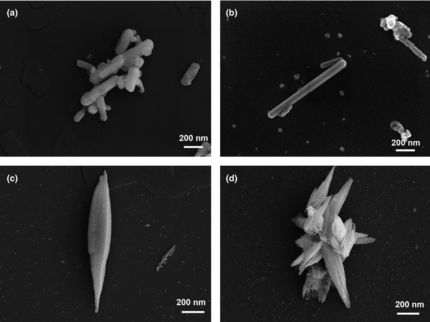
Although samples #1–#4 are all crystalline, SEM characterisation shows that their morphology varies considerably (Fig. 2). In the absence of both Ca2+ and egg white (sample #1), the crystals are regular rods with an average diameter of ~200 nm and some small particles adhering to their surface (Figs 2a, S3a). When Ca2+ is absent but egg white is present (sample #2), the crystals are still rod-like, but their average diameter is only ~50 nm, and the proportion of non-rod-like small particles increases significantly (Figs 2b, S3b). When Ca2+ is present but egg white is absent (sample #3), the crystals are no longer rod-shaped. Instead, they have a non-isodiametric spindle-like shape, with a diameter at their centre of ~200 nm decreasing gradually towards their ends (Figs 2c, S3c), until they dwindle to a point. When both Ca2+ and egg white are present (sample #4), the same non-isodiametric spindle-like shape results, but with a central diameter that is comparable to that of sample #2 (Figs 2d, S3d).
SEM images of all four samples: (a) sample #1 (blank control); (b) sample #2 (only egg white added); (c) sample #3 (only calcium added); (d) sample #4 (both egg white and calcium).

Given the above, two laws affecting mineralisation in this case can be proposed: (1) the presence of Ca2+ leads to non-isodiametric mineral growth; and (2) the presence of egg white restricts mineral growth, the crystal surface is rough, and the assembly trace of small particles can be seen clearly. Thus, one can expect that egg-white proteins will restrict the formation of rod-like crystals in the absence of Ca2+, with a consequent significant increase in the number of nanoparticles (Fig. S3b), as HRTEM analysis indeed shows (Fig. 3). By contrast, in the presence of Ca2+, the nanoparticles disappear completely whether or not egg white is present, resulting in pure spindle-like minerals, which is consistent with the XRD results (Fig. 1a). The peak intensity of face (002) of samples #3 and #4 prepared with Ca2+ is significantly stronger than that of samples #1 and #2 without Ca2+, meaning that Ca2+ can enhance crystallinity.
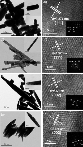
TEM, HRTEM and SAED images of samples #1–#4: (a and b), sample #1 (blank control); (c and d), sample #2 (only egg white added); (e and f), sample #3 (only calcium added); (g and h), sample #4 (both egg white and calcium added). The insets give the SAED patterns of their respective samples.

In order to investigate further the growth habit of mineral crystals, TEM, HRTEM and SAED characterisations were performed. The results in Fig. 3 show that the morphology of the crystals (Fig. 3a,c,e,g) is consistent with the SEM image (Fig. 2), but there are obvious nodes in the rod-like crystals of sample #2 (Fig. 3c), and partial fractures occur between the nodes. This may be caused by adsorption of egg-white protein molecules on the surface of the initial witherite nanoparticles, preventing their further elongation. This is probably also an important factor contributing to the large number of nanoparticles visible in sample #2 (Fig. 2b). The attachment of egg white around the spindle-like minerals can also be observed in sample #4 (Fig. 3g). On the one hand, these adsorbed proteins restrict the further growth of spindle-like minerals, and on the other hand, the hydrogen bonds between the protein molecules cause the minerals to further agglomerate. The clear diffraction fringes and SAED lattice patterns (Fig. 3b,d,f,h and their insets) for samples #1–#4 indicate that the minerals are all nanocrystals. The interplanar distances for samples #1 and #2 (Fig. 3b,d) are 0.374 nm and 0.368 nm, respectively, corresponding to the (111) crystal plane. Therefore, the rod-like nanocrystals of samples #1 and #2 are formed by the growth of minerals along the [111] direction (c axis). The interplanar distances for samples #3 and #4 (Fig. 3f,h) are 0.321 nm and 0.320 nm, respectively, corresponding to the (002) crystal plane. This indicates that spindle-like nanocrystals grow along the c axis in the [002] direction, which is in good agreement with the XRD results. In short, the introduction of egg white leaves the growth habit of the nanocrystals unaffected, while the introduction of Ca2+ affects it significantly.
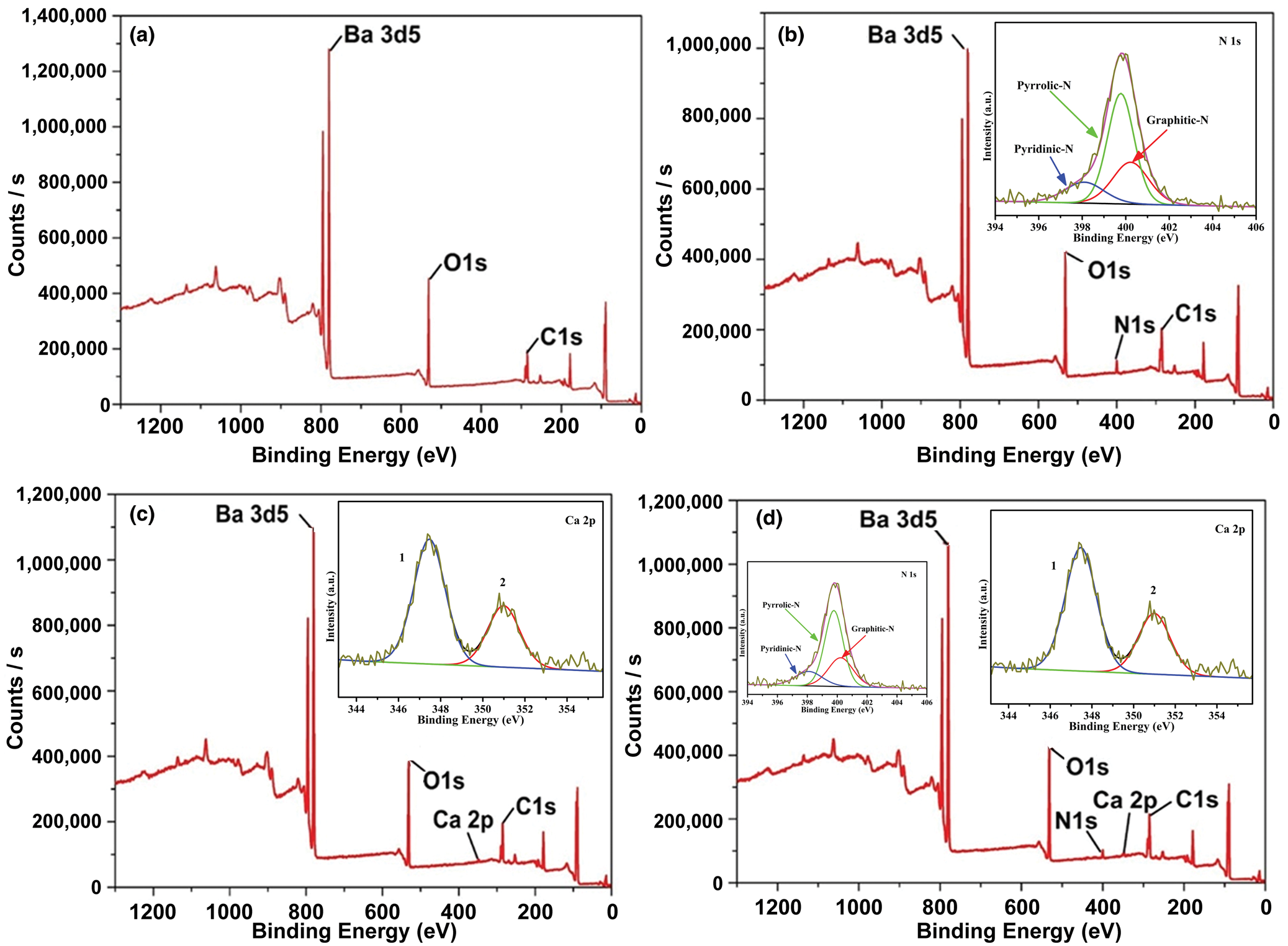
To detect the distribution of Ca2+ and egg-white protein molecules in minerals and confirm the chemical states of each element, XPS and EDS characterisation of samples #1–#4 were conducted, and the results appear in Figs 4 and S4–7. In the survey spectrum of Fig. 4a, the three strong peaks at 285 eV, 531.8 eV and 780 eV reflect the chemical bonding energy of C1s, O1s and Ba3d5, respectively, which indicates that the main elements in these minerals are C, O and Ba. In contrast to Fig. 4a, Fig. 4b has a peak at 398.4 eV that corresponds to the binding energy of N1s, thereby confirming that sample #2 contains egg-white protein molecules. The energy spectrum (Fig. S5d) shows that N is uniformly distributed in the nanocrystals. Sample #3 has a new peak at 346.6 eV (Fig. 4c) that is caused by the binding energy of the Ca2p orbital, indicating that Ca2+ has entered into the crystal structure. Figure S6d shows the distribution of Ca2+ in the minerals, which will be discussed further later. Figure 4d has two more binding peaks than Fig. 4a, at 398.4 eV and 346.6 eV, which correspond to the binding energies of N1s and Ca2p, respectively, and further confirms the presence of egg white and Ca2+ in the minerals. Figure S7d and e reflect the distribution of Ca and N in the minerals. The atomic contents of all elements in samples #1–#4 as determined by XPS appear in Table S1. Results from XPS show that egg white and Ca2+ not only regulate the witherite mineralisation process but also enter into the mineral itself through adsorption and substitution. The ion radius of Ca2+ is 0.1 nm and that of Ba2+ is 0.135 nm (Shannon, Reference Shannon1976), therefore, it is possible that the formation of shuttle-like non-isodiametric mineral particles is due to Ca substitution for Ba, which changes the growth habit of the crystal.
XPS survey spectra of samples #1–#4 and the high resolution spectra of Ca and N (inset graphs): (a), sample #1 (blank control); (b), sample #2 (only egg white added); (c), sample #3 (only calcium added); (d), sample #4 (both egg white and calcium added).

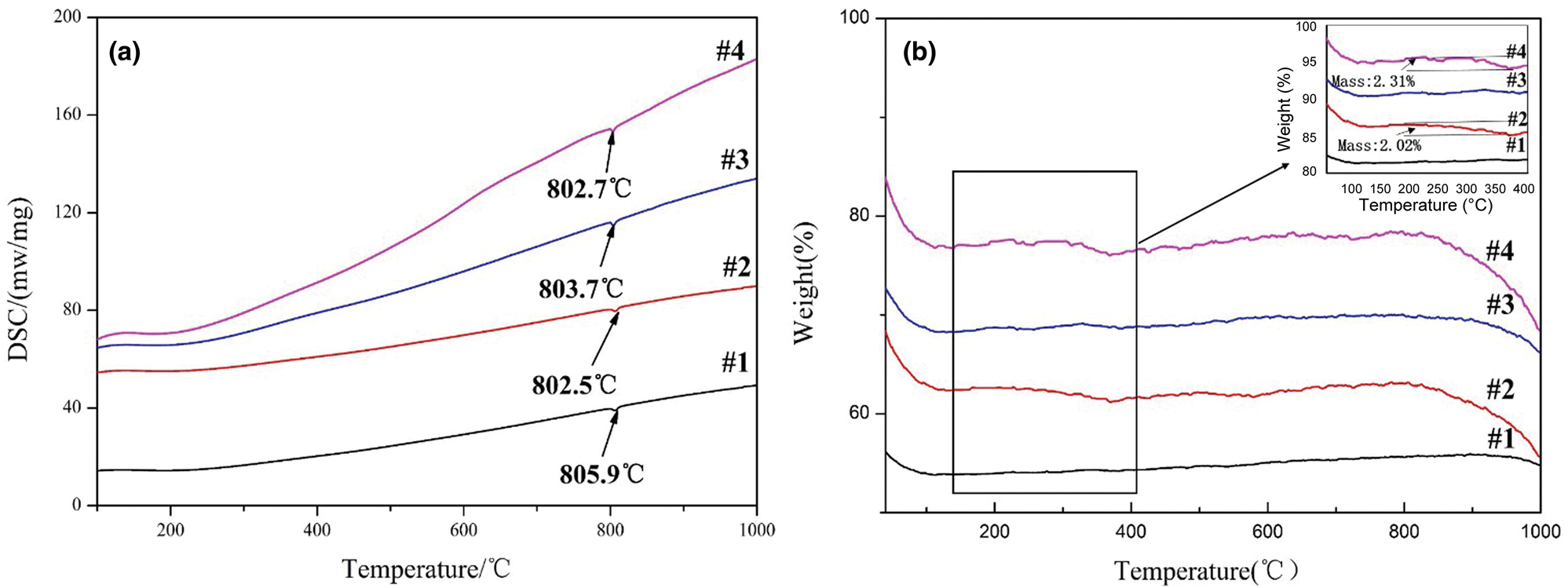
To investigate the effects of Ca2+ and egg-white protein molecules on the thermal properties of witherite particles, and to measure the egg-white protein content more accurately, TG and DSC characterisations were conducted for samples #1–#4. The results appear in Fig. 5. The DSC analysis curves (Fig. 5a) show that the four samples have endothermic peaks at 805.9°C, 802.5°C, 803.7°C, and 802.7°C, respectively, all of which reflect the phase transition of BaCO3 from γ phase to β phase (Chen et al., Reference Chen, Chang, Chen, Zhao and Peng2015). The phase transition temperature of sample #1 is the highest, with that of samples #2 and #4 lower (~3°C) as a result of their smaller particle size, which is caused by the addition of egg white. Sample #3 also shows a lower phase transition temperature, in its case due to the larger specific surface area of spindle-like crystals as compared to rod-like ones. As one would expect, particle shape affects specific surface area less than particle size does, so the phase transition temperature of sample #3 is higher than that of samples #2 and #4. The DSC results show that the addition of Ca2+ has no direct effect on the phase transition temperature of the BaCO3.
(a) DSC curves and (b) TG curves of samples #1–#4. Sample #1 is blank control, sample #2 had only egg white added, sample #3 had only calcium added, and sample #4 contains both egg white and calcium. The detail in b is an enlargement of the 200–400°C range.

The results of TG characterisation (Fig. 5b) show obvious weight loss in all four samples below 100°C and above 900°C, but no significant change between 100°C and 900°C. Below 100°C, the weight loss is caused by the loss of adsorbed water; above 900°C, it is caused by thermal decomposition. However, if one enlarges the TG curves over the range from 200°C to 400°C, one sees that samples #2 and #4, both prepared with egg white, experience weight losses over this temperature range of 2.02% and 2.31%, respectively. Comparison to the TG curve for pure egg white (Fig. S8) indicates that the weight loss in this temperature range is caused by combustion of the egg white and therefore that the egg-white content of samples #2 and #4 is ~2%.
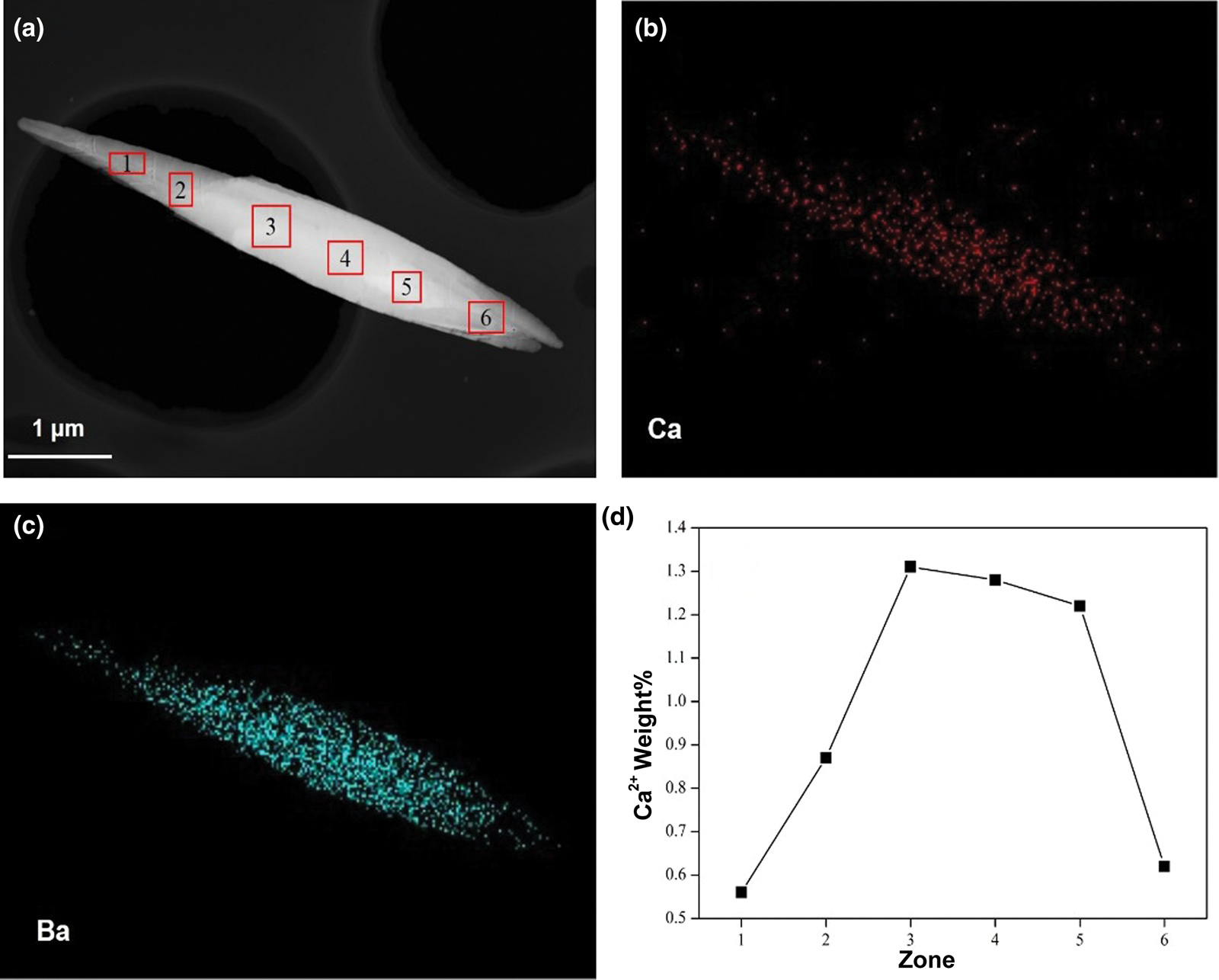
To investigate the non-isodiametric growth mechanism of witherite particles modulated by Ca2+, more detailed and in-depth TEM selective electron spectroscopy and elemental spectroscopy measurements were conducted for samples #3 and #4, as shown in Figs 6 and S9. The energy spectrum mapping of Ca2+ (Fig. 6b) shows clearly that the distribution of Ca2+ in the spindle-like crystals decreases from the middle to the ends. In other words, the Ca2+ content is higher in the larger-diameter region. This trend is still more apparent when the Ca2+ content is measured in different regions (Fig. 6d and Table S2). The same Ca2+ distribution pattern also appears in the test results for sample #4 (Fig. S9 and Table S3). This indicates that the non-isodiametric growth of witherite particles is regulated by a Ca2+ gradient.
(a) The selected regions, using the TEM image of sample #3 (only calcium added); (b) and (c), mapping images of the Ca2+ and Ba2+ electron energy spectra, respectively; (d) the Ca2+ content of the six selected regions in image (a).

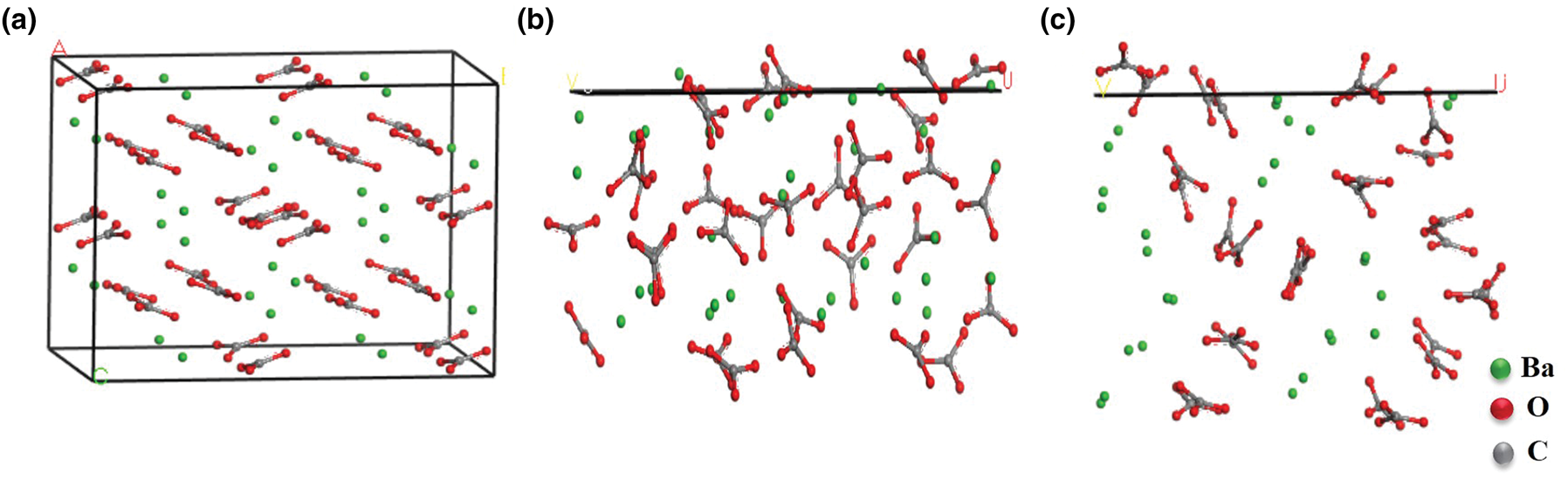
The crystal structure of witherite and the atomic composition of faces (111) and (002) appear in Fig. 7. These data are obtained from crystal data of the CCDC (Cambridge Crystal Data Centre), and simulated by calculation software of Materials Studio 8.0. Energy optimisation and molecular dynamics are calculated by the general force field and implemented in the predict calculation module. It shows that face (111) is composed of B, O and C, while face (002) is composed of C and O. To ascertain the intrinsic relationship between the Ca2+ gradient and the diameter of the crystals, molecular dynamic simulation was used to calculate the surface energy of faces (111) and (002) of witherite at 1.025 kJ/mol and 0.9288 kJ/mol, respectively. The energy optimisation and molecular dynamics were accounted for by the universal force field. The different substitution rates along with their corresponding surface energy calculations are listed in Table S4. Generally, the larger the surface energy, the greater the growth rate (Hu et al., Reference Hu, Dong, Zhang, Liu, He, Wu, Yang and Deng2018). In the absence of other factors, face (111) has more surface energy than face (002) does. This means that growth will be faster along [111] than along [002], producing a nanorod-like shape. Given the atomic composition of faces (111) and (002), Ca2+, if present, can substitute for Ba2+ on face (111), thereby altering the surface energy of that plane. At a substitution rate of Ca2+ for Ba2+ of 5%, the surface energy of face (111) will drop to 0.9188 KJ/mol. As the Gibbs free energy change (ΔG) in the substitution process will then be <0, the substitution can proceed spontaneously thereafter. By contrast, as plane (002) contains no Ba2+, substitution of Ca2+ for Ba2+ is impossible, and therefore the presence of Ca2+ has no direct effect on the growth of face (002). As the surface energy of face (111) decreases due to Ca2+/Ba2+ substitution, its growth advantage and therefore its influence on the direction of crystal growth will also decrease. The decrease in the (111) face ratio also means that the reaction area for Ca2+/Ba2+ substitution will dwindle, further reducing the substitution rate. Therefore, as a crystal grows, its Ca2+ content gradually decreases, creating a Ca2+ gradient. In addition, as the growth advantage for face (111) diminishes, the growth advantage of the other faces increases. Given this analysis, plane (002) will become the favoured growth plane, and so the direction of crystal growth will be along [002], resulting in non-isodiametric growth and finally a crystal with a spindle-like shape. The full width at half maximum (FWHM) of the (111) crystal plane and (002) crystal plane of all samples are shown in Table S5. Generally, FWHM is inversely proportional to the grain size. That is, the smaller the grain diameter, the larger the FWHM. It can be seen from the table that the FWHM of planes (111) and (002) of samples #2 and #4 containing egg white is larger than samples #1 and #3 prepared without egg white, indicating that the particle size of samples #2 and #4 decreases. The FWHM of plane (002) increases more than that of plane (111), which is due to the change of crystal growth habit caused by the substitution of Ca2+ for Ba2+.
(a) The crystal structure of witherite, and the atomic composition of (b) face (111) and (c) face (002).

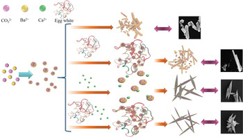
Based on above results and analysis, it can be seen that the regulation of witherite mineralisation by egg-white proteins and Ca2+ involves two relatively independent processes. As shown in Fig. 8, egg-white proteins are mainly adsorbed onto the surface of the witherite crystals, coating them and therefore limiting their growth, but Ca2+ can change the growth habit of the crystals by interacting with different crystal faces. The interaction between the growth advantages of the different crystal planes and the Ca2+/Ba2+ substitution rate leads to a Ca2+ gradient and non-isodiametric growth of the crystals.
Schematic diagram of crystal growth in samples #1–#4. Sample #1 is blank control, sample #2 had only egg white added, sample #3 had only calcium added, and sample #4 contains both egg white and calcium.

Conclusion
The use of egg white and Ca2+ to simulate the organic and inorganic environments of witherite mineralisation, respectively, produce a confinement effect and non-isodiametric growth of witherite particles. Research into the mechanism of this process shows that egg-white proteins and Ca2+ act to regulate the process in two relatively independent ways. The main role of egg-white proteins is to limit crystal growth, while Ca2+ is the key to non-isodiametric growth because of its effect on the growth advantage of different crystal faces as it substitutes for Ba2+. The growth advantages of the different crystal faces and the Ca2+/Ba2+substitution rate interact, thereby creating a Ca2+ gradient and isodiametric growth. This study shows the different roles of organic materials and inorganic ions in the formation of witherite and offers insight into the genesis of various morphologies of witherite. It needs to be emphasised, however, that our research is only an initial exploration into the subject, and the environment of witherite mineralisation is far more complex than in the model used in this paper. Future research needs to incorporate more types of organic matrices (e.g. humic acid and polysaccharide) and inorganic ions (e.g. Na+, K+ and Fe3+) at different concentrations in order to give a fuller picture of how witherite is mineralised in Nature.
Acknowledgements
This work is supported by the National Natural Science Foundation of China (11572270), the Scientific Research Fund of Sichuan Provincial Education Department (18ZA0493 and 07ZD1118), the Project of State Key Laboratory of Environment-friendly Energy Materials, Southwest University of Science and Technology (18FKSY0217) and the Scientific Research Fund of the Southwest University of Science and Technology (17ZX9104 and 18LZX556). The data produced within this work are available in the references, tables and supporting information.
Supplementary material
To view supplementary material for this article, please visit https://doi.org/10.1180/mgm.2020.54