Acute myocardial infarction from left main coronary artery occlusion is an atypical cause of chest pain among the paediatric population in the absence of identified risk factors. Primary cardiac tumours are rare in the paediatric population and are usually benign, with rhabdomyomas, myxomas, and fibromas being the most common.Reference Chan, Sonley, Moes, Daneman, Smith and Martin 1 Cardiac inflammatory myofibroblastic tumour is a rare tumour consisting of inflammatory cells and myofibroblastic spindle cells with a benign potential. Although benign in nature, these intra-cardiac tumours can compromise blood flow causing variable degrees of obstruction and can potentially have serious complications.
Case
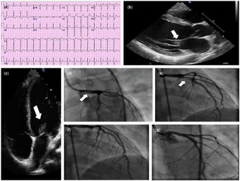
An 11-year-old African–American girl with a history of bronchial asthma presented to the emergency department with acute onset of chest pain. She woke up to severe, retrosternal, pressure-like chest pain radiating to the back, associated with mild shortness of breath. There was no reported family history of any CHD, coronary artery disease, sudden cardiac death, or familial dyslipidaemia. On presentation, she appeared to be in moderate distress with blood pressure of 122/52 mmHg, heart rate of 120 beats/minute, respiratory rate of 20 breaths/minute, and temperature of 37.2°C. There was no jugular venous distention and the lung examination was normal. Cardiac examination revealed no murmur, clicks, or gallops. The 12-lead electrocardiogram showed ST-segment elevation in lead augmented vector right (aVR) with associated ST-segment depression in leads II, III, and augmented vector foot (aVF) (Fig 1a). The chest radiograph was normal. The initial set of cardiac enzymes showed an elevated troponin I level of 2.5 ng/ml, which eventually peaked at >40 ng/ml. Transthoracic echocardiography revealed moderately depressed left ventricular systolic function with hypokinesis of the anterior, anteroseptal left ventricular wall from base to apex. A small hyperechoic mass of 6×12 mm was observed on the anterior leaflet of the mitral valve (Fig 1b and c). No mitral valve insufficiency or stenosis was observed. Cardiac catheterisation was urgently performed and revealed 80% obstructive lesion in the left main coronary artery extending to the left anterior descending artery (Fig 2d and e). Mechanical embolectomy with a 6-Fr Export Aspiration Catheter (Medtronic, Inc., Minneapolis, USA) was attempted twice unsuccessfully. The embolic material was successfully removed using a 7-Fr Priority One Aspiration Catheter (Terumo Interventional Systems (Somerset, NJ)) with 0% residual stenosis after the procedure (Fig 1g). Distal embolic protection technique was utilised to prevent distal embolisation using a Spider filter distal protection device (Covidien, Mansfield, USA) (Fig 1f). The removed tissue was sent for pathological examination, and the initial analysis revealed undifferentiated malignant neoplasm with epitheliod features, suggestive of sarcoma. No evidence of metastatic disease or cerebral infarction was suggested by further imaging workup. The oncology service consultation recommended surgical excision of the tumour to prevent further embolic events.
( a ) Electrocardiogram on presentation showing ST-segment elevation in lead aVR with widespread ST-segment depression in leads II, III, aVF and leads V4, V5, V6. Transthoracic echocardiography showing a 6×12 mm mass on the anterior mitral valve, ( b ) two-dimensional echocardiogram, in the parasternal long-axis view, of the mitral valve tumour (white arrow), ( c ) two-dimensional echocardiogram, in the apical four-chamber view, of the mitral valve tumour (white arrow), ( d ) right anterior oblique view, obtained during cardiac catheterisation, showing the embolised tumour in the left main coronary artery (white arrow), ( e ) right anterior oblique view showing the tumour extension to the long anterior descending coronary artery (white arrow), ( f ) right anterior oblique view of the aspiration catheter with the Spider filter distal protection device, ( g ) left main coronary artery and long anterior descending coronary artery with 0% residual occlusion after successful aspiration of the tumour. aVF=augmented vector foot; aVR=augmented vector right.

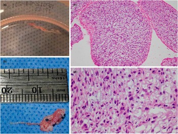
( a ) The tumour embolus aspirated from the left main coronary artery, ( b ) macroscopic examination of the excised tumour, revealing a gelatinous mass 9×5 mm, ( c ) microscopic examination 10× with haematoxylin and eosin stain showing spindle-shaped cells arranged in storiform pattern in a myxoid background, ( d ) microscopic examination 40× showing hyperchromatic cells with frequent mitotic figures and areas of necrosis, are strongly immunoreactive for vimentin, have cytoplasmic dot-like immunoreactivity for CD99, and are negative for desmin, CD34, CD163, S-100, actin, and ALK-1, with scattered histiocytes and few vessels.

Cardiac surgical resection of the tumour, using cardiopulmonary bypass, was performed 6 days after the transcatheter embolectomy. The final pathology report for the tumour removed at the time of cardiac catheterisation was still pending at the time of the cardiac surgery. In the operating room, the tumour appeared as a fimbriated mass involving the entire medial half of the anterior leaflet with involvement of the free edge and extended to the chordae tendineae. Mitral valve repair was not considered feasible. The mitral valve leaflets were excised, and the native mitral valve was replaced with a 27-mm St. Jude prosthetic valve (St. Jude Medical, Inc., Minnesota, USA) with a subsequent uncomplicated post-operative course. The tumour specimen was sent for histopathological examination. Macroscopic examination revealed a villous, rubbery mass measuring 0.7×0.5×0.4 cm (Fig 2a and b). Histological examination revealed spindle-shaped cells arranged in a storiform pattern within a myxoid background. The cells were hyperchromatic with frequent mitotic figures. The final histopathological analysis concluded that this was a benign inflammatory myofibroblastic tumour (Fig 2c and d). At the 6- and 12-month follow-up outpatient visits, the left ventricular ejection fraction was mildly reduced with global hypokinesis and without stenosis or regurgitation of the prosthesis.
Discussion
Inflammatory myofibroblastic tumour is a rare benign tumour characterised by the proliferation of myofibroblasts and inflammatory cell infiltration. There have been few case reports of inflammatory myofibroblastic tumour with the exact incidence unknown.Reference Krishna, Ng and Chachlani 2 , Reference Kelly, Lambie and Singh 3 It occurs most commonly in the lungs and usually presents as a solitary pulmonary nodule.Reference Coffin, Watterson, Priest and Dehner 4 Inflammatory myofibroblastic tumour has been rarely reported in the heart.Reference Burke, Li, Kling, Kutys, Virmani and Miettinen 5 The most accepted pathogenesis is thought to be related to the upregulation of the immune system with resulting symptoms of fever, weight loss, and laboratory abnormalities that include an elevated sedimentation rate, iron-deficiency anaemia, thrombocytosis, and hypergammaglobulinaemia. Patients with cardiac involvement usually present with syncope, chest pain, or shortness of breath. The distinction between inflammatory myofibroblastic tumour and low-grade sarcoma can be challenging with lack of significant pleomorphism and atypical mitotic figures favouring the diagnosis. Cardiac inflammatory myofibroblastic tumours are biologically benign, but due to their location inside the heart they can be fatal. Burke et al reported a case series of 10 patients with one case reporting coronary artery embolisation resulting in sudden cardiac death. In our case, we report an inflammatory myofibroblastic tumour of the mitral valve presenting with an acute myocardial infarction as a result of embolic occlusion of the left main coronary artery. Our patient was successfully rescued with percutaneous coronary intervention. Benign tumours involving valves should be excised with reasonable attempts made to preserve and repair the involved valve; however, in our case, excision of the tumour and reconstruction of the mitral valve were not feasible and the mitral valve was replaced.
To the best of our knowledge, this case is the first reported case in the paediatric population of acute myocardial infarction due to left main coronary artery occlusion in the absence of identifiable risk factors that was successfully rescued by percutaneous coronary intervention. Changes in aVR are sometimes ignored and considered as reciprocal changes from the left lateral side – augmented vector left (aVL), V5, V6 – however, as shown in our patient, changes in aVR can be very useful in identifying acute left main coronary artery occlusion.
Acknowledgements
None.
Financial Support
This research received no specific grant from any funding agency, commercial, or not-for-profit sectors.
Conflicts of Interest
None.