Left ventricular pseudoaneurysms may occur when cardiac rupture is contained by adherent pericardium. The true incidence of left ventricular pseudoaneurysm is unknown and rarely seen in children.Reference Frances, Romero and Grady1,Reference Ercan, Senkaya, Semizel and Cil2 Untreated pseudoaneurysms may lead to sudden cardiac death. Mortality has been reported up to 48% in untreated patients.Reference Sakai, Nakamura and Ishizuka3 Although open surgery is conventional, more recently, percutaneous closure of pseudoaneurysms has been described.Reference Kumar, Alli, Bjarnason, Hagler, Sundt and Rihal4 Here, we present a child who underwent transcatheter ventricular septal defect closure and who 27 months later developed a left ventricular pseudoaneurysm which was closed percutaneously.
Case
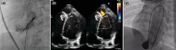
A 3-month-old boy with Down syndrome presented with murmur. Echocardiography revealed a 5-mm apical ventricular septal defect, 7-mm atrial septal defect, and patent ductus arteriosus. Five months later, transcatheter closure of ventricular septal defect and patent ductus arteriosus was performed. “A 5Fr JR4 diagnostic catheter along with guidewire was advanced from the inferior vena cava into the right atrium, the atrial septal defect was crossed and the catheter positioned in the left atrium. The catheter was subsequently advanced through the mitral valve into the left ventricle. Then a Glidewire® (Terumo, Somerset, NJ, USA) was guided slowly and gently through the ventricular septal defect into the right ventricle. Another venous access was used for diagnostic imaging. By this way ventricular septal defect was closed without forming an arteriovenous loop.” (Fig 1a). A 6/6-mm Amplatzer duct occluder II device was used. Once fully deployed, both echocardiography and left ventriculography showed a stable device position with minimal residual defect. The patent ductus arteriosus was closed from the pulmonary side in a standard manner by another Amplatzer duct occluder II device.
(a) Left ventriculography showing the Amplatzer ductal occluder II device closing the ventricular septal defect and minimal residual shunt proximal to the device. (b) Echocardiography showing an apical left ventricular pseudoaneurysm adjacent to the Amplatzer septal occluder device. (c) Left ventriculography showing the pseudoaneurysm.

His post-operative course was unremarkable. Follow-up echocardiography showed regression in left ventricular dilatation and pulmonary artery pressure with minimal residual shunt from the ventricular septal defect.
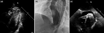
Twenty-seven months after the initial procedure, routine echocardiography revealed an apical left ventricular pseudoaneurysm adjacent to the device (Fig 1b). The atrial septal defect had closed spontaneously. Since open surgery carried a high risk, we decided on percutaneous closure. Left ventriculography demonstrated a large pseudoaneurysm, measured 21 × 17-mm with an 8-mm connecting neck without any residual shunt from the ventricular septal defect (Fig 1c and Supplementary video S1). A 0.035-in angled-tip Glidewire® hydrophilic-coated guidewire was gently passed across the stalk of the pseudoaneurysm to prevent perforation. Subsequently, a 0.035-in J-tipped Amplatz Super Stiff exchange wire was substituted for the Glidewire®. A 6-F delivery sheath was introduced over the guidewire to enter the defect. A 9-mm Amplatzer® atrial septal defect occluder (St. Jude Medical, an Abbott company, Saint Paul, MN, USA) was deployed in the standard manner across the myocardial wall. To prevent device embolisation, both discs were embedded in the pseudoaneurysm cavity. An echocardiography and left ventriculography confirmed total occlusion of the pseudoaneurysm (Fig 2a and b and Supplementary video S2). The following day, echocardiography was uneventful with complete closure of communication between the left ventricle and pseudoaneurysm. Three days after the procedure, echocardiography revealed effusion measuring 12 mm at the maximal point. Anti-inflammatory treatment with ibuprofen was initiated. On post-operative day 10, the pericardial effusion increased up to 24 mm (Fig 2c). Pericardiocentesis was performed. The pericardial fluid analysis was consistent with transudate. Medical treatment was changed to intravenous prednisolone administration. Starting on second day of the treatment, pericardial effusion slowly regressed. The patient was discharged on day 10 of prednisolone treatment without any effusion. An echocardiogram 2 months later showed closed ventricular septal defect and pseudoaneurysm without any residual shunt.
(a) Echocardiography showing Amplatzer septal occluder device occluding left ventricular pseudoaneurysm. (b) Left ventriculography confirmed total occlusion of the pseudoaneurysm. (c) Echocardiography showing post-procedural pericardial effusion.

Discussion
Muscular ventricular septal defects affecting haemodynamics pose a surgical challenge, particularly when their location is close to the ventricular apex. In this patient group, percutaneous ventricular septal defect closure is an alternative option. Complications of this procedure are usually periprocedural, such as ventricular perforation, unsatisfactory device position, device embolisation, and residual septal defects. Late complications are confined to a few case reports. There are two case reports in which left ventricular pseudoaneurysm developed after perventricular ventricular septal defect device closure. In one of them, it developed periprocedurally and was treated by surgery.Reference Ozyilmaz, Saygi, Yildiz, Erek and Guzeltas5 In the other, the pseudoaneurysm was noticed 10 months after the hybrid procedure.Reference Breinholt, Rodefold and Hoyer6 These complications suggest that even in hybrid procedures, the risk of perforation continues. The introduction of guidewires, delivery sheath, and its dilator increases perforation risk. Even if there was no direct perforation during the procedure, the manipulations may predispose the ventricular free wall to pseudoaneurysm development. The leading edge of the ventricular septal defect device may perforate this weakened portion of the ventricle in time. Even though Amplatzer duct occluder II is a low-profile device, it may still damage the infant heart. We hypothesise that the triggering event in this case was manipulations during the initial procedure.
Ventricular pseudoaneurysms may result in catastrophe; fortunately, the patient was asymptomatic and the pseudoaneurysm was recognised incidentally during routine echocardiographic follow-up. Nevertheless, we could not determine the exact time for development of pseudoaneurysm. Probably, it was recognised only after it became large enough to be visualised on echocardiography. Awareness of the complication and closer echocardiographic follow-up might increase its diagnosis.
The rarity of this condition in children poses a challenge for treatment. Although surgery is an option, there is limited experience with high mortality rates.Reference Abe, Yamazato and Munakata7 Transcatheter treatment is less prevalent.Reference Matsumoto, Okuyama, Cheng, Makkar and Kar8 To the best of our knowledge, this is the first case where a patient developed left ventricular pseudoaneurysm after percutaneous ventricular septal defect device closure and was treated by a second percutaneous method. Device selection needs to be individualised depending on the location and size of the pseudoaneurysm. Septal occluder devices, coils, and vascular plugs have all been used successfully.Reference Baspinar, Mete and Davutoglu9 To reduce device instability and the associated risk of device dislodgement, oversize ratios of 1.2–2.0 have been reported for the Amplatzer septal occluder in previous cases.Reference Madan, Juneja, Raval and Thakkar10 We preferred a device large enough to fully fill the aneurysmal sac to prevent residual leak. The diameter of the atrial septal defect closure device, including its rims, was same as the widest diameter of the sac. Instead of the usual closing pattern, we opened both discs of the device within the pseudoaneurysm. Although this may increase the risk of aneurysmal rupture periprocedurally, once positioned properly, we propose that this is safer to prevent device dislodgement.
Post-procedural pericardial effusion development was concerning with the risk of left ventricular free wall perforation. We hypothesised that it was either an inflammatory reaction or developed secondary to manipulations. We claim that its improvement with high-dose steroid suggests an inflammatory reaction.
Conclusion
Left ventricular pseudoaneurysm may be seen as a late complication of percutaneous ventricular septal defect closure, which has not been previously reported. Transcatheter occlusion might be an alternative option to conventional surgery in selected cases.
Supplementary Material
To view supplementary material for this article, please visit https://doi.org/10.1017/S1047951120000815
Acknowledgements
None.
Financial Support
This research received no specific grant from any funding agency, commercial or not-for-profit sectors.
Conflicts of Interest
None.
Ethical Standards
All procedures performed in this case were in accordance with the ethical standards of the institutional and national research committee and with the 1975 Helsinki declaration and its later amendments or comparable ethical standards.
Informed Consent
Informed consent was obtained from patients’ parent.