I. INTRODUCTION
Bioactive glasses have a wide range of clinical applications in the field of orthopedics for the repair and regeneration of bone, dentistry for teeth repair, and treatment against diseases like osteoporosis and osteomyelitis. Reference Hench and Jones1 Bioactive glasses bond to bone due to their ability to form an interface that helps in biological fixation with the natural bone. Reference Hench2 The interface is formed when the surface of the bioactive glass interacts with the physiological environment inside the body and is found to be made of biologically active hydroxyl carbonate apatite (HCA), which is chemically and structurally similar to the apatite mineral of natural bone. Reference Hench2 Hench developed first bioactive glass in SiO2–Na2O–CaO–P2O5 system known as 45S5 (Bioglass®) which has been commercially used for many bone-related treatments. Other glasses like S53P4 (BonAlive®) Reference Andersson, Liu, Karlsson, Niemi, Miettinen and Juhanoja3 and 13-93 Reference Brink, Turunen, Happonen and Yli-Urpo4 were commercially developed for synthetic bone graft applications.
Bioactive glasses when added with ions exhibiting antibacterial properties can be advantageous in treatments of bacterial infected bones. Osteomyelitis is a bone disease caused by infectious microorganisms and occurs in the form of inflammation around the bone resulting in bone destruction. Reference Lew and Waldvogel5 Usually, treatment of osteomyelitis involves removal of the infected part and treating it with antibiotics like penicillin, methicillin, oxacillin, and amoxicillin. But, recently these bacteria have shown resistance to these antibiotics and emerged as methicillin-resistant Staphylococcus aureus (MRSA) making the treatment of osteomyelitis difficult. Reference Lew and Waldvogel5,Reference Sakoulas and Moellering6 A material that can heal the bone defect as well as prevent the bacterial infections in the infected zone could be a better choice for the osteomyelitis treatment. In this regard, a bioactive glass showing bone bonding, bone regeneration, and antibacterial properties could be a better material for treating osteomyelitis. Reference Jia, Fu, Huang, Zhang and Rahaman7
The bioactive glasses (e.g., S53P4) without the addition of any antibacterial ions can show antibacterial properties either due to the effect of increase in pH in the surrounding medium resulting from the release of Na+ and Ca2+ ions. Reference Allan, Newman and Wilson8 To get a long-term and effective antibacterial effect, the glasses have to be doped with ions exhibiting broad spectrum antibacterial action. Reference Bellantone, Williams and Hench9 Many research groups have studied the effect of antibacterial ions like silver (Ag+), cerium (Ce3+), and copper (Cu+) ions doped in the bioactive glass systems against various bacteria like Escherichia coli, Staphylococcus aureus, Pseudomonas aeruginosa, and Streptococcus sanguis. Reference Bellantone, Williams and Hench9–Reference Goh, Alshemary, Akram, Kadir and Hussain15
Though, the antibacterial effects of various ions in bioactive glasses have been studied by many research groups, the effect of bismuth ions for improving antibacterial properties in bioactive glasses has not yet been explored even after the wide usage of bismuth compounds in gasteroenterology Reference Bierer16 in treating stomach-related diseases and its antimicrobial properties in the treatment against bacterial infections caused by H. pylori in the gut. Reference Lambert and Midolo17
Lambert and Midolo have discussed various mechanisms by which bismuth ions exhibit bactericidal effects against the H. pylori bacteria. Reference Lambert and Midolo17 Bismuth-added calcium silicate-based mineral trioxide aggregate (MTA) had been widely used as the dental filling. This MTA has shown biocompatibility, radiopacity, cell proliferation, and antibacterial properties. Reference Parirokh and Torabinejad18,Reference Torabinejad and Parirokh19 Bismuth ions replacing calcium ions in substituted hydroxyapatite has shown better osteoblast proliferation and increased alkaline phosphatise activity. Reference Webster, Massa-Schlueter, Smith and Slamovich20 Inhibition toward both Gram positive S. aureus and Gram negative E. coli was observed in bismuth-substituted HAp. Reference Ciobanu, Bargan and Luca21 Bi2O3 was successfully added to nano-bioactive glass powders of 45S5 glass composition using flame spray process by 20 and 50 wt%, and these were found to be radiopaque to X-rays along with improved apatite formation. Reference Heid, Stoessel, Tauböck, Stark, Zehnder and Mohn22,Reference Mohn, Zehnder, Imfeld and Stark23
The present work aims to elucidate the effect of progressive bismuth oxide incorporation substituting SiO2 and Na2O in S53P4 glass composition on the structural, thermal, and physical properties. The prepared glasses were also studied for their surface apatite formation in in vitro bioactivity studies, cell proliferation, and antibacterial properties.
II. MATERIALS AND METHODS
A. Glass synthesis using melt-quench technique
The glasses were prepared in the system SiO2–Na2O–CaO–P2O5–Bi2O3 using melt-quench technique. The compositions of glasses in both wt% and mol% for the present study is given in Table I which has base glass (B0) and four glasses B1, B2, B4, and B8 having bismuth oxide equally substituting SiO2 and Na2O together in 1 wt%, 2 wt%, 4 wt%, and 8 wt%, respectively.
Composition of base glass and bismuth oxide substituted glasses in wt% and mol%.

The glasses were prepared from a batch of fine chemicals such as SiO2 (99.8%, Sipur A1 Bremtheler Quartz-itwerk, Usingen, Germany), Na2CO3 (99.5%, Merck, Mumbai, India), CaCO3 (99%, Sigma Aldrich, Tokyo, Japan), CaHPO4·2H2O (98%, Sigma Aldrich, Steinheim, Germany) and Bi2O3 (99.9%, Sigma Aldrich, Steinheim, Germany). The batches were prepared for 100 g glass from each composition, and weighed chemicals were mixed thoroughly. These batches were charged in a platinum crucible at 1300 °C, and melting was done at 1450 °C in a raising hearth furnace. The molten batches were homogenized by stirring manually twice using a fused silica rod with a 45 min time interval. The molten batches were then casted on a stainless steel mold of length 88 mm and width 30 mm to get glass blocks. These glass blocks were annealed in the annealing furnace at 520 °C for 2 h and cooled slowly to room temperature. The as-prepared glass samples were cut into cuboid pieces having 10 mm length, 10 mm width, and a thickness of 2 mm and also circular discs of 10 mm diameter and thickness of 2 mm followed by rough and fine polishing. Glass powders were prepared by grinding crushed glass pieces using mortar and pestle and sieved through a 45 μm mesh. These powdered glass samples were characterised for X-ray diffraction (XRD), Fourier-transform infrared (FTIR) spectroscopy, mechanical, physical, and thermal properties followed by in vitro bioactivity, ion release studies, cell proliferation, and antibacterial studies. After immersing the samples in a simulated body fluid (SBF) solution, the surfaces of the glasses were investigated for phase and microstructural analysis, while the SBF supernatant was used for ion release studies.
B. X-ray diffraction (XRD)
The amorphous nature of the as-prepared glass powders and crystalline phase identification in glass powders subjected to immersion in SBF solution were examined on X-Ray Diffractometer (Ultima IV, Rigaku, Tokyo, Japan) with X’Celerator operating at 40 kV and 30 mA using Ni-filtered Cu Kα radiation having a wave length of 1.5418 Å.
C. Fourier-transform infrared (FTIR) spectroscopy
FTIR transmission spectra of as-prepared glass powders as well as glass powders subjected to immersion in the SBF solution were recorded in mid-infrared (MIR) (4000–400 cm−1) using the FTIR/FIR spectrophotometer (Frontier IRL 1280119; Perkin Elmer, Waltham, MA). The sample powders were mixed with KBr (ratio 1:100) and pelletised followed by analysis in transmission mode.
D. Differential scanning calorimetry
The differential scanning calorimetry (DSC) analysis of glass powders was performed using a simultaneous thermal analyzer (model: STA 449F3, Netzsch GmbH, Selb, Germany) at a heating rate of 10 K/min. 20–30 mg of glass powders were taken in Pt–Ir crucibles for DSC analysis.
E. Density measurement
The density (ρ) values of glass samples were measured on a Mettler-Tolledo analytical balance (Model: AB-204-S/FACT, Mettler Toledo, Greifensee, Switzerland) fitted with a density measurement kit by applying the Archimedes’ buoyancy principle using double distilled water as an immersion liquid.
F. In vitro bioactivity studies
In vitro bioactivity studies were carried out using the SBF solution having pH 7.4 at a temperature of 37.4 °C. The SBF was prepared using the standard method described by Kokubo et al. using the standard reagents. Reference Kokubo, Kushitani, Sakka, Kitsugi and Yamamuro24 The powder samples were studied in the SBF solution in the ratio of 150 mg per 100 mL in polymer bottles and kept inside an incubator maintaining a uniform temperature of 37.4 °C. These samples were analyzed for 1, 3, 7, and 14 days immersion for apatite formation through XRD and FTIR characterization techniques. To understand the microstructure of the surface layer, field emission scanning electron microscope (FESEM) measurements were carried out on bulk samples using instrument Carl Zeiss FESEM Supra 35 VP (ZEISS, Oberkochen, Germany). The bulk glass samples were prepared by covering all surfaces except the top surface with wax such that the surface area of 100 mm2 will be exposed to the SBF solution. These samples were placed in polymer tubes containing 15 mL of SBF solution with the uncoated surface facing up which was kept inside the incubator maintaining a uniform temperature of 37.4 °C and studied for 7 and 14 days. It is important to mention that to understand the comparative apatite forming tendency of different glasses, the immersion time in SBF was extended up to 14 days, while the actual pH (7.4) of the SBF solution is expected to alter with the increase in immersion time. The blank SBF solution was estimated for the pH values and Si4+, Na+, P5+, and Bi3+ ion concentrations. The pH value measurements were done using a pH meter (L1-1122pH, LABARD, Kolkata, India) at 37.4 °C in triplicates, and the average values are reported. For each ion, samples were done in duplicates, and the average values were reported. The Si4+ and P5+ ion concentration in the blank SBF solution and SBF supernatant were estimated by colorimetric methods using UV-Vis double beam spectrophotometer (AU 2701, Syntronics, Ahmedabad, India). The concentration of Na+ ions in the blank SBF solution and SBF supernatant was estimated using a flame emission photometer (Mediflame 127, Syntronics, Ahmedabad, India). The Bi3+ ion concentration in the SBF supernatant was measured using the inductively coupled plasma—mass spectroscopy (ICP-MS) instrument (NexIon 300X ICP-MS, Perkin-Elmer, Waltham, Massachusetts). In determining the Bi3+ ion concentration in the ICP-MS instrument, calibration was done using the standard Bi3+ ion containing solution.
G. In vitro cell proliferation studies
In vitro cell proliferation test of the bulk glass samples was evaluated for 3, 7, and 11 day culture periods using the mouse embryonic fibroblast cell line (NIH3T3). The dimension of the samples used for this test was 10 × 10 × 2 mm3. All the samples were sterilized in an autoclave at 15 psi pressure and at 121 °C before cell culture study. For comparison, experiments were carried out on triplicate samples and placed in a 24-well plate, seeded with 5 × 10³ cells/well. After 4 h of cell seeding, 1 mL of aliquot growth media containing Dulbecco’s modified Eagle’s medium cell media supplemented with 10% fetal bovine serum and 0.1% penicillin and streptomycin (1:1 ratio) antibiotic solutions was added to each well and placed in an incubator at 37 °C under an atmosphere of 95% humidified air and 5% CO2. The culture medium was changed for every three days throughout the duration of the experiment. The in vitro cytotoxicity of bioactive glass samples was assessed using an MTT assay following the standard protocol. Reference Bernard, Balla, Davies, Bose and Bandyopadhyay25,Reference Bodhak, Balla, Bose, Bandyopadhyay, Kashalikar, Jha and Sastri26 The optical density (OD) absorbance was measured by an ELISA plate reader (Bio rad, iMark™, Tokyo, Japan) at 595 nm. The mean, standard deviation (SD), and standard error (SE) values for the obtained OD values were calculated, and the mean ± SE plotted with time (days) for each glass sample and base glass (B0) was considered as the control. For each sample, the statistical significance was set at P < 0.05 and validated using the P value from the Student’s t-test with respect to the base glass (B0) considering the one-tailed hypothesis with two independent samples having equal variance and 95% confidence level.
H. Antibacterial studies using broth microdilution method
The broth microdilution method was modified from the standard method provided by the European committee for antimicrobial susceptibility testing (EUCAST) to test the glass samples. Reference Andrews27 The glass powder samples were used to study the antibacterial effects against the Gram-negative bacterium E. coli (ATCC 25922) strain. The stock suspension was prepared by mixing 1 mL E. coli with 9 mL of autoclaved Mueller-Hinton Broth (MHB) and incubated at 37 °C with continuous shaking at 200 rpm till it reached the OD at 595 nm (OD) of 0.3. The stock suspension was again diluted with sterile MHB to obtain the OD value of 0.03, and the glass powder samples were autoclaved. 0.02 g from each autoclaved powder sample was poured (in triplicates) in the 96-well micro titer plate. Base glass powder was assigned as positive control, while the E. coli suspension without any glass powder sample was assigned as negative control in the template. 200 µL of the E. coli suspension was pipetted and mixed with the glass powder samples in each well to achieve 100 mg/mL concentration, and the 0 h-OD reading was taken. The plate was placed in an incubator at 37 °C and subsequent OD readings after 1 and 2 h were recorded. After 24 h of incubation, 100 µL of the mixture was pipetted and spread out on Mueller-Hinton agar (MHA) Petri-plates in triplicates to verify the bactericidal property of the glass powder samples. The MHA plates were then incubated overnight at 37 °C, and the colony forming units (CFUs) in each agar plate were counted next day. The bactericidal activity of the glass samples was studied from the reduction in the CFU on the agar plates compared to the initial CFU in the bacterial suspension. To check the inhibition of bacterial growth (bacteriostatic property of the glass powder samples) of the samples, ∆OD values for each of the glass powder mixed bacterial suspension were calculated by subtracting the 0 h-OD readings from the obtained OD reading. The mean, standard deviation (SD), and standard error (SE) values for the ∆OD values were calculated and the mean ± SE are plotted with their time of incubation (h) for each glass powder samples along with the positive and negative controls for comparison. Each sample was tested with respect to the negative control for statistical significance (P < 0.05) determined from the Student’s t-test (one-tailed hypothesis, two independent samples having equal variance and 95% confidence level).
III. RESULTS
A. X-ray diffraction (XRD) analysis
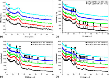
The XRD patterns obtained for all the powder samples of the melt-quenched glasses as shown in Fig. 1(a) exhibit typical broad halos indicating the complete amorphous nature. The XRD patterns of the glasses after immersion in the SBF solution for 1 day exhibited broad halos showing no indication of any crystalline phases (not shown in Fig. 1). After immersion in the SBF solution for 3, 7, and 14 days, the XRD patterns of certain glasses have shown sharp diffraction peaks as presented in Figs. 1(b)–1(d), respectively, depending on the glass composition. The peaks have been identified and assigned to standard hexagonal HAp (JCPDS: 09-0432) and HCA (JCPDS: 04-0697) crystalline phases. The presence of a broad halo occurring at around 2θ = 22.5° along with the sharp crystalline peaks of all the samples matches with the XRD pattern of the Si–O–Si gel layer Reference Ou, Xu, Warnett, Holmes, Zaheer, Garforth, Williams, Jiao and Fan28 confirming the formation of the silica gel layer along with HAp and HCA. From Figs. 1(b) and 1(c), it can be observed that XRD patterns of B0, B1, and B2 samples have shown well-defined peaks of HAp and HCA phases after immersing in the SBF solution for 3 and 7 days, while XRD patterns of the B4 and B8 samples are dominated by broad halos indicating the lack of formation of HAp and HCA. In Fig. 1(d), after 14 days immersion, well-defined peaks of HAp and HCA phases are observed for all the glass samples except in the B8 sample indicating the lack of apatite formation.
The XRD pattern of (a) melt-quenched glass powders; the XRD pattern of glass powders after immersion in the SBF solution for (b) 3 days, (c) 7 days, and (d) 14 days.

B. Fourier-transform infrared (FTIR) spectroscopy studies
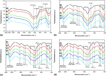
The FTIR transmission spectra of the melt-quenched glass samples in Fig. 2(a) show various vibrational bands corresponding to Si–O–Si bond vibrations. The bands at 1060 and 922 cm−1 are assigned to Si–O–Si asymmetric stretching and Si-NBO stretching, respectively. The band at around 755 cm−1 corresponds to Si–O–Si symmetric stretching vibrations. The bands occurring at 590 and 464 cm−1 correspond to the bending modes of O–Si–O and Si–O–Si bonds, respectively. Reference El Badry, Moustaffa, Azooz and El Batal29
FTIR transmission spectra of (a) melt-quenched bulk glass samples, glass powders after immersion in the SBF solution for (b) 3 days, (c) 7 days, and (d) 14 days.

The FTIR transmission spectra of different glass powders after immersion studies in SBF solution for 3, 7, and 14 days are shown in Figs. 2(b)–2(d), respectively. The vibrational bands corresponding to Si–O–Si bonds, (PO4)3−, (CO3)2,− and molecular water were identified in the FTIR spectra Reference El Badry, Moustaffa, Azooz and El Batal29–Reference Ren and Leng31 of certain samples depending on the composition. The band occurring at 3448 cm−1 corresponds to molecular water vibrations. The bands occurring at 1645 and 1750 cm−1 are due to the presence of molecular water. The bands occurring at 1413 and 1470 cm−1 are due to asymmetric stretching vibrations of the (CO3)2− unit while 873 cm−1 represents bending vibrations of the (CO3)2− unit. This confirms that the apatite is of B-type HCA in which (CO3)2− units partially substitute (PO4)3− units in the HAp lattice. The bands at 1095 and 1035 cm−1 are assigned to asymmetric stretching of the P–O bond in the (PO4)3− units, while the band at 960 cm−1 is assigned to the symmetric stretching mode of the P–O bond in the (PO4)3− units. The bands at 606 and 566 cm−1 are assigned to P–O bond bending in the (PO4)3− units.
The bands occurring at 1249, 1064, 800, and 468 cm−1 correspond to out-of-plane asymmetric stretching, in-plane asymmetric stretching, symmetric stretching, and bending of Si–O–Si, respectively. From Fig. 2(b), it is observed that all the samples after 3 days of immersion show similar bands corresponding to Si–O stretching and bending as well as molecular water vibrations. But the presence of P–O bending and C–O bond stretching is more dominant in the base glass (B0) sample and decreases with the gradual incorporation of Bi2O3. However, the asymmetric stretching mode of the P–O bond cannot be well defined as it is overlapped with the vibrational bands of Si–O–Si asymmetric stretching. In Fig. 2(c), it is observed that after immersion in the SBF solution for 7 days, the intensity of all the bands related to the P–O bond is found to be increased compared to that of the samples after immersion in the SBF solution for 3 days. Also, the P–O asymmetric stretching band in samples immersed in the SBF solution for 7 days has become distinct. Furthermore, the bands corresponding to P–O bending and stretching, Si–O–Si symmetric stretching, and C–O bond bending stretching are more dominant in the base glass (B0) and gradually reduces with the incorporation of bismuth oxide. From Fig. 2(d), the glass powders after immersion in the SBF solution for 14 days, it is observed that the vibrational bands are quite similar to those of the glass powders which were immersed in the SBF solution for 7 days.
C. Thermal properties
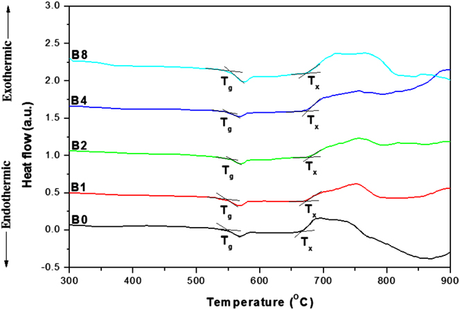
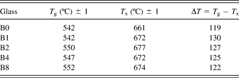
The DSC thermograms for all the glasses recorded at 10 K/min of rate of heating are shown in Fig. 3. It can be observed that all samples show a distinct endothermic signal corresponding to the glass transition temperature (T g) followed by a broad exothermic peak corresponding to crystallisation. The glass transition temperature (T g), crystallisation onset temperature (T x), and glass stability factor (ΔT = T x − T g) for all the glasses are summarised in Table II.
DSC thermogram of the glass powders recorded at a heating rate of 10 K/min.

The values of glass transition temperature (T g), crystallisation onset temperature (T x) and glass stability factor (ΔT) for different glasses obtained from their respective DSC thermogram at heating rate of 10 K/min.

D. Density
The measured density values of the glasses are 2.68, 2.69, 2.71, 2.77, and 2.85 g/cc for B0, B1, B2, B4, and B8 samples, respectively. Thus, the variation in the density values indicates that it increases with the gradual substitution of Bi2O3. As the density of Bi2O3 (8.90 g/cc) is much higher than that of SiO2 (2.196 g/cc) and Na2O (2.127 g/cc), the density of glasses increases with the increase in substitution of Bi2O3 replacing SiO2 and Na2O.
E. Microstructural studies
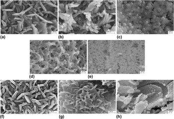
FESEM micrographs of all five glass samples immersed in the SBF solution for 7 days are shown in Figs. 4(a)–4(e) which corresponds to B0, B1, B2, B4, and B8 glass samples, respectively. The B0 glass surface shows a flake-like fine microstructure with high porosity, while the average thickness of the flakes is around 60 nm. The surfaces of B1 and B2 glasses show slightly modified rod-like and spherical morphology, respectively. The average diameter of the rod-like structure in the B1 glass surface is around 70 nm, while the average diameter of the spherical particles on the surface of the B2 glass is around 120 nm. However, from FESEM micrographs, it is apparent that porosity decreases in the case of B1 and B2 samples compared to the B0 glass. Even though there is a difference in the surface morphology of B0, B1, B2 glasses, the formation of HAp and HCA was confirmed on the surface of B1 and B2 samples similar to that of the B0 glass as previously observed in Sec. III.A (XRD analysis). The change in morphology of the surface apatite layer may be attributed to the gradual substitution of Bi2O3 in the glass. The B8 glass surface after immersion in the SBF solution for 7 days does not seem to contain a precipitated layer of HAp or HCA and may be only having some polymerised silica gel layers on its surface. Section III.A (XRD analysis) also showed similar evidences of formation of the Si–O–Si gel layer.
FESEM micrographs of glass samples immersed in SBF for 7 days, (a) B0, (b) B1, (c) B2, (d) B4, and (e) B8 and for 14 days, (f) B0, (g) B2, and (h) B8.

FESEM images of three selected glasses (B0, B2, and B8) immersed in the SBF solution for 14 days are shown in Figs. 4(f)–4(h), respectively. The surface of the B0 glass sample shows similar morphology to that of immersion in the SBF solution for 7 days, while the morphology changes from spherical to flake-like crystals in case of the B2 glass sample surface.
F. pH and ion release studies
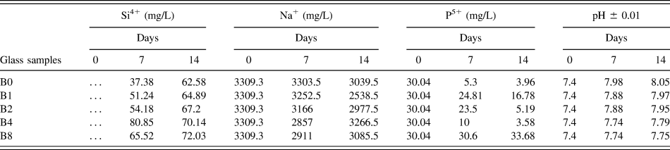
The concentration of Si4+, Na+, and P5+ ions released from glasses during immersion in SBF solution has been analyzed from the supernatant collected after 7 and 14 days, and the data is given in Table III. From Table III, it can be observed that the concentration of Si4+ ions increases in the SBF supernatant as Bi2O3 substitution is gradually increased in glass. Generally, Si4+ ions are released in small concentrations from the glasses along with the formation of the Si–O–Si gel layer on the glass surface. Also, some Si4+ ions in the form of (SiO4)4− may replace few (PO4)3− ions in the apatite lattice. Reference Ibrahim, Mostafa and Korowash32 Thus, it is expected that with an increase in apatite formation on the surface, there will be an increase in the Si4+ ion substitution in apatites. As reported in Sec. III.A (XRD analysis), it was observed that with gradual increase in Bi2O3 substitution in the glass composition, apatite formation was gradually reduced. Thus, when the rate of Si4+ ion release from glass becomes higher than the rate of Si4+ ion substitution in apatites, there might be an increase in Si4+ ion concentration in the SBF supernatant. After immersion in the SBF solution for 14 days for the B4 glass, Si4+ ion concentration decreases in comparison to the Si4+ concentration after 7 days among all glasses studied due to slower surface apatite formation and Si4+ substitution in apatites. The highest concentration of Si4+ ion is evidenced in the B8 sample after 14 days which is also attributed to the lack of apatite formation.
Concentration of Si4+, Na+, P5+, and Bi3+ ions and pH of SBF supernatant for different immersion time of all glasses.

From Table III, it can be observed that for B0, B1, and B2 glasses, the Na+ ions in the supernatant after 14 days is lower compared to the concentration of Na+ ions after 7 days. It can be concluded that Na+ ions may participate into an apatite structure forming substituted apatites. Reference Ibrahim, Mostafa and Korowash32 However, due to the poor apatite forming ability of B4 and B8 glasses, an increase in the concentration of Na+ ions in the supernatant is observed after 14 days in comparison to the supernatant after 7 days.
The concentration of P5+ ions in the SBF supernatant of all glasses has decreased in comparison to the prepared SBF due to the utilization of these ions for the formation of HAp and HCA layers except the B8 sample. Least apatite formation has been evidenced for the B8 glass resulting in a higher P5+ ion concentration in the supernatants.
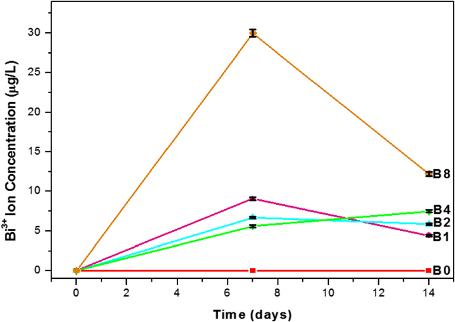
Figure 5 shows the concentration of Bi3+ ions in the supernatant collected after immersing the glass samples in the SBF solution for 7 and 14 days. Interestingly, Bi3+ ion release is very low (in ppb range) for all the samples in comparison to other ions due to the comparatively lower concentration of Bi2O3 in the glass composition. After immersion in the SBF solution for 7 days, the highest release of Bi3+ ions is observed in case of the B8 sample. For other Bi2O3-substituted glasses, their SBF supernatant shows a lower Bi3+ ion concentration than that of the B8 glass sample. However, after immersion for 14 days, the Bi3+ ion concentration increases in the supernatant with the gradual increase of Bi2O3 in the glass composition. Glasses B1 and B2 show higher Bi3+ ion concentration in the supernatant after 7 days compared to that of 14 days. This may occur due to the participation of some released Bi3+ ions into the apatite lattice during immersion for 7 days. Reference Webster, Massa-Schlueter, Smith and Slamovich20,Reference Ciobanu, Bargan and Luca21
Concentration of Bi3+ ions in the supernatent with different immersion time of all glasses studied in SBF solution.

The pH values measured from the SBF supernatant collected after 7 and 14 days are reported in Table III. Compared to the pH values of SBF supernatants after 7 days, there is an increase in the pH values with that of 14 days. It can also be observed that the pH values gradually decrease with the increase in Bi2O3 substitution in glasses after both 7 and 14 days.
G. Cell proliferation studies
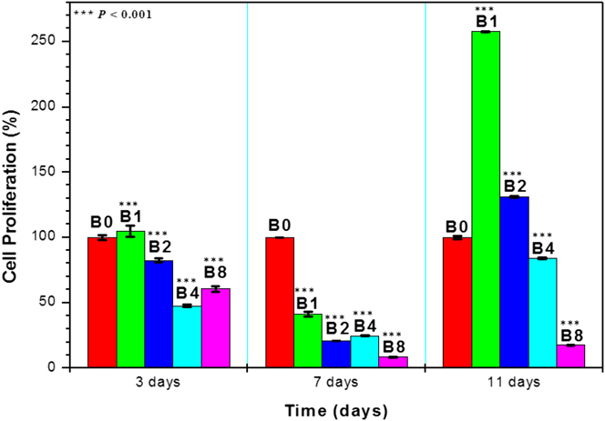
The cell proliferation (%) of NIH3T3 mouse fibroblast cells on the surface of B0, B1, B2, B4, and B8 glass samples for 3, 7, and 11 days has been estimated from the OD values obtained through an MTT assay, and the plotted data is shown in Fig. 6. The base glass (B0) was considered as the control. All calculated P values showed statistically highly significant results (P < 0.001) compared to the base glass (B0). It can be observed that among Bi2O3-substituted glasses, cell proliferation is higher for 1 wt% Bi2O3 substitution and decreases with further increase in Bi2O3 substitution in all the time periods studied. After 3 days, the B1 glass shows a marginally higher cell proliferation (%) than that of cells on the base glass (B0), whereas cell proliferation is less in B2 and B4 glasses compared to the base glass. The least cell proliferation (%) was evidenced on the B8 sample indicating the adverse effect of Bi3+ ions on cell proliferation. After 7 days, cell proliferation rates on all the Bi2O3-substituted glasses are lower than those of the base glass (B0). But after 11 days, the cell proliferation (%) is higher for B1 and B2 glasses and gradually decreases in B4 and B8 glasses. However, there is an increase in cell proliferation after 11 days compared to 7 days for all the Bi2O3-substituted glasses.
Cell proliferation (%) of NIH3T3 mouse fibroblast cells on the surface of different glass samples plotted using MTT assay results and taking the base glass (B0) as control. Data are expressed as mean cell proliferation (%) ± SE. Statistical significance was determined by using the P value from Student’s t-test (one-tailed hypothesis with two independent samples having equal variance). Samples show statistical differences for all the samples relative to the base glass (B0) as control at ***P < 0.001.

H. Antibacterial studies
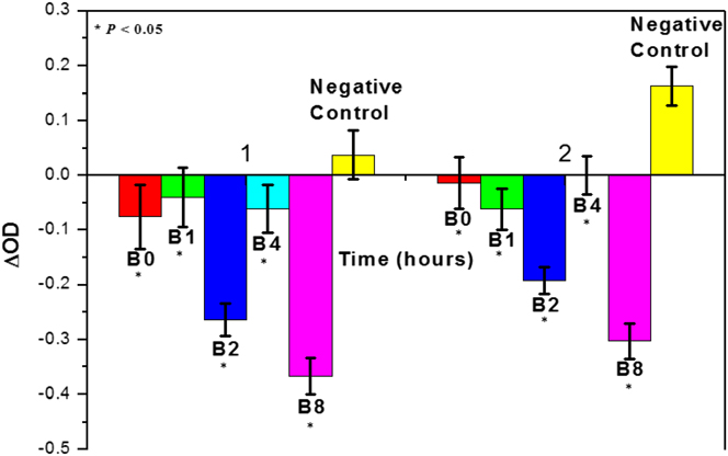
The change in optical density (ΔOD) values obtained for all the glass powders-added E. coli bacterial suspensions from the broth microdilution method was used to understand the bacteriostatic effects of the glass samples. Figure 7 shows the comparison of ΔOD values of glass powders-added Escherichia coli (E. coli) bacterial suspensions after 1 and 2 h of incubation with ΔOD values of the base glass (B0) as the positive control and ΔOD of the only E. coli bacterial suspension as the negative control. From the Student’s t-test analysis, it is ensured that all the samples exhibited statistically significant results (P < 0.05) compared to the negative control. It can be observed that all the Bi2O3-substituted glasses as well as the base glass (B0) has shown a bacteriostatic effect on the E. coli bacteria after 1 and 2 h of incubation when compared with the negative control.
Plot of ΔOD values of the different glass powders added bacterial suspension after incubation for 1 and 2 h with base glass (B0) as positive control and only bacterial suspension as negative control. Data are expressed as mean ΔOD values ±SE. Statistical significance was determined by using the P value from Student’s t-test (one-tailed hypothesis with two independent samples having equal variance). Samples show statistical differences for all the samples relative to the negative control at *P < 0.05.

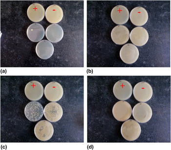
The bactericidal properties of the glass powders were studied by counting the CFU on cultured agar plates spread with the glass powders-added bacterial suspension. Figure 8 shows the cultured agar plates spread with different glass powders-added bacterial suspensions incubated for 24 h. Each image in Fig. 8 contains one cultured agar plate for positive control (+) and another cultured agar plate for negative control (−) along with the cultured agar plates for Bi2O3-substituted glasses in triplicates. On observing the CFU present on the respective agar plates of Bi2O3-substituted glasses, it can be concluded that B1 glass powders having 1 wt% Bi2O3 substitution have caused bactericidal activity on the E. coli bacteria. There are no CFUs present in the cultured agar plates applied with 24 h incubated B1 glass powders-added bacterial suspension while “too numerous to count” (TNTC) CFUs are present on agar plates applied with glass samples having Bi2O3 substitution above 1 wt%. The positive-control and negative-control agar plates in the entire images show TNTC colony-forming units (CFU) as observed in Fig. 8.
Overnight cultured agar plates: (+) denotes positive control, (−) denotes negative control and three agar plates applied with 24 h incubated glass powders [(a) B1, (b) B2, (c) B4, and (d) B8] added E. coli bacterial suspension.

IV. DISCUSSION
The XRD and FTIR results of as-prepared glasses have shown the amorphous nature of the glasses and absence of significant structural changes in the glasses, respectively. However, it should be noted that in FTIR spectra of bismuth oxide containing glasses, the stretching vibrations related to Bi–O bond in distorted BiO6 units (490–500 cm−1) could be overlapped with Si–O–Si and O–Si–O bending vibrational bands. Also, the symmetric stretching vibrations of Bi–O bond in BiO3 units at 840–880 cm−1 may also be overlapped with the broad and intense vibrational bands associated with Si-NBO stretching. Reference Ardelean, Cora and Ioncu33,Reference He, He, Xie and Li34 After SBF immersion studies, XRD, FTIR and FESEM studies showed similar results where substitution of Bi2O3 upto 2 wt% had no adverse effect on surface apatite forming ability of modified glasses but significant reduction in surface apatite formation could be seen for the glasses with Bi2O3 substitution higher than 4 wt%. With the gradual increase in Bi2O3 concentration in glass, the poor apatite forming ability is attributed to the increased network connectivity on account of the reduction in non-bridging oxygen associated with the decrease in Na2O content in the glasses. The results obtained during present bioactivity studies contradict with the results obtained by Mohn et al. who reported improved apatite formation for Bi2O3 added nano-bioactive glass powders. Reference Mohn, Zehnder, Imfeld and Stark23 However, in their studies, the actual role of Bi3+ ions in the glass network was not reported.
To understand the glass stability, the crystalline phase formed during heating was investigated for which selected glass samples were heat treated at 725 °C, and the corresponding phase was identified as Na2Ca2Si3O9 (JCPDS 22-1455) from their XRD patterns (not shown). Similar phase formation upon heat treatment was reported by Fujikura et al. and Massera et al. Reference Massera, Fagerlund, Hupa and Hupa35,Reference Fujikura, Karpukhina, Kasuga, Brauer, Hill and Law36 The glass stability factor plays a significant role in scaffold processing or sintering as the glassy phase should not get devitrified when heated above T g. Reference Wang, Fagerlund, Massera, Södergård and Hupa37 The well-known 45S5 glass exhibits a ΔT value of 88 °C and is prone to crystallisation forming the Na2CaSi2O6 crystalline phase while scaffold processing. Reference Massera, Fagerlund, Hupa and Hupa35 Bismuth oxide-modified glasses have higher ΔT values than that of base glass (B0) as well as 45S5 glass, indicating their suitability in scaffold processing without devitrification.
The concentration of Bi3+ ions released from the Bi2O3-substituted glasses is in the ppb range, which could be due to lower concentration of Bi2O3 in glasses and participation of the Bi3+ ion as the network former through [BiO3] and [BiO6] polyhedral structural units instead of the network modifier.
The fibroblast cells were used in the present cell proliferation studies due to their wide acceptability in cell viability and toxicity studies for several decades. Reference da Silva Gasque, Al-Ahj, Oliveira and Magalhães38 The results obtained in ion release studies and pH measurements could be correlated to understand the cell compatibility and antibacterial properties of the glasses. The pH values decrease with a gradual increase in Bi2O3 substitution in glass, and improved cell proliferation has been observed at a slightly increased pH. Reference Kruse, Singh, Targosinski, Sinha, Sørensen, Eriksson and Nuutila39,Reference Galow, Rebl, Koczan, Bonk, Baumann and Gimsa40 However, after 3 days of cell growth, the B1 glass showed a slightly higher cell proliferation compared to the base glass (B0). This could be due to the formation of Bi-substituted HAp, as it has been reported that the incorporation of Bi3+ ions substituting the Ca2+ ions in the apatite improves the cell viability. Reference Webster, Massa-Schlueter, Smith and Slamovich20 Though, Bi-substituted HAp formation was similar in B1 and B2 glasses, the B1 glass showed better cell proliferation than the B2 glass due to a slightly higher pH value. Also, there is a possibility that the high concentration of Bi3+ ions can also affect the cell proliferation. For B4 and B8 glasses, lower pH changes with that of the growth media (7.4), reduced the surface apatite-forming ability, and higher Bi3+ ion concentration could have resulted in lower cell proliferation compared to B0, B1, and B2 glasses. After 7 days, there is a decrease in cell proliferation on all the Bi2O3-substituted glasses though the surface apatite and pH values are expected to increase compared to that of 3 days. This could be only due to the increase in Bi3+ ion concentration in the surrounding medium. After cell proliferation studies for 11 days, the higher cell proliferation in B1 and B2 glasses compared to the base glass (B0) is related to the decrease in the Bi3+ ion concentration in the SBF supernatant between 7 and 14 days as revealed in the ion concentration studies. It should be noted that bismuth compounds on excessive intake are associated with neurological toxicity like encephalopathy and nephrological problems like renal failures. Reference Thomas, Bialek and Hensel41,Reference Reynolds, Abalos, Hopp and Williams42 However, it has been reported that the blood level of the Bi3+ ion in humans below 50 ppb was unlikely to induce toxicity. Reference Reynolds, Abalos, Hopp and Williams42 The Bi3+ ion concentrations measured from SBF supernatants after 7 and 14 days are lower (4.4–9.1 ppb) for all the bismuth-oxide substituted glasses except B8 (12.24–30 ppb), corroborating the lowest cell compatibility of the B8 sample.
To evaluate the antibacterial action of the developed glasses, a Gram-negative Escherichia coli (E. coli) was selected instead of the Gram-positive bacterium for the preliminary study to understand the efficacy of bismuth ions. As the Gram-positive bacterium (e.g., S. aureus) possesses a thicker cell wall and more peptidoglycan molecules; the action of the antibacterial ions is more susceptible in the Gram-negative bacterium compared to the Gram-positive bacterium. Reference Feng, Wu, Chen, Cui, Kim, Kim, Kim and Kim43 The bacterial growth inhibition is expected to occur due to the increase in the pH value of the surrounding medium for bioactive glasses. Reference Allan, Newman and Wilson8 Also, Bi2O3-substituted glasses release Bi3+ ions into the surrounding medium which can bring in possible inhibition to the bacterial cells. The base glass (B0) showed higher inhibition than the B1 glass after 1 h, which is attributed to the increased pH change as shown in Table III. However, it is important to mention that in case of the B2 sample, though the change in pH value was decreased, a stronger inhibition is observed compared to B0 and B1 glasses due to the probable antibacterial action of Bi3+ ions rather than the pH effect. In case of the B8 glass, though it exhibits the least pH change compared to the pH of MHB, the higher Bi3+ ion release influenced the bacterial inhibition. Moreover, except the B1 glass, all the other glass samples exhibited a decrease in the ΔOD value of 2 h compared to 1 h. This could be due to the simultaneous influence of pH changes as well as the antibacterial action of Bi3+ ions, creating the strongest inhibitory effect toward the bacteria in the B1 glass. Interestingly, the B1 sample also demonstrated bactericidal properties as revealed in agar plate studies. Further, this can be evidenced from the fact that only the B1 glass showed continuous increase in bacterial inhibition while for other glasses, the bacterial inhibition was decreased with time.
As the B1 glass showed encouraging bacteriostatic and bactericidal effects on Gram-negative E. coli, the evaluation of bacteriostatic and bactericidal action of this glass is intended to be evaluated against the Gram-positive bacteria (e.g., S. aureus) in future, which may also give an insight about the possible antibacterial action of penetration through the cell wall.
V. CONCLUSIONS
In the present work, Bi2O3 modified glasses substituting SiO2 and Na2O equally with 1, 2, 4, and 8 wt% Bi2O3 have been synthesized using the conventional melt-quenching technique. XRD analysis of the as-cast glasses confirmed the amorphous nature of the glasses, while FTIR spectroscopy does not reveal any noticeable changes among the glasses due to the possible overlapping of the bands related to the stretching of BiO3 and BiO6 units with that of Si–O–Si stretching and bending bands. From DSC analysis, it could be observed that the glass transition temperature does not show major changes, while the glass stability factor (ΔT) for all the Bi2O3-containing glasses is higher than that of the base glass. Density of the glasses increases with a gradual increase in Bi2O3 substitution due to the higher density of Bi2O3 compared to SiO2 or Na2O. After SBF immersion for 7 and 14 days, it is observed that the surface apatite forming ability decreases with the gradual substitution of Bi2O3 in glasses as evidenced from XRD, FTIR, and FESEM studies. The B8 glass having the highest Bi2O3 content did not show any apatite formation up to 14 days of SBF immersion, which is attributed to the increased network connectivity. The release of Bi3+ ions into SBF for all the glasses is limited in the ppb level (4–30 µg/L). All the Bi2O3 modified glasses exhibit a bacteriostatic effect on the E. coli bacteria after 1 and 2 h of incubation. In particular, the 1 wt% Bi2O3-containing glass shows better cell proliferation rates as well as bactericidal properties compared to the base glass. This study infers that the inclusion of bismuth ions (1 wt%) into commercial glasses like (S53P4) could be beneficial with a desired combination of optimized apatite formation on the surface, cell compatibility and antibacterial properties. Finally, it can be concluded that Bi2O3 being a widely used radiopacifier can be a potential additive to bioactive glasses along with its antibacterial properties.
ACKNOWLEDGMENTS
Authors would like to express their grateful thanks to Dr. K. Muraleedharan, Director, CSIR-CGCRI and Dr. Ranjan Sen, Head, Glass division for their encouragement and support toward this work. The financial help from P 81-113 of the CSIR Extramural Research Fund for Young Scientists and CSIR-HRDG (Extramural Research Group) for providing GATE-JRF fellowship to SPS for pursuing Integrated M.Tech Ph.D. program is gratefully acknowledged. The authors gratefully acknowledge the suggestions and experimental help offered by Dr. Biswanath Kundu, Senior Scientist and Dr. Vamsi K. Balla, Senior Principal Scientist, Bioceramics and Coating Division, CSIR-CGCRI.