Introduction
Endoscopic ear surgery (EES) has evolved from a complementary adjunct to the operating microscope into a well-established operative modality for a range of otologic procedures.Reference Presutti, Gioacchini, Alicandri-Ciufelli, Villari and Marchioni1, Reference Li, Zhou, Wang, Wang and Zou2 Compared with conventional microscopic techniques, EES delivers a high-definition, wide-angled view of the operative field including recesses that are otherwise obscured.Reference Guy, Muzaffar and Coulson3 These advantages translate into improved visualisation of challenging anatomy and facilitate less invasive approaches. In addition, the endoscope is comparatively inexpensive, portable and accessible.
Competency in EES presents a technical challenge and, with any skill, is associated with a learning curve. Surgeons must operate single-handedly while the contralateral hand stabilises the endoscope, adapt to an altered sense of depth perception including a loss of stereopsis and adapt to altered haptic feedback.Reference Wilhelm, Reiser, Kohn, Witte, Leiner and Mühlbach4 These limitations can be partially mitigated through motion parallax cues and stepwise exposure. Notably, prior microscopic experience may be associated with a slower initial adaptation to EES, reflecting the need to recalibrate existing visuomotor patterns for a different technique.Reference Lucidi, Fernandez, Botti, Amorosa, Alicandri-Ciufelli and Villari5 Traditional Halstedian training remains the cornerstone of surgical education, grounded in an apprenticeship-style model.Reference MacCallum6 Simulation has emerged as a valuable adjunct, enabling deliberate and structured practice of core EES skills within a risk-free environment.
A range of physical simulators for EES has been described, from cadaveric temporal bones to patient-specific three-dimensional (3D)-printed models.Reference Anschuetz, Bonali, Ghirelli, Mattioli, Villari and Caversaccio7–Reference Molinari, Emiliani, Cercenelli, Bortolani, D’Azzeo and Burato11 Despite this diversity, no systematic review has undertaken a focused evaluation of physical simulation models for EES, specifically with respect to their validity and educational utility. This review aims to address this.
Methods
Search strategy
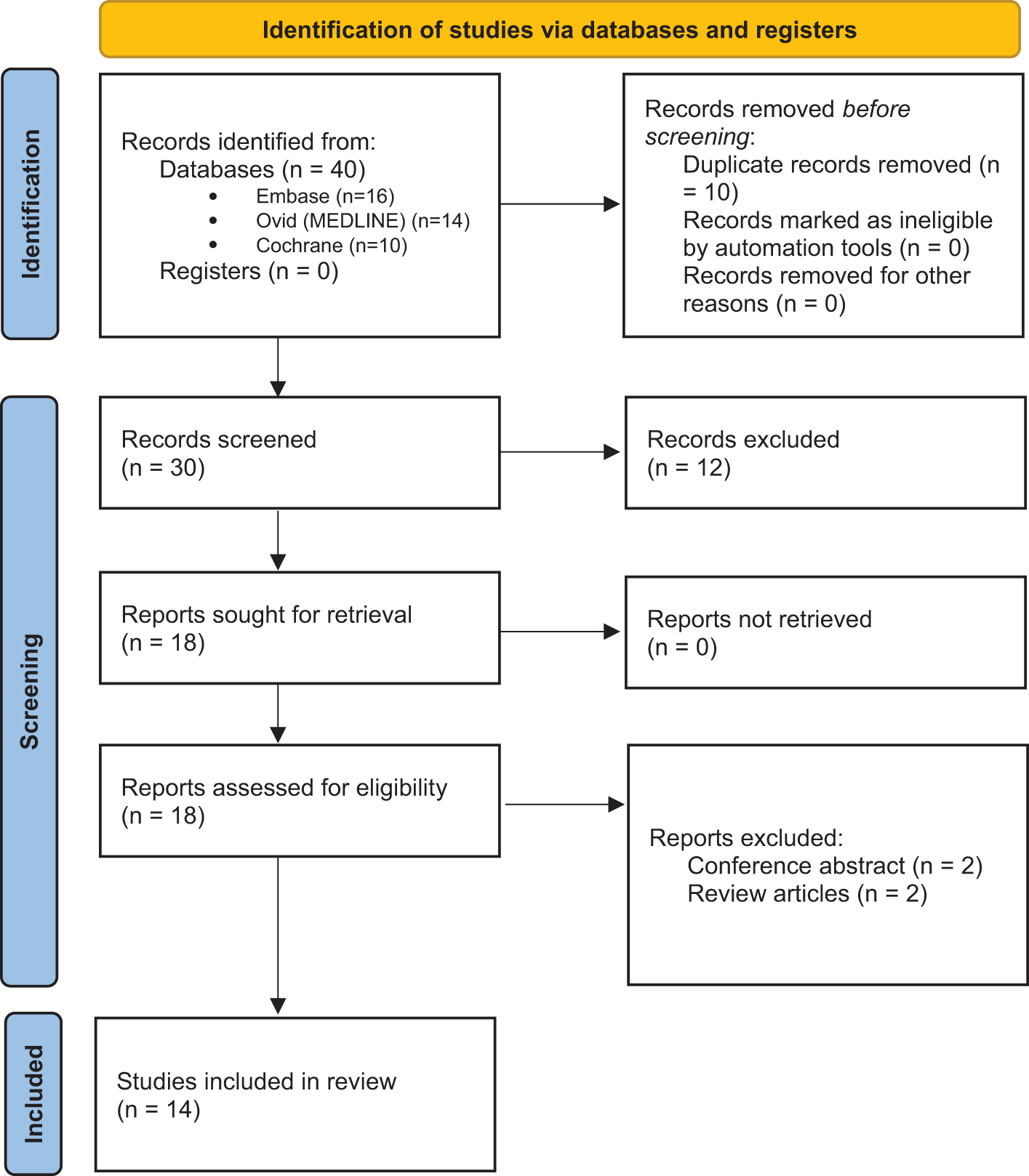
This systematic review was conducted in accordance with the Preferred Reporting Items for Systematic Reviews and Meta-Analyses (PRISMA 2020) statement (Figure 1). A protocol was prepared in advance but not registered on PROSPERO, as methodology-only reviews of simulation devices are currently ineligible for inclusion.
Preferred Reporting Items for Systematic Reviews and Meta-Analyses (PRISMA) 2020 flow diagram.

The electronic databases PubMed (MEDLINE), Embase (Ovid) and the Cochrane Library were searched from database inception to 23 June 2025. No date filters were applied. Searches were limited to English-language publications. The following Boolean strategy was adapted for each platform: (endoscopic ear surgery OR EES OR transcanal endoscopic ear surgery OR TEES OR ear endoscopy) AND (simulation OR simulator OR surgical training model OR surgical education) AND (model OR training model OR phantom OR cadaveric OR synthetic OR 3D-printed OR 3D printing OR 3-dimensional) AND (training OR education OR teaching OR validation OR assessment OR competency). The search syntax was modified as necessary to meet the indexing requirements of each database.
Study selection and eligibility (PICOS)
Eligible studies were primary research describing physical simulators for EES (Population). The intervention was defined as the use of a simulator for skills acquisition, educational assessment or validation (Intervention). While a comparator was not required, studies including participants of varying expertise, comparisons between alternative devices or no-intervention controls were eligible (Comparator). Outcomes were defined as validation domains including face, content and construct validity, the latter often assessed via Objective Structured Assessment of Technical Skills (OSATS), as well as other educational end-points such as task completion time, error rate, knowledge change and participant satisfaction (Outcome). Eligible study designs comprised randomised controlled trials, non-randomised studies, cohort studies, before–after comparisons and descriptive validation reports.
Validation domains
Prior to data extraction, operational definitions for validity evidence were pre-specified, adapted from established frameworks in surgical education and simulation methodology. The validation domains assessed included face, content, construct, task-specific and global validity. Face validity refers to the perceived realism of the simulator by users.Reference Cronbach and Meehl12 Content validity reflects the extent to which the simulator encompasses the essential components of the intended procedure.Reference Haynes, Richard and Kubany13 Construct validity denotes the ability of the model to distinguish between users of varying expertise, typically demonstrated by performance differences between novices and experts.Reference Cronbach and Meehl12 Task-specific validity assesses fidelity in simulating discrete procedural elements, such as anatomical landmarks or instrument handling.Reference McDougall14 Global validity evaluates the simulator’s capacity to replicate the entire operative workflow, including ergonomics and procedural integration.Reference McDougall14 Incorporating these domains enables systematic comparison across studies and reinforces the methodological distinction of this review from prior scoping reviews.
Data extraction and management
Title- and abstract-level screening was performed independently by two reviewers (G.R.H., P.S.), with full-text eligibility confirmed by the same reviewers and arbitration by the remaining authors (P.R.C., H.T.) where required. Deduplication and screening were carried out manually using Microsoft Excel (version 16.99); no automation tools were used due to the limited number of search results.
Studies were included if they described physical (non-virtual) simulation models for EES, involved any procedure performed using a transcanal endoscopic approach to the middle ear, reported at least one validation domain (e.g., face, content, construct) and/or an educational outcome and were original, peer-reviewed full-text articles published in English.
Exclusion criteria comprised studies involving virtual, augmented or mixed-reality simulators; simulators developed solely for pre- or intra-operative planning without a training purpose; studies unrelated to ear surgery or not involving an endoscopic approach; and all narrative reviews, conference abstracts, editorials or letters without original data.
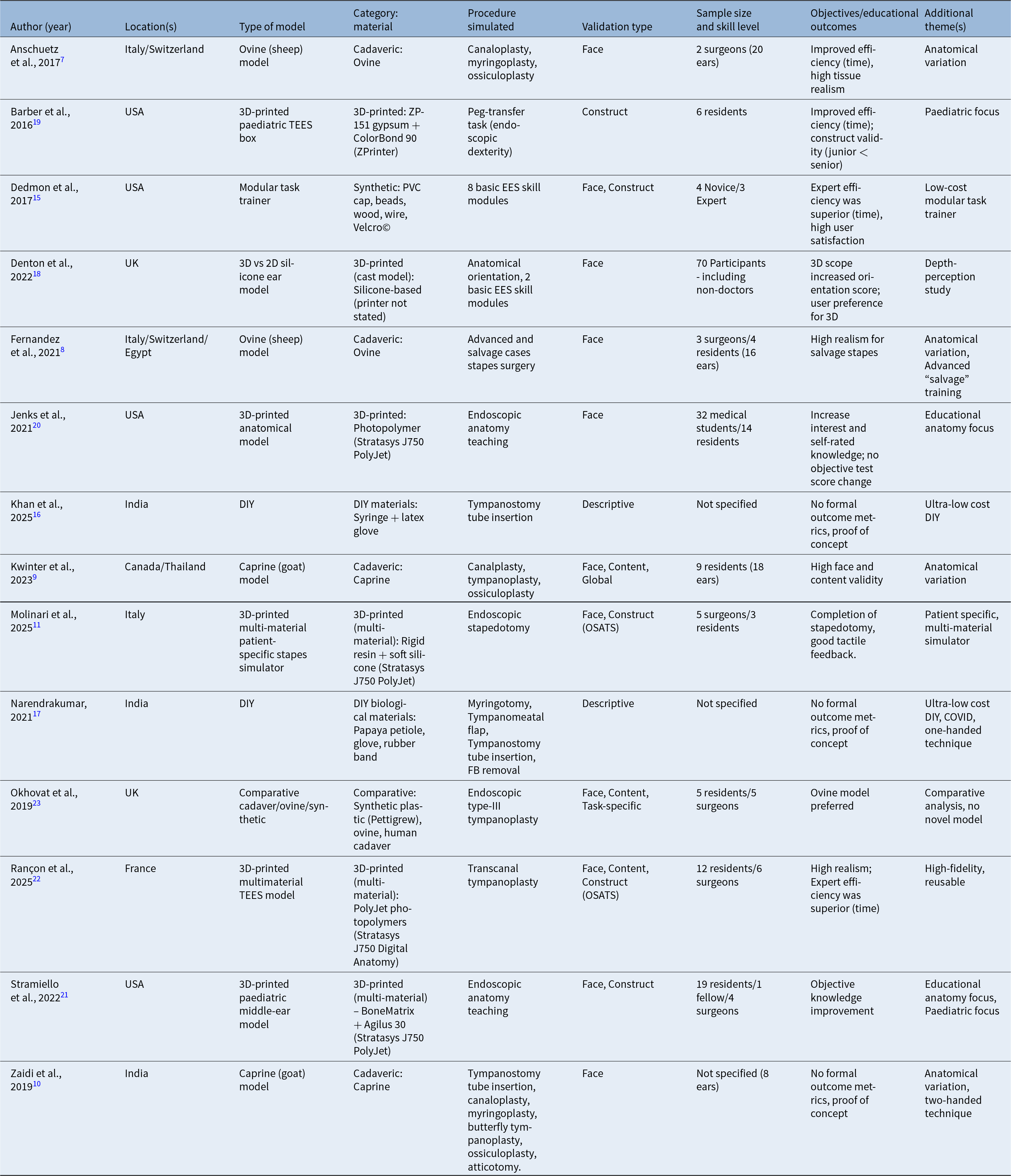
A standardised data extraction sheet was piloted on three studies and subsequently applied to all included articles. Extracted variables included: bibliographic details (author, year, country); simulator type, material(s) used and 3D printer model (if applicable); procedures simulated; reported validation domains; participant cohort (sample size, skill level and number of ear specimens, where available); main objectives or educational outcomes; and additional thematic findings. Data were tabulated using Microsoft Word (version 16.98) and cross-checked for transcription accuracy. As a result of heterogeneity in methodology and educational outcomes, meta-analysis was not carried out; instead, results are presented descriptively in a comparative table (Table 1). No universally accepted risk-of-bias tool exists for simulation validation studies; therefore, formal scoring was not performed.
Overview of simulators for endoscopic ear-surgery training included in this systematic review

Note: For every study the table summarises: author (year), geographic setting, simulator type, main construction material(s) ± 3D-printer model, procedure(s) simulated, reported validation domain(s), participant cohort (formatted “participants [ears]” when ear-counts were given), key educational outcome(s) and distinctive themes. Participant labels were harmonised: “residents” covers residents/registrars/trainees; “surgeons” covers consultants/attendings; where grade was absent, experience descriptors (novice, expert) were retained. Validation domains: Face = perceived realism/usefulness; Content = curricular/task relevance; Construct = discrimination between expertise levels or statistically significant learning gain (including OSATS or pre- and post-testing).
3D = three-dimensional; 2D = two-dimensional; DIY = do-it-yourself; EES = endoscopic ear surgery; FB = foreign body; OSATS = Objective Structured Assessment of Technical Skills; PVC = poly-vinyl chloride; TEES = transcanal endoscopic ear surgery.
Results
Study selection summary
A total of 40 records were retrieved. After removal of 10 duplicates, 30 titles and abstracts were screened. Twelve records were excluded because they were not ear-surgery–related, did not involve an endoscopic approach or described simulators intended solely for peri-operative planning. The remaining 18 full-text articles were assessed. Four further records were excluded because they were conference abstracts and narrative reviews. A total of 14 studies met the inclusion criteria and were entered into qualitative synthesis.Reference Anschuetz, Bonali, Ghirelli, Mattioli, Villari and Caversaccio7–Reference Molinari, Emiliani, Cercenelli, Bortolani, D’Azzeo and Burato11, Reference Dedmon, O’Connell, Kozin, Remenschneider, Barber and Lee15–Reference Okhovat, Milner and Iyer23 The selection process is shown in the PRISMA 2020 flow diagram (Figure 1).
Simulator typology and materials
Cadaveric animal heads, including ovineReference Anschuetz, Bonali, Ghirelli, Mattioli, Villari and Caversaccio7, Reference Fernandez, Bonali, Yacoub, Ghirelli, Fermi and Presutti8 and caprineReference Kwinter, Chayaopas, Ma and James9, Reference Zaidi, Khan and Parab10 specimens, provided high-fidelity tissue handling and anatomical realism and were used to simulate procedures including endoscopic canaloplasty, tympanoplasty and ossiculoplasty. Synthetic modular trainers enabled low-cost, task-specific rehearsal of fundamental EES skills and basic psychomotor training.Reference Dedmon, O’Connell, Kozin, Remenschneider, Barber and Lee15 Do-it-yourself (DIY) models constructed using readily available materials offered accessible, home-based simulation options.Reference Khan, Patel, Parab, Ingale and Shinde16, Reference Narendrakumar17 Single-material 3D-printed models predominantly replicated osseous anatomy using rigid substrates, facilitating orientation and instrumentation practice.Reference Denton, Brahmabhatt, Ahmed and Sanu18–Reference Jenks, Patel, Bennett, Dunham and Devine20 In contrast, multi-material 3D-printed simulators integrated both hard- and soft-tissue analogues to enhance anatomical and tactile fidelity which enabled simulation of transcanal tympanoplasty and stapedotomy.Reference Molinari, Emiliani, Cercenelli, Bortolani, D’Azzeo and Burato11, Reference Stramiello, Wong, Good, Tor, Ryan and Carvalho21, Reference Rançon, Prebot, Denoyelle, Khonsari and Simon22
Procedures simulated
The simulation models reviewed varied in complexity, ranging from basic dexterity trainers to high-fidelity procedural simulators. Three studies focused on the development of core technical skills, including endoscope control, depth perception and bimanual coordination. These employed modular tasks such as peg transfer, hoop stacking and elastic band placement to support psychomotor training.Reference Dedmon, O’Connell, Kozin, Remenschneider, Barber and Lee15, Reference Denton, Brahmabhatt, Ahmed and Sanu18, Reference Barber, Kozin, Dedmon, Lin, Lee and Sinha19 In addition, three studies incorporated orientation or anatomy-focused components to familiarise trainees with middle ear structures and spatial relationships.Reference Denton, Brahmabhatt, Ahmed and Sanu18, Reference Jenks, Patel, Bennett, Dunham and Devine20, Reference Stramiello, Wong, Good, Tor, Ryan and Carvalho21 The remaining studies replicated defined surgical procedures. Cadaveric animal heads were used to simulate multi-step interventions requiring soft-tissue dissection and anatomical navigation. Three studies employed ovine and caprine models for endoscopic canaloplasty, tympanoplasty and ossiculoplasty, offering tissue handling while requiring adaptation to non-human anatomy.Reference Anschuetz, Bonali, Ghirelli, Mattioli, Villari and Caversaccio7, Reference Kwinter, Chayaopas, Ma and James9, Reference Zaidi, Khan and Parab10 Additionally, Fernandez et al. described a model for complex salvage procedures in endoscopic stapes surgery using ovine heads, incorporating bone cement to mimic a fixed stapes footplate.Reference Fernandez, Bonali, Yacoub, Ghirelli, Fermi and Presutti8 Several high-fidelity models were made using multi-material 3D printing techniques. These platforms allowed simulation of complex procedures such as transcanal tympanoplasty and endoscopic stapedotomy, with anatomically differentiated soft and osseous components designed to replicate operative challenges more accurately.Reference Molinari, Emiliani, Cercenelli, Bortolani, D’Azzeo and Burato11, Reference Rançon, Prebot, Denoyelle, Khonsari and Simon22
Validation evidence
Face validity was the most frequently reported validation type and was assessed in 11 studies.Reference Anschuetz, Bonali, Ghirelli, Mattioli, Villari and Caversaccio7–Reference Molinari, Emiliani, Cercenelli, Bortolani, D’Azzeo and Burato11, Reference Dedmon, O’Connell, Kozin, Remenschneider, Barber and Lee15, Reference Denton, Brahmabhatt, Ahmed and Sanu18, Reference Jenks, Patel, Bennett, Dunham and Devine20–Reference Okhovat, Milner and Iyer23 Most authors used 5-point Likert scales and consistently obtained mean scores ≥ 4/5. Rançon et al. were the sole group to use a 7-point scale, still achieving 6.3/7.0 vs. 6.0/7.0 for residents and experts respectively, further confirming strong face validity across simulator types.Reference Rançon, Prebot, Denoyelle, Khonsari and Simon22
Content validity was formally evaluated in only three studies.Reference Kwinter, Chayaopas, Ma and James9, Reference Rançon, Prebot, Denoyelle, Khonsari and Simon22, Reference Okhovat, Milner and Iyer23 Again, most authors used 5-point Likert scales and consistently obtained mean scores ≥ 4/5.Reference Kwinter, Chayaopas, Ma and James9, Reference Okhovat, Milner and Iyer23 Rançon et al. reported 6.3/7.0 vs. 6.0/7.0 for residents and experts, respectively.Reference Rançon, Prebot, Denoyelle, Khonsari and Simon22 Expert faculty assessment was used to confirm alignment with procedural requirements.
Construct validity was demonstrated in five studies. Barber et al. and Dedmon et al. demonstrated significantly faster task-completion times for senior versus junior residents.Reference Dedmon, O’Connell, Kozin, Remenschneider, Barber and Lee15, Reference Barber, Kozin, Dedmon, Lin, Lee and Sinha19 Similarly, Molinari et al. and Rançon et al. showed higher OSATS scores in expert cohorts.Reference Molinari, Emiliani, Cercenelli, Bortolani, D’Azzeo and Burato11, Reference Rançon, Prebot, Denoyelle, Khonsari and Simon22 Stramiello et al. added a pre- and post-knowledge test and documented a statistically significant improvement after model use.Reference Stramiello, Wong, Good, Tor, Ryan and Carvalho21 Collectively, these studies confirm that well-designed platforms can distinguish novice from expert performance or detect measurable learning gain.
Educational outcomes
Seven groups recorded task-completion time as their primary metric, and four demonstrated statistically significant reductions either with repeated attempts or when comparing novices with experts.Reference Anschuetz, Bonali, Ghirelli, Mattioli, Villari and Caversaccio7, Reference Dedmon, O’Connell, Kozin, Remenschneider, Barber and Lee15, Reference Barber, Kozin, Dedmon, Lin, Lee and Sinha19, Reference Rançon, Prebot, Denoyelle, Khonsari and Simon22 OSATS further discriminated between experience levels in two studies, again reinforcing construct validity.Reference Molinari, Emiliani, Cercenelli, Bortolani, D’Azzeo and Burato11, Reference Rançon, Prebot, Denoyelle, Khonsari and Simon22 Improvements in timed performance were therefore used to support construct validity. Knowledge gain was less frequently addressed. Jenks et al. reported higher self-rated anatomical understanding after model use, whereas Stramiello et al. documented an increase in objective anatomy-quiz scores.Reference Jenks, Patel, Bennett, Dunham and Devine20, Reference Stramiello, Wong, Good, Tor, Ryan and Carvalho21 Denton et al. demonstrated improved spatial orientation when using a 3D endoscope.Reference Denton, Brahmabhatt, Ahmed and Sanu18 Finally, learner confidence and overall satisfaction were high across all validated platforms,Reference Kwinter, Chayaopas, Ma and James9, Reference Molinari, Emiliani, Cercenelli, Bortolani, D’Azzeo and Burato11, Reference Dedmon, O’Connell, Kozin, Remenschneider, Barber and Lee15, Reference Barber, Kozin, Dedmon, Lin, Lee and Sinha19, Reference Rançon, Prebot, Denoyelle, Khonsari and Simon22 supporting accepted face validity and perceived educational value.
Cost and practical considerations
Three studies provided numeric cost estimates. Dedmon et al. reported that their modular dexterity trainer could be constructed for under US $15.Reference Dedmon, O’Connell, Kozin, Remenschneider, Barber and Lee15 Jenks et al. stated that each 3D-printed paediatric middle-ear model cost US $136 to manufacture, excluding initial design work.Reference Jenks, Patel, Bennett, Dunham and Devine20 Kwinter et al. purchased caprine heads for CAD $45 each.Reference Kwinter, Chayaopas, Ma and James9 Other simulators were qualitatively described as low-cost and accessible.Reference Khan, Patel, Parab, Ingale and Shinde16, Reference Narendrakumar17 In contrast, multi-material 3D-printed simulators were acknowledged to have high fidelity but were limited by cost and access to advanced printing equipment.Reference Molinari, Emiliani, Cercenelli, Bortolani, D’Azzeo and Burato11, Reference Rançon, Prebot, Denoyelle, Khonsari and Simon22 Cadaveric animal models required biohazard handling and storage, while 3D-printed simulators offered repeatability and the potential for patient-specific customisation, although costs were not consistently quantified.
Discussion
Simulator typology and training application
Five simulator categories were identified: cadaveric animal heads, DIY models, synthetic modular trainers, single-material 3D-printed models and multi-material 3D-printed simulators. Each simulator type offers distinct pedagogical advantages and limitations. Animal cadaveric models (ovine and caprine) offered excellent soft-tissue realism and were well suited to complex procedures such as ossiculoplasty.Reference Anschuetz, Bonali, Ghirelli, Mattioli, Villari and Caversaccio7–Reference Zaidi, Khan and Parab10 However, they were limited by anatomical discrepancies, hygiene requirements and reduced repeatability. DIY models provided a low-cost, accessible platform for basic skills such as endoscope handling but lacked anatomical accuracy, potentially limiting their use beyond novice training.Reference Khan, Patel, Parab, Ingale and Shinde16, Reference Narendrakumar17
Single-material 3D-printed models enabled repeatable training and anatomical orientation but lacked soft-tissue fidelity, reducing their value for procedural rehearsal.Reference Denton, Brahmabhatt, Ahmed and Sanu18–Reference Jenks, Patel, Bennett, Dunham and Devine20 In contrast, multi-material 3D-printed simulators offered enhanced realism through differentiated soft- and hard-tissue components, supporting advanced skill acquisition and more robust assessment (e.g., OSATS). These benefits were offset by high production costs and technical demands.Reference Molinari, Emiliani, Cercenelli, Bortolani, D’Azzeo and Burato11, Reference Stramiello, Wong, Good, Tor, Ryan and Carvalho21, Reference Rançon, Prebot, Denoyelle, Khonsari and Simon22 Notably, 3D-printed simulators have the potential to be tailored to specific pathologies, anatomical variants, or demographic groups, enhancing their curricular alignment. A tiered training approach may optimise educational value, beginning with low-cost DIY models, progressing through single-material 3D-printed models and culminating in high-fidelity or cadaveric models for advanced competencies.
Validation approaches and methodological quality
Face validity was the most frequently assessed domain and generally scored highly reflecting broad agreement on procedural realism, although often using unblinded Likert-style questionnaires prone to subjective bias. Interestingly, trainees rated the simulator more realistic than experts, possibly due to differences in anatomical familiarity and tactile expectations based on lived experience.Reference Rançon, Prebot, Denoyelle, Khonsari and Simon22 Realism and perceived usefulness were not always consistent as although experts were more critical in face validity scoring, their content validity ratings remained comparable to those of residents.Reference Rançon, Prebot, Denoyelle, Khonsari and Simon22
Content validity was less frequently assessed and typically relied on expert opinion rather than curriculum mapping or task analysis, revealing a methodological shortfall. In studies with non-significant results, low participant numbers and limited repetitions suggest a risk of type II error, potentially underestimating true differences between expertise levels.Reference Rançon, Prebot, Denoyelle, Khonsari and Simon22
Construct validity, arguably the most robust validation measure, included timed performance metrics, OSATS scoring and pre- and post-testing of anatomical knowledge, which reliably discriminated between experience levels or demonstrated learning gain. Two studies included assessor blinding during OSATS scoring, improving rigour.Reference Molinari, Emiliani, Cercenelli, Bortolani, D’Azzeo and Burato11, Reference Rançon, Prebot, Denoyelle, Khonsari and Simon22 Nonetheless, overreliance on task completion time as a sole metric risks oversimplifying competence, particularly in the absence of qualitative performance appraisal. Crucially, no studies assessed predictive or transfer validity, limiting conclusions on clinical translation and highlighting a persistent gap in simulation research.Reference McDougall14
Educational utility
Tools for evaluating the educational effectiveness of physical models for surgical skill acquisition remain limited.Reference Van Nortwick, Lendvay, Jensen, Wright, Horvath and Kim24, Reference Cook, Brydges, Zendejas, Hamstra and Hatala25 Learning outcomes have been assessed using structured tools such as OSATS, aligning with Level 2 of Kirkpatrick’s evaluation model. However, no study addressed behavioural change, Level 3, or impact on patient outcomes, Level 4.Reference Kirkpatrick26 Skill transfer has not yet been demonstrated in EES simulators, though early evidence from endoscopic sinus and mastoid surgery supports feasibility.Reference CV27, Reference Gawęcki, Węgrzyniak, Mickiewicz, Gawłowska, Talar and Wierzbicka28
Limitations of the evidence
The strength of conclusions drawn from this review is constrained by methodological heterogeneity and overall study quality. Most included studies were small, single-centre investigations relying on convenience sampling and unblinded assessments, which limits generalisability and increases the risk of bias. Definitions of expertise varied considerably, impairing cross-study comparability. Outcome measures frequently centred on subjective ratings or task duration, with limited adoption of validated tools such as OSATS or objective performance metrics. Although no universally accepted validation framework exists, future studies may benefit from more rigorous instruments, including modified OSATS protocols for construct validation and broader methodological appraisal tools such as the Medical Education Research Study Quality Instrument (MERSQI).Reference Reed, Cook, Beckman, Levine, Kern and Wright29, Reference Martin, Regehr, Reznick, MacRae, Murnaghan and Hutchison30 While multicentre and randomised simulator validation trials have been conducted in related otologic fields,Reference Youner, Chillakuru, Xu, Dedmon, Labadie and Djalilian31 the practical constraints of EES make such designs unlikely. While challenging to implement, multi-institutional studies using blinded, objective assessments are likely to provide more robust evidence which is achievable in the field.
Future research directions and implications for training pathways
Future studies should aim to standardise validation methodology using established frameworks, incorporate objective assessment tools such as blinded OSATS scoring and evaluate longer-term outcomes including clinical transfer. Advances in 3D printing and sensor integration offer opportunities to simulate pathology, monitor performance and support competency-based progression aligned with curriculum objectives. However, in the absence of robust validation evidence, simulators should be regarded as adjuncts rather than substitutes for supervised operative experience.
• Physical simulators for endoscopic ear surgery (EES) include cadaveric, DIY, synthetic, single-material 3D-printed and multi-material 3D-printed models
• Existing literature has consisted mainly of broad scoping reviews across otolaryngology simulators with limited critical evaluation of validation methodology or educational outcomes specific to ESS
• This review provides a systematic review focused on validation frameworks and educational utility of physical EES simulators
• Face validity was consistently high, although typically assessed with subjective rating scales
• Construct validity was demonstrated in some models using Objective Structured Assessment of Technical Skills (OSATS) scores and timed tasks, while content validity was assessed less frequently and inconsistently
• Transfer validity and clinical outcome data were not reported; future studies should standardise validation, employ objective metrics and evaluate clinical applicability to support integration into training
Conclusion
Physical simulators for EES demonstrate strong face validity and emerging construct validity, particularly in 3D-printed and cadaveric models. However, methodological variability, small sample sizes and the absence of transfer validity limit their generalisability. While these models offer educational value, their integration into training pathways remains constrained by gaps in validation and clinical outcome data. Future research should standardise validation protocols, incorporate objective metrics and evaluate clinical applicability to support their role in competency-based otologic training.