Introduction
Stossich (Reference Stossich1888) described, for the first time and with the name of Distoma brusinae, a digenetic trematode collected in Trieste, Italy, from the intestine of Oblada melanura. A detailed study of this parasite, particularly the observation of certain structures such as the strong lips of the ventral sucker or acetabulum and the presence of a sexual orifice which opened dorsally, allowed Looss (Reference Looss1901) to amend its diagnosis and rename it Distomum brusinai. The genus Diphtherostomum was created by Stossich (Reference Stossich1903) because of the affinity between the species Distomum betencourti Monticelli, 1893 and Distoma brusinae Stossich, Reference Stossich1888. Odhner (Reference Odhner1911) established the systematic position of the genus Diphterostomum Stossich, Reference Stossich1903 in the Zoogonidae family, which the author created in 1902. The life cycle of Diphtherostomum brusinae was described by Palombi (Reference Palombi1930, Reference Palombi1934), and Prévot (Reference Prévot1966) contributed with information related to the metacercaria.
Diphtherostomum brusinae was reported in Portugal, during a study conducted on the larval stages of digenean species that infect molluscs in the Aveiro estuary (Russell-Pinto, Reference Russell-Pinto1990; Russell-Pinto & Bowers, Reference Russell-Pinto and Bowers1998; Russell-Pinto & Bartoli, Reference Russell-Pinto and Bartoli2002; Russell-Pinto et al., Reference Russell-Pinto, Gonçalves and Bowers2006), namely Cerastoderma edule (Linnaeus, 1758) and Nassarius reticulatus (Linnaeus, 1758).
The complete life cycle of D. brusinae was described by Palombi (Reference Palombi1930, Reference Palombi1934) in the Mediterranean. This author reported that the miracidium gives origin to sporocysts, which develop in the visceral mass of the first intermediate hosts Nassa mutabilis (Linnaeus, 1758), Nassarius reticulatus (Linnaeus, 1758) and Natica poliana (Shiage, 1826), and the cercariae encyst directly inside the sporocysts. The definitive host is a fish that may belong to different families (Sparidae, Gobiidae, Labridae, Bleniidae), and is infected by consumption of the molluscs or the metacercariae adherent to the littoral flora and fauna.
In the Black Sea, the first intermediate hosts are N. reticulatus and Cyclope donovani (Risso, 1826), which may also play the role of second intermediate hosts. Other molluscs such as Venus gallina (Linnaeus, 1758) and Spisula subtruncata (Da Costa, 1778) lodge the metacercariae, functioning only as second intermediate hosts. Occasionally, the metacercariae encyst on vegetable supports (namely on the substrate of algae and aquatic plants) (Dolgikh & Naidjenovav, Reference Dolgikh and Naidjenovav1967).
The life cycles of most digenean species are complex, and frequently only a portion of the cycle is described, with only suggestions concerning the probable course of development. The recent use of molecular biology techniques has contributed to the extension of knowledge on the diversity of digenean life cycles (Schulenburg et al., Reference Schulenburg, Englisch and Wägele1997). Up to date, only 291 bases of the V4 region small subunit rRNA (18S rRNA) gene have been sequenced for D. brusinae (Hall et al., Reference Hall, Cribb and Barker1999). The 18S rRNA has been widely used to study the phylogenetic relations between the Platyhelminthes. However, the 18S gene was not informative about the earliest divergences among the digeneans (Blair et al., Reference Blair, Campos, Cummings and Laclette1996). The use of a highly variable molecular marker, such as the ITS1 (internal transcribed spacer), allows the establishment of a correspondence between different larval stages (Jousson et al., Reference Jousson, Bartoli, Zaninetti and Pawlowski1998), overcoming the limitations of the morphological approach. Moreover, the ITS1 makes the distinction among closely related digenean species possible (Morgan & Blair, Reference Morgan and Blair1995; Jousson et al., Reference Jousson, Bartoli, Zaninetti and Pawlowski1998, Reference Jousson, Bartoli and Pawlowski1999; Nolan & Cribb, Reference Nolan and Cribb2005).
The aim of this work was to identify the larval stages of D. brusinae life cycle, from different hosts collected in the north of Portugal. The search for the sporocysts, cercariae and metacercariae was conducted on the molluscs N. reticulatus and C. edule. For the study of the adult form, the known definitive host Diplodus sargus was examined. This work presents morphological data on the sporocyst, cercaria, metacercaria and adult. We used the nuclear ribosomal ITS1, as a molecular marker, in order to clearly prove that the three parasitic stages belong to the same species, i.e. D. brusinae.
Materials and methods
Collection of hosts and observation of larval stages
Nassarius reticulatus and Cerastoderma edule were collected at low tide, in Aveiro estuary (north-west of Portugal), from March to October 2004. Nassarius reticulatus was isolated in glass containers filled with 100 ml of filtered water obtained from the estuary, maintained at 22°C and exposed to about 10 h of light/day. The containers were examined daily under a stereomicroscope for evidence of spontaneous emission of cercariae. Several live specimens were placed in a small drop of filtered freshwater on a slide, pressed with a cover glass, studied and photographed. When the parasite emission began, the hosts were dissected and the live sporocysts, containing cercariae and encysted metacercariae, were isolated, studied under a microscope and photographed. Samples of the two larval stages were frozen in 50 μl of distilled water at − 20°C for DNA study.
Cerastoderma edule was maintained in laboratory tanks filled with aerated and filtered brackish water from the same location. Specimens were dissected in order to search for encysted metacercariae. The various organs were removed and pressed between two glass slides for cyst detection. Isolated cysts were observed, photographed and stored for further DNA analysis. In vitro excystment of the metacercariae was carried out at 41°C, for 20–60 min, in 10 ml of bicarbonate saline (0.8% w/v sodium chloride and 1.5% w/v sodium bicarbonate) containing 0.25% w/v sodium taurocholate and 0.25% w/v trypsin to which 0.2 ml of 0.02 m hydrochloric acid containing 0.8% w/v cysteine were added just before use (adapted from Irwin et al., Reference Irwin, McKerr, Judge and Moran1984).
Wild Diplodus sargus specimens were obtained, in November 2007, from local fishermen. A total of ten specimens were caught by gill netting in north-west Atlantic waters, refrigerated and examined in the laboratory on the same day. After dissection, the intestine was removed to search for D. brusinae adult stages. The adult description was based on several live and 12 stained flukes.
For light microscopy (LM), sporocysts, cercariae, metacercariae and adults were placed in a small drop of sea water on a slide, heat-killed over a flame and then immediately fixed in Bouin's fluid without flattening. After staining in borax carmine, they were dehydrated through a series of ethanol concentrations (50–100%), followed by a series of similar xylene solutions in 100% ethanol and mounted on a glass slide in Entellan (Merck, Darmstadt, Germany). Observations were made under a binocular microscope (Olympus BH2) and photographs were taken using the digital camera (Camedia C-5050 Zoom).
For scanning electron microscopy (SEM), cercariae and metacercariae were fixed for 2 h at 4°C in 2.5% glutaraldehyde, buffered with 0.1 m sodium cacodylate at pH 7.4 and washed for 2 h at 4°C in the same buffer. The specimens were post-fixed for 1 h in 1% osmium tetroxide in the same buffer at room temperature and dehydrated through a graded ethanol series (50–100%). The samples were critical point dried in CO2, mounted and coated with gold. Photomicrographs were obtained with a JEOL-JSM-6301 F scanning electron microscope operating at 15 kV.
Molecular analysis
DNA from 110 naturally emitted cercariae and 107 cysts found in N. reticulatus, as well as 118 cysts obtained from C. edule and nine adults found in naturally infected D. sargus was extracted using a Sigma (St. Louis, Missouri, USA) kit (GenElute™ mammalian genomic DNA kit) with a slight alteration to the recommended protocol which consisted of a complete digestion overnight at 55°C instead of an incubation at 70°C for 10 min (Pina et al., Reference Pina, Russell-Pinto and Rodrigues2007).
Polymerase chain reaction (PCR) amplifications were performed in a total volume of 50 μl with an amplification profile consisting of 40 cycles of 30 s at 94°C, 30 s at 54°C, 120 s at 72°C, followed by 10 min at 72°C for final extension. The ITS1 region of the rDNA was amplified using a digenean specific primer located at 195 base pairs (bp) from the 3′ end of the 18S rDNA (5′-GGT AAG TGC AAG TCA TAA GC-3′), and a universal primer located at 35 bp from the 5′ end of the 5.8S rDNA (5′-GCT GCG CTC TTC ATC GAC A-3′) (adapted from Bartoli et al., Reference Bartoli, Jousson and Russell-Pinto2000). Amplified PCR products were purified using Qiagen Qiaquick® gel extraction kit (Valencia, California, USA) and sequenced. ITS1 sequences were submitted to GenBank under the following accession numbers: EU979526 (cercaria), EF123271 (metacercaria), EU979527 (adult), and compared using Multalin software (available at http://bioinfo.genotoul.fr/multalin/multalin.html).
Results
Taxonomy
Hosts. Nassarius reticulatus Linnaeus, 1758 (Mollusca, Gastropoda, Nassaridae) as first and second intermediate host; Cerastoderma edule Linnaeus, 1758 (Mollusca, Bivalvia, Cardiidae) as second intermediate host; Diplodus sargus Linnaeus, 1758 (Chordata, Actinopterygii, Sparidae) as definitive host.
Site of infection. Sporocysts with cercariae in gonads and digestive gland of N. reticulatus; encysted metacercariae in gonads and digestive gland of N. reticulatus or in the foot, mantle border and especially the gills of C. edule; adults in the rectum of D. sargus.
Locality. S. Jacinto, Ria de Aveiro, north of Portugal (40°30′ N; 8°43′W).
Prevalence. 2.2% for cercariae (6 of 269 N. reticulatus) from March to October 2004; 44% for metacercariae (236 of 535 C. edule) from March to August 2004; 70.0% for adults (7 of 10 D. sargus) in November 2007.
GenBank accession numbers. EU979526 (cercaria), EF123271 (metacercaria), EU979527 (adult).
Description of developmental life cycle stages of Diphtherostomum brusinae (Stossich, Reference Stossich1888) Stossich, Reference Stossich1903
Sporocysts
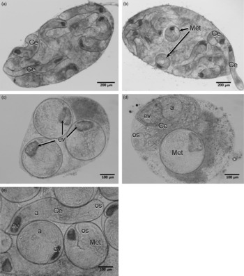
A large number of sporocysts (fig. 1a–e) were observed firmly attached to N. reticulatus tissues. They were motionless, yellowish in colour and with thin and transparent body walls. They presented variable sizes and shapes, from long (fig. 1a and b) to small, ovoid sacs (fig. 1c and d). Often three types of sporocysts were observed in the same individual snail: sporocysts filled exclusively with cercariae (fig. 1a), sporocysts containing only cysts (fig. 1c) and sporocysts filled with both (fig. 1b, d and e). Cercariae moved very rapidly inside the sporocysts and exited by rupture of the wall (fig. 1b). The cysts remained motionless with metacercariae rotating inside.
Larval stages of Diphtherostomum brusinae. Long sporocyst sacs (a,b) filled only with cercariae (a) and containing cercariae and encysted metacercariae (b). Small and ovoid sporocyst sacs (c, d) containing only encysted metacercariae (c) and cercariae and metacercariae (d). (e) Magnification of the sporocyst contents. Abbreviations: a, acetabulum; Ce, cercariae; ev, excretory vesicle; Met, metacercariae; os, oral sucker.

Cercariae
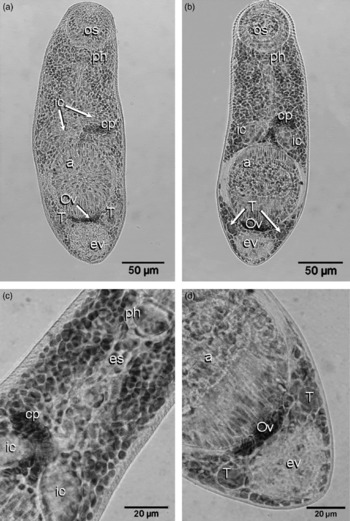
Tailless xiphidiocercaria (data not shown) with small and elongated, more or less cylindrical body, depending on its contraction state (fig. 2a). Three pairs of penetration glands, organized in two groups, were situated next to the bifurcation of the intestinal caeca, and the long ducts extended up to the edge of the oral sucker, where they opened. The digestive system consisted of a globose pharynx connecting to a long and straight oesophagus, which bifurcated into two short saccular intestinal caeca, terminating at the anterior edge of the ventral sucker. The prepharynx was absent. The excretory vesicle was spherical and filled with fine and refringent granules. An immature ovary was visible between the acetabulum and the excretory vesicle, with two oval masses, representing the emergent testes, on each side. An undeveloped cirrus pouch was located between the intestinal caeca.
The cercaria and metacercaria of Diphtherostomum brusinae. (a) Ventral view of the cercaria naturally emitted by Nassarius reticulatus. (b) Ventral view of the excysted metacercaria isolated from Cerastoderma edule. (c) Anterior dorsal region of the metacercaria body. (d) Detail of the metacercaria posterior region. Abbreviations: a, acetabulum; cp, cirrus pouch; es, oesophagus; ev, excretory vesicle; ic, intestinal caeca; os, oral sucker; Ov, ovary; ph, pharynx; T, testis.

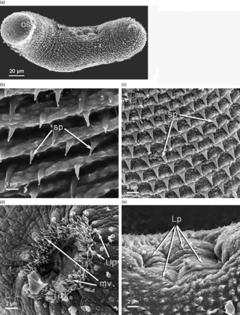
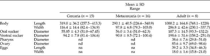
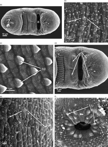
Observed by SEM, the body surface was folded into annular ridges bearing rows of long and pointed spines (fig. 3a–c). The oral sucker was terminal, circular and with thin lips. Surrounding the oral sucker was a ring of uniciliated papillae, each with a protruding long cilium (fig. 3d). The oral cavity was surrounded by a dense crown of long microvilli (fig. 3d). The ventral sucker, or acetabulum, was under two times larger than the oral sucker, and postequatorial. On the anterior and posterior edges of the ventral sucker, four protruding muscular masses, which constituted the lips, were seen (fig. 3a and e). Measurements are given in table 1. When naturally emitted by N. reticulatus, the cercariae moved quickly on the bottom of the containers by contraction and distension of the body.
SEM of Diphtherostomum brusinae cercariae: (a) lateral view; (b) tegument of the ventral face of the body; (c) tegument of the dorsal face of the body; (d) oral sucker; (e) ventral sucker or acetabulum. Abbreviations: a, acetabulum; Lp, lips; mv, microvilli; os, oral sucker; sp, spines; Up, uniciliated papillae.

Measurements (μm) of cercaria, metacercaria and adult stages of Diphtherostomum brusinae.
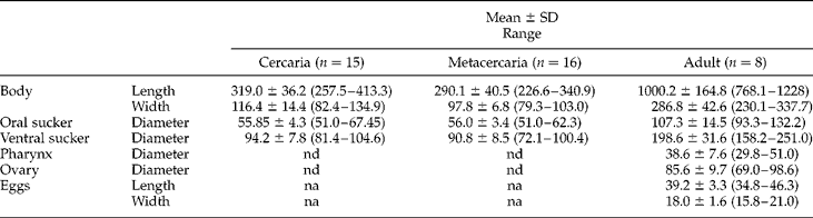
na, not applicable; nd, not determined; SD, standard deviation.
Metacercariae
Due to the fact that the cyst wall was very thin and transparent, the metacercariae could be observed coiled inside the cyst (fig. 1b–e). The cysts from N. reticulatus appeared rounder and more transparent than those from C. edule, which showed a thicker wall. The cysts from N. reticulatus had length 131.8–165.8 (146.3 ± 8.4) and width 123.6–170.0 (145.5 ± 10.7) and those from C. edule were 127.7–175.1 (146.6 ± 12.0) long and 113.3–137.0 (124.5 ± 7.1) wide (all values μm).
The excysted metacercariae (fig. 2b) were small, and the anterior region of the body was more elongated than the posterior. The digestive (fig. 2c) and excretory systems were similar to those of the cercariae (fig. 2b and d). Primordial gonads were located between the acetabulum and the excretory vesicle. Two symmetrical testes were observed surrounding a rudimentary ovary (fig. 2d). A sketch of the cirrus pouch was situated between the intestinal caeca (fig. 2b and c).
Using SEM, the metacercarial body surface was completely covered with spines (fig. 4a). However, the size, morphology and disposition of the spines changed with the body region. In the ventral region, the spines were short and rounded, with irregular distribution (fig. 4b). In the ventral and pre-acetabular region, a rectangular band of strong triangular pointed spines was seen, arising from a pearled tegument (fig. 4c and d). The dorsal spines were regularly distributed (fig. 4e) becoming longer and more pointed in the posterior region. The oral sucker was terminal, with thin lips. A ring of uniciliated papillae, each with a short cilium, was observed surrounding the oral sucker (fig. 4f). The ventral sucker or acetabulum was transversally elongate and under two times larger than the oral sucker (fig. 4a and d). The opening of the ventral sucker was circular, like the oral sucker, but presented an oval shape because of the lips, which were organized into four characteristic muscular masses (fig. 4a and d). Measurements are given in table 1.
SEM of Diphtherostomum brusinae metacercariae: (a) ventral view; (b) tegument of the ventral face of the body; (c) detail of the surface of the rectangular band situated in the ventral and pre-acetabular region; (d) ventral sucker or acetabulum; (e) tegument of the dorsal face of the body; (f) oral sucker. Abbreviations: a, acetabulum; Lp, lips; os, oral sucker; sp, spines; Up, uniciliated papillae.

Adults
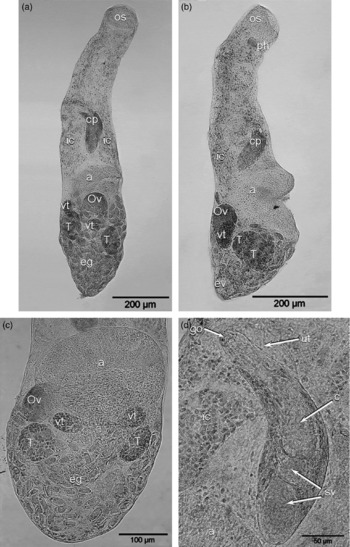
The tegument was densely spiny and the body presented a cylindrical to conical shape (fig. 5a and b). The posterior region was slightly wider and more globular than the anterior region. In specimens observed in profile, two small projections, which corresponded to the lips, were seen (fig. 5b). The two suckers had different sizes, and the ventral sucker, or acetabulum, had almost twice the diameter of the oral sucker. Moreover, the acetabulum presented a reddish colour. The digestive tract consisted of a small and muscular pharynx, without prepharynx, followed by a long and straight oesophagus which was divided into two small and saccular intestinal caeca, just before the ventral sucker (fig. 5a and b). The excretory vesicle was situated at the end of the posterior region and was spherical in shape (fig. 5b). Two equal and symmetrical testes, ovoid to spherical in shape, were observed disposed on each side of the posterior margin of the acetabulum (fig. 5a–c). The large cirrus pouch was pre-acetabular, with a curvature pointing to the dorsal region (fig. 5a, b and d). It contained the bipartite seminal vesicle and a long cirrus which occupied about half of the whole pouch. The genital pore was located on the dorsal and left margin of the body (fig. 5d). The ovary was situated between the two testes, and immediately posterior to the acetabulum. The vitelline glands were represented by two compact lobes, generally involved by the ovary and testes. Uterine loops filled with well-developed eggs were observed. The eggs were elliptic and large, with a very thin shell (fig. 5a–c). Measurements are given in table 1.
Adult of Diphtherostomum brusinae isolated from Diplodus sargus: morpho-anatomy of the adult in (a) dorsal view and (b) lateral view; (c) posterior ventral region of the body; (d) detail of the cirrus pouch. Abbreviations: a, acetabulum; c, cirrus; cp, cirrus pouch; eg, eggs; ev, excretory vesicle; go, genital pore; ic, intestinal caeca; os, oral sucker; Ov, ovary; sv, seminal vesicle; T, testis; ut, uterus; vt, vitellaria.

Molecular data
The sequence of the amplified ITS1 fragment showed a single product of length 1457 bp. After the sequence analysis, putative 18S and 5.8S regions were identified through comparison with identical regions of other digeneans and found to be 190 and 26 nucleotides long, respectively. The sequence encoding for the ITS1 region was 1241 nucleotides in length. No identical sequence was found in GenBank. The sequences encoding the ITS1 region of D. brusinae cercariae (collected in naturally infected N. reticulatus), encysted metacercariae (isolated from the N. reticulatus and C. edule) and adults (isolated from naturally infected D. sargus) were found to be identical.
Discussion
This work provides information regarding the life cycle of D. brusinae in the north of Portugal. The sporocysts of D. brusinae, containing cercariae and encysted metacercariae, were observed in the gonads and digestive gland tissues of N. reticulatus. We also observed that N. reticulatus naturally emitted cercariae, which moved quickly on the bottom of the containers by contraction and distension of the anterior region of the body. According to Palombi (Reference Palombi1930), the free cercariae present great mobility when they are close to encystment.
It is important to point out that the cysts of D. brusinae, found in C. edule, appear to have a thicker wall when compared with the cysts found in N. reticulatus. The formation of a new layer around the cysts, in addition to the layer of parasitic origin, seems to result from the direct contact of the cysts with the host tissues, unlike the cysts in N. reticulatus which were protected by the sporocyst wall.
LM studies of the larval stages of the cercariae (emitted by N. reticulatus) and the metacercariae (extracted from C. edule) seemed to indicate they were the same larval stage, as they presented a similar size and shape, and numerous identical morphological characters. These similarities have been a source of confusion for some authors, namely Prévot (Reference Prévot1966), who stated that the metacercariae were naturally emitted by N. reticulatus. In our work, the use of SEM techniques proved that it was not the metacercariae but the cercariae which were emitted by the gastropod. Important differences between these two larval stages were observed, mainly in the oral sucker and tegument. In the cercariae, a dense crown of microvilli was observed around the oral cavity which was absent in the metacercariae. Concerning the tegument, the body surface of the cercariae was uniformly covered by long and pointed spines, whereas the metacercariae showed a tegument covered by short and rounded spines with a random disposition in the ventral region. Moreover, the metacercariae presented a specific pre-acetabular rectangular band with conspicuous triangular spines. For the cercariae and metacercariae, the morphological measurements presented in table 1 are in accordance with the data described by Palombi (Reference Palombi1930, Reference Palombi1934) and Prévot (Reference Prévot1966).
Adults isolated from D. sargus showed the characteristics of the D. brusinae species by comparison with the descriptions of the adult stage by Stossich (Reference Stossich1889), Looss (Reference Looss1901), Stafford (Reference Stafford1905), Palombi (Reference Palombi1930, Reference Palombi1934), Yamaguti (Reference Yamaguti1958) and Prévot (Reference Prévot1966). The specific characters observed comprise a lip-like thickening on the anterior and posterior margins of the acetabulum, a large and pre-acetabular cirrus pouch containing a bipartite seminal vesicle, the presence of an intertesticular ovary, compact vitellaria and uterus filled with large and elliptical eggs, occupying all available space of the hind body. The morphometric data presented in table 1 are in agreement with those described by Stossich (Reference Stossich1889), Looss (Reference Looss1901), Stafford (Reference Stafford1905) and Palombi (Reference Palombi1930, Reference Palombi1934).
In this study, the ITS1 sequence of ribosomal DNA was used in order to identify and establish a correspondence between the cercariae naturally emitted by N. reticulatus, the encysted metacercariae from N. reticulatus and from C. edule, and the adults isolated from D. sargus. A match was found among the sequences of the three parasitic stages, indicating that they belong to the same species, i.e. D. brusinae.
It is known that the life cycles of digenetic trematodes typically require three hosts for successful completion. However, uncommon deviations such as one-, two- and four-host cycles, are known to be derived from the ancestral one (Esch et al., Reference Esch, Curtis and Barger2001).
After identification of the adult form of D. brusinae and the respective larval stages, we were able to clearly demonstrate two distinct parasitic strategies adopted by this parasite to complete its life cycle. The first strategy consists of using the first intermediate host, N. reticulatus, also as second intermediate host. Instead of cercariae emerging from the sporocysts of the molluscan host, they form metacercarial cysts within the same mollusc and await ingestion by the definitive host. In this case the parasite has simplified its life cycle.
The second strategy is the typical three-host cycle. The first intermediate host, N. reticulatus, emitted cercariae that penetrated the second intermediate host, C. edule, in which they encysted. Metacercarial cysts were found infecting various tissues, such as the foot, mantle border and especially the gills of the cockle. The free cercariae could also encyst in the neighbouring flora and fauna. The life cycle is completed when the definitive host D. sargus ingests C. edule or the free cysts in the environment.
According to Poulin & Cribb (Reference Poulin and Cribb2002), both the three-host cycle and the abbreviated cycle could be displayed by conspecific worms. The authors suggested that the parasitic shorter life cycles could be adopted by several species under certain environmental conditions. This explanation looked plausible in the case of the life cycle of D. brusinae in the north of Portugal. During this study, the levels of prevalence of infection observed for C. edule, which acts as second intermediate host, increased considerably in the warmer months (data not shown).
In conclusion, by using a combination of morphological and molecular characteristics, our results led to the identification of D. brusinae in the north of Portugal and contributed to its life cycle characterization. Thus, the first intermediate host, N. reticulatus, is parasitized by sporocysts containing cercariae which may develop into metacercarial cysts. In this case, the gastropod acts as second intermediate host. Encysted metacercariae are found in C. edule, which constitutes an alternative second intermediate host. The adult form infects the definitive host, D. sargus.
Acknowledgements
We would like to thank João Villares Neves from IBMC, Porto University for helping in the molecular biology techniques. The authors are indebted to anonymous reviewers for pertinent suggestions and improvement of the manuscript. We declare that the experiments comply with current Portuguese laws.