Anomalous origin of the left coronary artery from the pulmonary trunk, also known as the Bland-White-Garland syndrome, is a rare congenital anomaly affecting 1 in 300,000 live births.Reference Bland, White and Garland1, Reference Perlof2 Up to nine-tenths of children thus afflicted, if untreated, die during the first year of life because of myocardial ischaemia and left ventricular failure.Reference Matherne3 Early recognition is important, since surgical correction should be undertaken prior to the onset of any left ventricular dysfunction. Cardiac catheterization with aortography remains the standard means of diagnosis.Reference Donaldsson, Raphael, Smith, Yacoub and Ross4 Because infants or children with idiopathic dilated cardiomyopathy may also present with acute and severe congestive heart failure, cardiac catheterization, if attempted, carries a much higher risk for morbidity and mortality than other elective catheterizationds in children.Reference Vitiello, McCrindle and Nykanen5 Recently, significant advances in echocardiographic technology have permitted definitive diagnosis based on cross-sectional transthoracic echocardiographic interrogation combined with Doppler colour flow mapping.Reference Holley, Sell, Hougen and Martin6, Reference Frommelt, Miller, Williamson and Bergstrom7 This can allow quick recognition of anomalous origin of the left coronary artery from the pulmonary trunk. In this study, we describe our experience of this rare congenital abnormality, emphasizing the importance also of assessing the electrocardiogram.
Patients and methods
From June, 1999, to March, 2007, we admitted 9 infants and children, 6 males and 3 females to our department with either poor feeding, dyspneoa, chest pain, heart murmurs, or shortness of breath after exercise due to the suspicion of anomalous origin of the left coronary artery from the pulmonary trunk raised by transthoracic echocardiography. Their age at presentation ranged from 2 months to 10 years, with a median of 3.02 years. In all patients, the cross-sectional transthoracic echocardiogram and Doppler colour-flow mapping had shown a broad jet of retrograde flow into the pulmonary trunk, along with a dilated right coronary artery, intraseptal coronary collateral flow, and/or the left coronary artery arising from the pulmonary trunk.
All patients were studied retrospectively by electrocardiography and cardiac catheterization. We reviewed the details of the clinical data, investigations, and treatment.
Electrocardiogram
Standard 12-lead electrocardiograms were performed preoperatively in all patients. All studies were recorded with the paper speed set at 25 mm per second, using a standard amplitude of 10 mm.
Echocardiogram and Doppler colour-flow mapping
All studies were performed with a VingMed machine (General Electric Company), a Vivid 7 machine (General Electric Company) or a 7500 Philips machine. Either 7.5-MHz or 5-MHz transducers were used to obtain the images in the neonates and infants, with 5-MHz or 3-MHz transducers used for older children. We obtained standard subcostal, apical, parasternal, and suprasternal notch views as used in our routine investigations. Mitral regurgitation was assessed by Doppler colour flow in the apical four-chamber view and parasternal long-axis views, ranking any regurgitation as none, trivial, mild, moderate, or severe.
Results
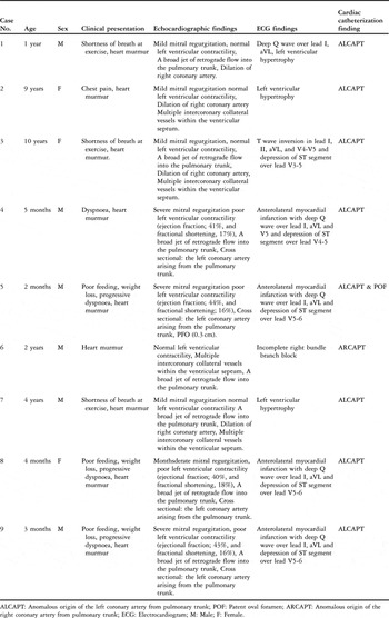
We studied 9 patients, 6 males and 3 females, having a mean age of 3.02 years, with a range from 2 months to 10 years. Of the patients, 5 were between the ages of 2 and 12 months, and had presented with a history of poor feeding, recent weight loss, and dyspnoea. The remaining patients were children, 3 being aged between 4 and 10 years, and who had a history of shortness of breath at exercise and repeated chest pain. Only 1 patient, who was 2 years of age, had no clinical symptoms. All had a cardiac murmur. Individual demographics and clinical presentations are given in Table 1.
Demographics and clinical presentations of the patients.

ALCAPT: Anomalous origin of the left coronary artery from pulmonary trunk; POF: Patent oval foramen; ARCAPT: Anomalous origin of the right coronary artery from pulmonary trunk; ECG: Electrocardiogram; M: Male; F: Female.
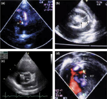
Among the 5 infants, the cross-sectional echocardiogram and Doppler colour flow showed a broad jet of retrograde flow into the pulmonary trunk in 4 (Fig. 1a), moderate-to-severe mitral regurgitation with poor left ventricular contractility, and an enlarged left atrium and left ventricle. The cross-sectional echocardiogram in the modified parasternal short-axis view showed the left coronary artery arising from the pulmonary trunk (Fig. 1b). In the remaining infant, the echocardiogram and Doppler colour flow showed a broad jet of retrograde flow into the pulmonary trunk, mild mitral regurgitation with normal left ventricular contractility, and dilation of the right coronary artery. The left coronary artery in this patient was not seen arising from the pulmonary trunk. Among the 4 children, the echocardiograms and Doppler colour flow in 2 showed mild mitral regurgitation with left ventricular dilation and normal left ventricular contractility, a broad jet of retrograde flow into the pulmonary trunk, dilation of the right coronary artery (Fig. 1c), and multiple intercoronary collateral vessels within the ventricular septum (Fig. 1d). In 1 patient, the broad jet of retrograde flow into the pulmonary trunk was not seen in the echocardiographic study. In the remaining patient, the echocardiographic interrogation, including Doppler colour flow, showed only multiple intercoronary collateral blood vessels within the ventricular septum, along with abnormal diastolic flow into the pulmonary trunk. This patient had suffered from proximal esophageal atresia with distal tracheoesophageal fistula, and had undergone corrective surgery when 3 days old. Our findings showed that the sensitivity of the transthoracic echocardiogram in making the diagnosis was 100%, the sensitivity for establishing mitral regurgitation was 50%, that for showing origin of the left coronary artery from the pulmonary trunk on cross-section was 62.5%, finding retrograde flow into the pulmonary artery on Doppler colour flow mapping was 37.5%, and finding dilation of right coronary artery, with intercoronary collateral vessels within the ventricular septum, was also 37.5%.
Echocardiography with Doppler color-flow mapping of an infant at 4 months in the parasternal short axis view (a) showed a broad jet of retrograde flow (arrow) into the pulmonary trunk. Ao: aorta; PA: pulmonary trunk. In the same patient, the cross sectional echocardiogram in high parasternal short axis view (b) showed the anomalous left coronary artery (arrow) arising from the pulmonary trunk. Ao: aorta; PA: pulmonary trunk; RV: right ventricle. A cross-sectional echocardiogram of a 10 year old in the parasternal short axis view (c) showed dilation of the right coronary artery. Ao: aorta; RCA: right coronary artery. In the same patient, echocardiography with Doppler color-flow mapping in apical four-chamber view (d) showed multiple intercoronary collateral vessels within the ventricular septum. IC: intercoronary collateral vessels; LA: left atrium; LV: left ventricle; RA: right atrium; RV: right ventricle.

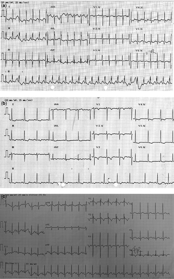
We also performed a standard 12-lead electrocardiogram in all patients. In all the infants, we found deep Q waves in leads I and aVL, and/or depression of the ST segments over leads V4 through 6, along with left ventricular hypertrophy (Fig. 2a). Among the 4 children, the electrocardiogram revealed left ventricular hypertrophy in 2 cases, while in another, the electrocardiogram showed inversion of the T waves in leads I, II, aVL, and V4 and V5, and depression of the ST segment over leads V3 through V5, along with left ventricular hypertrophy (Fig. 2b). In the remaining patient, the electrocardiogram only showed incomplete right bundle branch block (Fig. 2c). In this patient, therefore, the electrocardiographic findings did not match our provisional diagnosis of anomalous origin of the left coronary artery from the pulmonary trunk. We later found, at cardiac catheterization, that this patient had anomalous origin of the right coronary artery from pulmonary trunk. When assessing the value of the electrocardiogram in diagnosis, finding deep Q waves over leads I and aVL had a sensitivity of 75%, while depression of the ST segments in leads V5 and V6 carried a sensitivity of 62.5%.
Electrocardiogram of an infant at 5 months old with anomalous left coronary artery (a) shows mean QRS axis +14 degrees, a deep Q wave in I, aVL, and V5. There is marked ST depressions in leads V4-5. An electrocardiogram of a child of 10 years (b) shows T wave inversion in I, II, aVL, and V4–V5. There is ST depression in leads V3–V5, and signs of left ventricular hypertrophy. The electrocardiogram of a child at 2 year old with anomalous origin of the right coronary artery (c), in contrast, shows incomplete right bundle branch block.

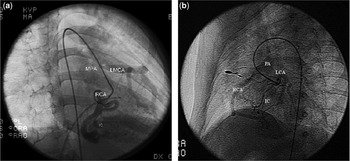
Cardiac catheterization with angiography confirmed the diagnosis of anomalous origin of the left coronary artery from the pulmonary trunk in 8 patients (Fig. 3a), but revealed anomalous origin of the right coronary artery from the pulmonary trunk in the remaining patients (Fig. 3b). In all patients with anomalous origin of the left coronary artery, surgical repair was performed by reimplantation of the anomalously arising artery, or by construction of a transpulmonary baffle. The patient with anomalous origin of the right coronary artery, however, was referred at this stage for regular follow-up.
Cardiac catheterization and angiography in a 10 year old patient with an anomalous left coronary artery (a) shows the right coronary angiography in frontal projection, which has normal aortic origin. The inferior interventricular artery gives rise to numerous intercoronary collaterals. These course through the ventricular septum, opacifying in retrograde fashion the left coronary arterial system, and filling the pulmonary trunk. AA: ascending aorta; IC: intercoronary collateral vessels; LMCA: left main coronary artery; MPA: pulmonary trunk; RCA: right coronary artery. In panel b, left coronary angiography is shown in a 2 year old patient with anomalous origin of the right coronary artery. The injection fills the left coronary artery, and then the right coronary artery via a number of collaterals, with subsequent opacification of the pulmonary trunk. IC: intercoronary collateral vessels; LMCA: left main coronary artery; MPA: pulmonary trunk; RCA: right coronary artery.

Discussion
Anomalous origin of the left coronary artery from the pulmonary trunk is an uncommon, but frequently lethal, lesion for both children and adults.Reference Ogden8, Reference Moodie, Fyfe and Gill9, Reference Hekmat, Rao, Chhabra, Chiavarelli, Anderson and Nudel10 Although it was first described by BrooksReference Brooks11 in 1885, the first report describing clinical and autopsy findings was that from Bland and colleagues.Reference Bland, White and Garland1 Subsequent to that description, the entity has also become known as the Bland-White-Garland syndrome.Reference Moodie, Fyfe and Gill9 It most often presents as an isolated defect, but in one-twentieth of cases, it may be associated with other cardiac anomalies, including atrial and ventricular septal defects, and aortic coarctation.Reference Bland, White and Garland1
Clinical diagnosis still remains a challenge. Cardiac catheterization with ascending aortography is the standard means of clinical diagnosis. But, because infants or children with this anomaly may suffer from acute and severe congestive heart failure, cardiac catheterization carries a much higher risk for morbidity and mortality than do other elective catheterizations in children.Reference Vitiello, McCrindle and Nykanen5 To aid clinical decision-making, and avoid high-risk catheterization, many investigators have sought to achieve diagnosis using noninvasive tools.Reference Karr, Parness, Spevak, van der Velde, Colan and Sanders12
Echocardiographic diagnosis in recent years has improved dramatically, largely because of refinements in cross-sectional and colour flow Doppler imaging, and also because careful inspection of the coronary arteries is now routinely performed.Reference Frommelt, Miller, Williamson and Bergstrom7 Cross-sectional imaging is now accepted as showing the abnormal origin of the left coronary artery. Reference Caldwell, Hurwitz, Girod, Weyman and Feigenbaum13–Reference Henry, Maron and Griffith16 Measurement of the diameter of the right coronary artery may also be helpful in diagnoss.Reference Koike, Musewe, Smallhorn and Freedom17 Use of colour flow Doppler interrogation, showing abnormal flow into the pulmonary trunk and multiple intercoronary collateral vessels within the ventricular septum, has also been demonstrated to add value to the echocardiographic examination.Reference Hildreth, Junkel, Allada, Sintek and Sapin18, Reference Hirota, Hiraishi and Nakahata19 We found that the cross-sectional study, using a modified parasternal short-axis view, showed the direct communication between the pulmonary trunk and the left coronary artery in all our infants, and revealed abnormal diastolic flow into the pulmonary trunk in all cases except one. In all our older patients, we found a broad jet of retrograde flow into the pulmonary trunk, along with dilation of the right coronary artery and multiple intercoronary collateral vessels within the ventricular septum. The echocardiogram, therefore, is not only suggestive, but is now demonstrative for the abnormal coronary artery.Reference Pelliccia, Spataro and Maron20, Reference Pisacane, Pinto and De Gregorio21 Differentiation between anomalous origin of the left as opposed to the right coronary artery from the pulmonary trunk, however, remains time-consuming and uncertain.Reference Stefanelli, Stevenson, Jones, Lester and Cecchin22 In our sixth case, we found multiple intercoronary collateral vessels within the ventricular septum along with abnormal diastolic flow into the pulmonary trunk. Our initial diagnosis, therefore, was anomalous origin of the left coronary artery from the pulmonary trunk. Analysis of the electrocardiogram, however, cast doubt on this diagnosis. And catheterization revealed that, in this patient, it was the right coronary artery which arose anomalously from the pulmonary trunk.
A 12-lead electrocardiogram, therefore, may be most helpful in diagnosis. Broad and deep Q waves, and associated inversion of the T waves in leads I and aVL, have already been recognized as characteristic for anomalous origin of the left coronary artery from the pulmonary trunk.Reference Perlof2, Reference Chu and Cheitlin23. We found deep Q waves over leads I and aVL, and depression of the ST segment over leads V4 and 5, with signs of left ventricular hypertrophy, in all our infants. Among our 4 children, only 3 had findings suggestive of myocardial infarction. In the exceptional patient, the electrocardiogram showed only incomplete right bundle branch block. This, of course, was the patient with anomalous origin of the right coronary artery from the pulmonary trunk. By combining transthoracic echocardiography with electrocardiography, therefore, we submit that it is possible to diagnose anomalous origin of the left coronary artery from the pulmonary trunk with accuracy, and also to distinguish those patients with the right coronary artery arising from the pulmonary trunk. In this respect, Chang and AlladaReference Chang and Allada24 distinguished 23 patients with anomalous origin of the left coronary artery from pulmonary trunk from those having idiopathic dilated cardiomyopathy by means of the electrocardiogram combined with cross-sectional transthoracic echocardiography. They also created a scoring system to establish the diagnosis. In their study, the scoring system had a much higher specificity and positive predictive value in differentiating anomalous origin of the left coronary artery from the pulmonary trunk and dilated cardiomyopathy. Use of their system would also have excluded our patient with anomalous origin of the right coronary artery from the pulmonary trunk.
It remains a fact, nonetheless, that although many noninvasive diagnostic means have been evaluated for diagnosing anomalous origin of the left coronary artery from the pulmonary artery, cardiac catheterization and aortography remain the definitive diagnostic tests. Surgical correction, with reimplantation of the left coronary artery, or creation of a transpulmonary baffle,Reference Takeuchi, Imamura and Katsumoto25 should be undertaken as soon as the diagnosis is established, since sudden death resulting from myocardial infarction is possible. Our own study is limited by the lack of control of the quality of the echocardiographic studies. Since the echocardiograms were performed by different operators, using different echocardiographic systems, the investigation of necessity varied between them.
In conclusion, nonetheless, it is our opinion that accurate diagnosis of anomalous origin of the left coronary artery from the pulmonary trunk in children can be made by combining cross-sectional and colour-flow Doppler echocardiographic interrogation with the electrocardiogram. Even though a very rare malformation, it is important to exclude this diagnosis in children suspected of having dilated cardiomyopathy. By combining the transthoracic echocardiographic studies with an electrocardiogram, we have shown that it is also possible to distinguish those having anomalous origin of the right as opposed to the left coronary artery from the pulmonary trunk.
Acknowledgements
We are grateful to Wen-Won Chang for providing help with this report. In addition, the excellent technical assistance given by Ms Li-Hui Chang is very much appreciated.