Introduction
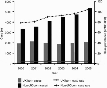
Tuberculosis is a leading cause of morbidity and mortality globally. It is estimated that nearly two million people die of the disease every year, despite significant advances in its treatment in the twentieth century.1 The number of tuberculosis cases has been increasing in England and Wales since the late 1980s, and has continued to do so in recent years (Figure 1).1
Number of tuberculosis cases and prevalence rates by place of birth (UK-born vs non-UK-born) in England, Wales and Northern Ireland, 2000–2005.1 Reproduced courtesy of the Health Protection Agency, Centre for Infections, Enhanced Tuberculosis Surveillance (ETS).

There is a well known association between tuberculosis and malignancy, with cancer patients having a nine times greater risk of developing tuberculosis (TB) compared with the general population.Reference Karnak, Kayacan and Beder2, Reference Libshitz, Pannu, Elting and Cooksley3 Immunological studies have increased our understanding of this association; the process of carcinomatosis has been shown to exert immunosuppressive effects extending from the primary tumour site to secondary lymphoid organs and peripheral vessels.Reference Kim, Emi and Tanabe4 This is mediated by tumour-derived soluble factors such as interleukin-10, transforming growth factor-β and vascular endothelial growth factor.Reference Kim, Emi and Tanabe4
In patients with TB, tuberculous lymphadenitis occurs most frequently in the head and neck region; in addition, the highest incidence of tuberculosis associated with malignancy is seen in cancers of the head and neck.Reference Solak, Sayar, Metin, Erdoğu, Çuhadaroğlu and Turna5, Reference Kumar, Shafiulla and Sridhar6 One may therefore anticipate that coexistence of both diseases in the head and neck region is common.
Herein, we present a previously undocumented case of synchronous, tuberculous cervical lymphadenitis in a Caucasian patient with T1a squamous cell carcinoma of the right vocal fold; we also present a thorough review of the relevant literature.
Case report
A 66-year-old ex-miner was referred with a nine-month history of hoarseness. He had an alcohol intake of 20 units per week, and had smoked 40 cigarettes per day up until nine years prior to presentation. His co-morbidities included renal stone disease, anxiety, Ménière's disease, hypertension and chronic obstructive pulmonary disease.
Flexible naso-endoscopy performed at presentation revealed an exophytic lesion over the posterior half of the right vocal fold. Both vocal folds were fully mobile. A palpable right jugulodigastric lymph node was also noted. The patient had no respiratory symptoms or indeed any other systemic manifestations. There was no previous history of TB, and chest auscultation was clear.
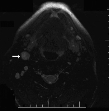
Panendoscopy and biopsy under general anaesthesia were performed. Subsequent histological analysis revealed an invasive squamous cell carcinoma. An ultrasound-guided fine needle aspiration cytology (FNAC) examination of the neck node was performed but was inconclusive. A computed tomography scan of the neck demonstrated the vocal fold lesion and the obviously enlarged right jugulodigastric node (measuring 18 mm in diameter), but also showed multiple smaller, suspicious cervical neck nodes bilaterally. This nodular disease was more than would be expected for an isolated T1a lesion. The CT scan also included the thorax and abdomen for staging purposes. This showed some small, nodular pulmonary thickening too minute to characterise. There was no mediastinal or hilar lymphadenopathy. Bilateral simple renal cysts and a calculus within the mid pole of the right kidney were incidentally noted. A magnetic resonance imaging scan of the neck was also performed (Figure 2). Radiological examination also showed some small, nodular pulmonary thickening too minute to characterise. There was no mediastinal or hilar lymphadenopathy. Bilateral simple renal cysts and a calculus within the mid pole of the right kidney were incidentally noted.
Axial, T2-weighted, fast spin section magnetic resonance imaging scan of the patient, showing marked lymphadenopathy with a prominent right jugulodigastric lymph node measuring 18 mm (arrow).

The patient was taken to the operating theatre for excision biopsy of the right jugulodigastric node. Intra-operatively, several matted nodes were seen which had a caseating appearance.
The post-operative period was complicated by a haematoma.
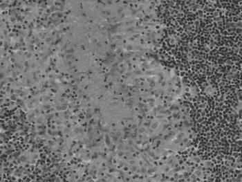
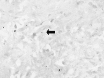
Histological analysis of the biopsy material revealed granulomatous lymphadenitis with features highly suggestive of mycobacterial disease (Figure 3). On examination with Ziehl–Neilson staining, one acid-fast bacillus was identified (Figure 4).
Photomicrograph of a section of the right jugulodigastric lymph node, showing a large aggregate with central caseation, areas of hyalinisation and scanty multi-nucleate giant cells, all features highly suggestive of mycobacterial infection (H&E; ×200).

Photomicrograph of a section of the right jugulodigastric lymph node stained with Ziehl–Neilson stain, showing a single acid-fast bacillus (arrow). (×600)

There was no previous history of bacillus Calmette–Guérin (BCG) vaccination. An urgent chest physician consultation was arranged, and sputum samples were sent for acid-fast bacilli staining, the result of which was negative. An urgent review of the original vocal fold histology was undertaken to exclude the possibility of laryngeal TB; this also proved negative.
The patient was referred urgently to the regional head and neck multidisciplinary team. A decision was made to offer the patient radiotherapy for his glottic lesion. He was concurrently commenced on anti-tuberculosis chemotherapy.
At six month's follow up, the patient had finished five months of anti-tuberculosis treatment, but continued to have a productive cough, shortness of breath on exertion and uncontrollable night sweats. However, there were no symptoms or signs of laryngeal cancer recurrence.
Discussion
This patient presented a diagnostic dilemma: faced with an obvious laryngeal primary squamous cell carcinoma, cervical lymphadenopathy would invariably appear to represent metastatic spread. The need for a histological diagnosis via nodal excision biopsy was tempered by the realisation that, should clinical suspicion of concomitant TB prove incorrect, then there was a real risk of seeding metastatic neck disease. Similarly, one must be vigilant as to the original diagnosis of the primary cancer, and we would advocate the good practice of reviewing the histology to ensure that a diagnosis of TB has not been misinterpreted.
Clearly, had a high index of suspicion of TB not been aroused by the patient's radiology, it is not inconceivable that, in the face of an unhelpful FNAC, an assumption of metastatic neck disease would be made. Any subsequent surgical and/or oncological intervention based on this assumption would be highly inappropriate, with potentially catastrophic effects; neck dissection and laryngeal surgery, or indeed localised radiotherapy to the area (leading to possible dissemination of TB as a consequence of immunosuppressive effects), would clearly both be highly inappropriate. Due to this complication, some have contemplated undertaking active tuberculosis investigation in children who develop malignancy in areas of high TB prevalence, prior to starting immunosuppressive therapy.Reference Wessels, Hesseling, Gie and Nel7
• There is a well known association between tuberculosis and malignancy, with cancer patients having a nine times greater risk of developing tuberculosis than the general population
• This paper describes an extremely rare presentation of concomitant tuberculous cervical lymphadenitis in a patient with carcinoma of the larynx
• This case highlights the importance of proper diagnosis, and emphasises the fact that tuberculosis should always be borne in mind in the differential diagnosis
Other, similar diagnostic difficulties have been described. Karnak et al. have described the coexistence of pulmonary TB in patients with lung carcinoma; in this situation, the radiological findings of either disease can mimic the other.Reference Karnak, Kayacan and Beder2 Other cases have been reported of concurrent breast cancer and axillary tuberculous lymphadenopathy.Reference Tulasi, Raju, Damodaran and Radhika8 In such cases, microscopic detection of acid-fast bacilli by Ziehl–Nielson staining has been found to be insufficiently sensitive to confirm the histopathological diagnosis; the use of polymerase chain reaction based assays has been suggested as a fast, cheap and reliable alternative.Reference Khurram, Tariq and Shahid9 Indeed, in our case only one acid-fast bacillus was demonstrated on Ziehl–Nielson staining.
This case highlights the importance of proper diagnosis, and emphasises the fact that TB should always be borne in mind in the differential diagnosis. We would also emphasise the need to consider routine TB screening prior to radiotherapy or chemotherapy, using a quick, cheap test such as polymerase chain reaction, in consideration of the background, rising tuberculosis prevalence in the UK.
Acknowledgements
We would like to thank Dr Phil Brumwel, Consultant Pathologist at the Princess of Wales Hospital, Bridgend, Wales, for producing the high quality images presented as Figures 2 and 3, for his active involvement in the histological diagnosis of this patient, and for his valuable advice.