INTRODUCTION
The largest planting area among all vegetable crops in Taiwan is given over to watermelon [Citrullus lanatus (Thunb.) Matsum & Nakai] and it is the third most important vegetable crop in the world (FAO 2010). Fusarium wilt caused by Fusarium oxysporum f. sp. niveum (E. F. Smith) Snyder and Hansen has become one of the major limiting factors in watermelon production because of the long-term monoculture system in Taiwan (Chen et al. Reference Chen, Liou, Chang and Huang2003b ). Fusarium oxysporum is a soil-borne plant pathogen that invades watermelon roots and causes wilt symptoms by colonizing the xylem tissues (Lin et al. Reference Lin, Chen, Liou, Huang and Chang2009b , Reference Lin, Chen, Chang, Wan, Hsu, Huang and Chang2010). In addition, the disease occurs in every period of watermelon cultivation and seriously decreases production (Martyn Reference Martyn, Zitter, Hopkins and Thomas1996; Michielse & Rep Reference Michielse and Rep2009). There are several ways to control Fusarium wilt in the field such as using chemical compounds, physical controls such as hot water treatment of seeds, or cultivation management, but the best strategy is to plant resistant cultivars (Chen et al. Reference Chen, Liou, Chang and Huang2003b ; Lin et al. Reference Lin, Chang, Liu, Chao, Huang and Chang2009a ). Recently, a Fusarium wilt-resistant watermelon line, JSB, derived from a spontaneous mutation of the susceptible watermelon cultivar Sugar Baby (SB) was obtained in Taiwan (Chen et al. Reference Chen, Chang, Liou and Huang2003a ). Chang et al. (Reference Chang, Hsu, Lin, Chen, Huang and Liou2008) compared the histological differences between resistant JSB and susceptible SB after inoculation with F. oxysporum and suggested that phenylalanine ammonia lyase (PAL) may be involved in the resistance to Fusarium wilt.
Phenylalanine ammonia lyase is the first enzyme involved in the phenylpropanoid pathway (Bate et al. Reference Bate, Orr, Ni, Meromi, Nadler-Hassar, Doerner, Dixon, Lamb and Elkind1994; Boudet Reference Boudet2007). An increase in PAL activity is one of the indicators of a resistance reaction in some of the plant and pathogen interactions. Way et al. (Reference Way, Kazan, Mitter, Goulter, Birch and Manners2002) reported that lesion expansion caused by the oomycete pathogen Phytophthora parasitica pv. nicotianae and the fungal pathogen Cercospora nicotianae were significantly reduced in transgenic tobacco with increasing PAL activity. Resistance of oats to Erysiphe graminis was reduced by using a PAL inhibitor α-amino-β-phenylpropionic acid (AOPP) (Carver et al. Reference Carver, Zhang, Zeyen and Robbins1996). Chang et al. (Reference Chang, Hsu, Lin, Chen, Huang and Liou2008) also reported that the resistance of JSB to F. oxysporum could be suppressed by pre-treating the watermelon seedlings with another PAL inhibitor, α-aminooxyacetate (AOA). Furthermore, PAL activity in banana lines that tolerate Fusarium wilt was induced after the banana lines were treated with F. oxysporum f. sp. cubense elicitor (Subramaniam et al. Reference Subramaniam, Maziah, Sariah, Puad and Xavier2006).
In addition, phenolic compounds are products of the phenylpropanoid pathway and are important secondary metabolites of plants (Peters & Verma Reference Peters and Verma1990). Plant phenolics may be synthesized in response to physical injury, infection or other stresses (Beckman Reference Beckman2000). Meanwhile, there are three known functions of phenolic compounds in plant defence responses: first, antimicrobial activity that inhibits the growth of pathogens (Treutter Reference Treutter2006); second, cell wall-bound phenolics that strengthen the cell wall structure to resist pathogen invasion (El Modafar et al. Reference El Modafar, Tantaoui and El Boustani2000); and finally, signal molecules that trigger the resistance responses (Nicholson & Hammerschmidt Reference Nicholson and Hammerschmidt1992).
Lignin, a major component of the cell wall, is a macromolecule particle derived from different types of monolignol monomers. Peroxidase and polyphenoloxidase are related to the biosynthesis of lignin and expressions of these enzymes are related to some xylem disease (Boudet Reference Boudet1998).
The purpose of the present study was to elucidate the possible resistance mechanisms in watermelon to Fusarium wilt by comparing the differences between the resistant line JSB and the related susceptible cultivar Grand Baby (GB), after F. oxysporum inoculation. Firstly, the colonization of different parts of JSB and GB seedlings was investigated and the shoot bases were found to be the likely areas for F. oxysporum resistance in watermelon seedlings. Secondly, the PAL activity and phenolics content in the shoot bases of JSB and GB seedlings were analysed after F. oxysporum inoculation. Lastly, enzyme activities of peroxidase and polyphenoloxidase were measured and histopathology for lignin deposition was observed to define the structure defence in the shoot bases of JSB and GB.
MATERIALS AND METHODS
Plant material
Twenty watermelon seeds each of resistant JSB and susceptible GB cultivars were sprouted in running water for 2 days. After germination, the seeds were then placed on a water-soaked filter paper on a plastic plate and seedlings were grown in the dark. Twenty two-day-old seedlings of each cultivar were planted in sterilized soil (BVB No. 5, Bas van Buuren B.V., Maasland, the Netherlands) and 20 in infested soil (soil from NCHU campus mixed with BVB No. 5 and homemade F. oxysporum-colonized soil).
Fungal culture and inoculation
Fusarium oxysporum f. sp. niveum H0103 from a preservative tube was grown in water agar (20 g/l) for 6–8 h. Single spores of the F. oxysporum-H0103 isolate were grown on half strength potato dextrose agar (extracts of 200 g/l of potato, 10 g/l dextrose, 20 g/l agar) at room temperature. The fungal mycelia and spores were rinsed with sterile water after 5 days and mixed into sterilized soil which was then used as infested soil. The inoculum concentration of F. oxysporum-infested soil was made to 104 spores per gram of soil. The shoot bases of watermelon seedlings were taken 3, 6 and 9 days after inoculation (DAI) with F. oxysporum for the following tests. At least three samples were used for each experiment and every experiment was replicated three times.
Examination of watermelon seedlings
Pathogen isolation
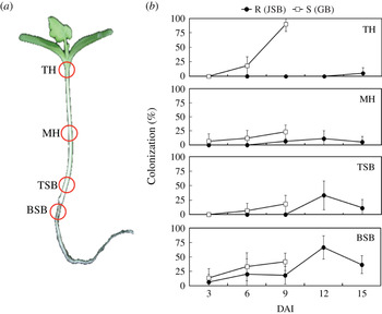
Watermelon seedlings were collected at 3, 6 and 9 DAI. Different parts of the seedlings, as shown in Fig. 1 a, were cut into pieces for isolation of the pathogen. Tissues were rinsed with sterile water, sterilized in 2·5% bleach (6% sodium hypochlorite) for 1 min, rinsed with sterile water again to wash out the bleach and placed on the pentachloronitrobenzene (PCNB) medium, which selectively allows the F. oxysporum pathogen to grow. Lastly, watermelon tissues with pathogen colonies were counted after cultivation and the pathogen-colonized ratio (%) was calculated.
Colonization of Fusarium oxysporum f. sp. niveum in different parts of resistant and susceptible watermelon seedlings after inoculation. (a) Different parts of watermelon seedlings. TH, top of the hypocotyls; MH, middle of the hypocotyls; TSB, top of the shoot base; BSB, bottom of the shoot base. (b) The colonization (%) of F. oxysporum f. sp. niveum in the different parts of resistant (R, JSB) and susceptible (S, GB) watermelon seedlings after inoculation. Watermelon seedlings planted in F. oxysporum-infested soil. At 3, 6, 9, 12 and 15 days after inoculation (DAI), different parts of seedlings were cut and then surface sterilized with 2·5% hypochlorite. The sterilized tissues were placed on PCNB selective medium for F. oxysporum growth and the colonization was analysed with Student's t test. P⩽0·05. Error bars were the s.e.m. (Colour version available online.)

Phenylalanine ammonia lyase activity assay
The PAL activity assay was performed with modifications from Edwards & Kessmann (Reference Edwards, Kessman, Gurr, Mcpherson and Bowles1993). Fresh samples (c. 100–200 mg of the cotyledon, 100–200 mg of hypocotyl, 50–80 mg of the shoot base or 40–60 mg of root) were homogenized with liquid nitrogen and the soluble proteins were extracted by adding 2 ml PAL extraction buffer (100 ml dH2O with 0·61 g Tris–HCl, 0·1 ml 2-mercaptoethanol and 5 g polyvinylpyrrolidone). The crude extracts were centrifuged at 15 000 g for 10 min and the supernatant was collected as soluble protein extracts. Soluble protein extract (0·1 ml) was then added to 0·9 ml reaction buffer (12·1 mM l-phenylalanine, 50 mM polyvinylpyrrolidone) and the mixture was incubated at 40 °C for 1 h before the absorbance values at 290 nm were measured using a spectrophotometer (Genesys 10 Scan Thermo Fisher Scientific Inc., Pittsburgh, PA, USA).
Phenolic compounds extraction and concentration measurement
Soluble phenolic compounds extract
Fresh plant samples as mentioned above were chopped into slices and extracted by adding 5 ml of methanol (0·8 g/ml) at 70 °C for 24 h. After extraction, the alcohol insoluble residues and soluble phenolic compound extracts were separated by filter paper (Whatman No. 1). The supernatant of extracts was collected and the concentration of soluble phenolics was estimated according to Thangavelu et al. (Reference Thangavelu, Palaniswami, Doraiswamy and Velazhahan2003), whereas the alcohol insoluble residues were dried in an oven and the concentration of cell wall-bound phenolics was analysed.
Cell wall-bound phenolics extract
The alcohol insoluble residues were treated with 5 ml 2 M sodium hydroxide (NaOH) at 70 °C for 1 day. After NaOH treatment, the alcohol insoluble residues solution was neutralized with 0·5 M H2SO4. These solutions were labelled as cell wall-bound phenolic extracts, the supernatant of which was analysed for the cell wall-bound phenolics content.
The protocol for the measurement of phenolics was modified from Thangavelu et al. (Reference Thangavelu, Palaniswami, Doraiswamy and Velazhahan2003). The 150-μl-extract solution was mixed with solutions of 750 μl sterile water and 37·5 μl 2 N Folin–Ciocalteau's phenol reagent (Sigma-Aldrich Inc., Stenheim, Germany). After a 2-min reaction, 150 μl saturated sodium carbonate (Na2CO3) and 75 μl sterile water solution was then added and reacted at room temperature for 2 h. The absorbance of the blue colour produced in the reaction was estimated with a spectrophotometer at 725 nm. The concentrations of phenolics were calculated according to the standard curve made using gallic acid.
Tissues stain of lignin deposition in the shoot bases
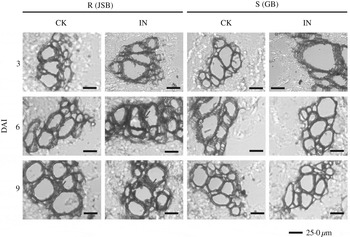
The lignin deposition was observed in the shoot base at 3, 6 and 9 DAI by paraffin section and tissue staining. The tissue samples were fixed with formalin, alcohol and acetic acid and dehydrated with tertiary-butyl alcohol. After that, the 7–8 μm paraffin sections were stained with 2 g/l phlorgulcinol–HCl to observe lignin deposition in the plant cell wall.
Peroxidase and polyphenoloxidase activity assays
The extraction and activity assays of peroxidase and polyphenoloxidase were performed according to Chen et al. (Reference Chen, Belanger, Benhamou and Paulitz2000) and De Ascensao & Dubery (Reference De Ascensao and Dubery2000), respectively. Proteins for enzyme activity were extracted from one watermelon shoot base for each time point with 1·5 ml extraction buffer (50 mM sodium phosphate buffer (pH 6·0) and polyvinylpyrrolidone at 12·5 g/l). After high-speed centrifugation of the protein crude extracts at 15 000 g for 10 min, the supernatant was estimated for protein concentration by the Bradford method (Bradford Reference Bradford1976) and the peroxidase and polyphenoloxidase activities were analysed.
The enzyme activity of peroxidase was estimated by mixing 10 μl protein extract and 890 μl peroxidase reaction buffer (5 mM sodium phosphate, 60 mM guaiacol). To this was added 100 μl hydrogen peroxide at 30 ml/l. Then, after 1 min reaction, the change of absorbance of the mixture at 470 nm wavelength (ΔOD470) was measured with a spectrophotometer. The activity was calculated by the formula:
 $$\displaystyle{{\Delta {\rm OD}_{{\rm 47}0}} \over {{\rm 26} {\cdot} {\rm 6}\,\left( {{\rm extinction}\,{\rm coefficient},\,/{\rm mM}/{\rm cm}} \right) \times {\rm 1}\,{\rm min} \times {\rm mg}\,{\rm protein}}}$$
The enzyme activity of polyphenoloxidase was estimated by mixing 200 μl extract and 800 μl polyphenoloxidase reaction buffer (100 μl 0·2 M catechol and 700 μl protein extract buffer). The change of absorbance at 405 nm wavelength (ΔOD405) in the mixture was measured with a spectrophotometer after a 1-min reaction. The activity was calculated by the formula:
 $$\displaystyle{{\Delta {\rm OD}_{{\rm 4}0{\rm 5}}} \over {{\rm 1} {\cdot} {\rm 28}\left( {{\rm extinction}\,{\rm coefficient},\,/{\rm mM}/{\rm cm}} \right) \times {\rm 1}\,{\rm min} \times {\rm mg}\,{\rm protein}}}$$
Statistical analysis
Data were tested for statistical significance analysis using the two-sample t test included in the R program.
RESULTS
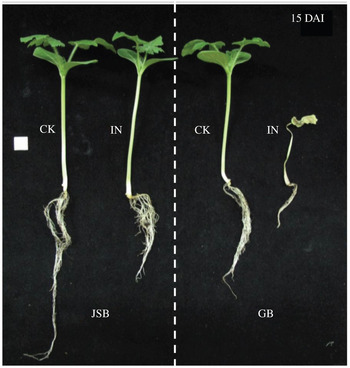
The cotyledons of the susceptible watermelon cultivar GB started to wilt at 8–10 DAI; these seedlings then wilted and died at 10–15 DAI. In contrast, JSB had no disease symptoms 15 DAI (Fig. 2). Therefore, tissues of GB and JSB seedlings were used for analysis at 3, 6 and 9 DAI in the present study.
Symptoms of the resistant JSB and susceptible GB watermelon seedlings at 15 days after inoculation (DAI) with Fusarium oxysporum f. sp. niveum. Watermelon seedlings planted in F. oxysporum-infested soil. At 15 DAI, whole seedlings of the resistant JSB and susceptible GB watermelon were washed and pictures were taken for comparison. The white square indicates 1 cm2. CK, control; IN, inoculation. (Colour version available online.)

Colonization of Fusarium oxysporum f. sp. niveum in resistant and susceptible watermelon seedlings
Colonization rate of F. oxysporum at the top of the hypocotyls in susceptible GB was significantly (P<0·05) higher than that in resistant JSB at 6 DAI (Fig. 1 b). At 9 DAI, colonization rates of F. oxysporum were significantly (P<0·05) higher in susceptible GB than in the resistant JSB at the top and the middle of the hypocotyls and on the top of the shoot base (Fig. 1 b). Colonization in resistant JSB seedlings was also observed at the bottom of the shoot base at 12 DAI (Fig. 1 b). These results suggested that F. oxysporum resistance may occur actively in the shoot bases of JSB seedlings. Therefore, the shoot bases of susceptible GB and resistant JSB watermelon seedlings were chosen for analysis of the resistance responses.
Phenylalanine ammonia lyase activity after Fusarium oxysporum inoculation
In the shoot bases of the resistant JSB seedlings, PAL activity was induced at 3 DAI, decreased at 6 DAI, then induced and increased again at 9, 12 and 15 DAI (Fig. 3 a). However, in the shoot bases of the susceptible GB seedlings, PAL activity was induced at 3 DAI, decreased at 6 DAI, and then stayed as low as that of the control at 9 DAI (Fig. 3 b).
Phenylalanine ammonia lyase (PAL) activity in the shoot bases of resistant and susceptible watermelon seedlings after inoculation with Fusarium oxysporum f. sp. niveum. Watermelon seedlings planted in F. oxysporum-infested soil were harvested at 3, 6, 9, 12 and 15 days after inoculation (DAI). The PAL activity in the shoot base of the (a) resistant (R, JSB) and (b) susceptible (S, GB) watermelon seedlings were analysed. The open symbols represent mock control (CK); the closed symbols represent the inoculated samples (IN). The error bars were the s.e.m.

Soluble and cell wall-bound phenolics contents after Fusarium oxysporum inoculation
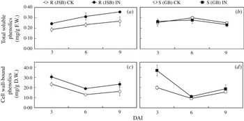
The total soluble phenolics concentrations in the shoot bases of resistant JSB seedlings increased at 3, 6 and 9 DAI (Fig. 4 a), but no significant difference was observed in the shoot bases of susceptible GB seedlings (Fig. 4 b). The content of cell wall-bound phenolics in the resistant JSB shoot bases increased at 3, 6 and 9 DAI (Fig. 4 c); however, it only increased at 3 DAI in the susceptible GB shoot bases (Fig. 4 d).
Total soluble and cell wall-bound phenolic contents in the shoot bases of resistant and susceptible watermelon seedlings after inoculation with Fusarium oxysporum f. sp. niveum. Watermelon seedlings planted in F. oxysporum-infested soil were harvested at 3, 6 and 9 days after inoculation (DAI). (a, b) Total soluble and (c, d) cell wall-bound phenolic contents in the shoot bases of the resistant (R, JSB; a, c) and susceptible (S, GB; b, d) watermelon seedlings were analysed. The open symbols represent mock control (CK); the closed symbols represent the inoculated samples (IN). The error bars were the s.e.m.

Lignin accumulation in xylem vessels of watermelon shoot bases
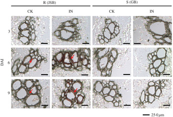
The results showed that the shoot bases of resistant JSB seedlings had significant lignin deposition on the cell wall of xylem vessels at 6 and 9 DAI (Fig. 5, indicated by the red arrows). Furthermore, inferior lignin deposition was observed in the shoot bases of control JSB seedlings. Nevertheless, no lignin deposition was observed in the shoot bases of susceptible GB seedlings.
Histology of lignin accumulation in xylem vessels of the shoot bases of the resistant and susceptible watermelon seedlings at 3, 6 and 9 days after inoculation (DAI) with Fusarium oxysporum f. sp. niveum. Paraffin sections were prepared from the shoot bases of F. oxysporum-inoculated resistant (R, JSB) and susceptible (S, GB) watermelon seedlings. The sections were stained with phlorglucinol–HCl for the observation of the lignin deposition (stained in red, as indicated by red arrows) in the xylem vessels. The bar indicates 25·0 μm. CK, control; IN, inoculation. (Colour version available online.)

Peroxidase and polyphenoloxidase activities
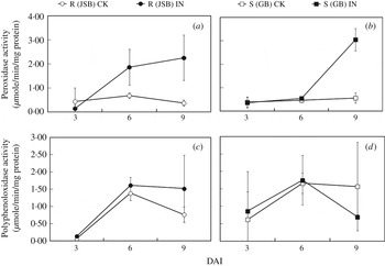
The peroxidase activity in the shoot bases of resistant JSB seedlings was induced at 6 and 9 DAI (Fig. 6 a), but the peroxidase activity was only induced in the shoot bases of susceptible GB seedlings at 9 DAI (Fig. 6 b). Nevertheless, no significant difference in polyphenoloxidase activity was observed in either resistant JSB (Fig. 6 c) or susceptible GB (Fig. 6 d) seedlings after F. oxysporum inoculation.
Peroxidase and polyphenoloxidase activities in the shoot bases of resistant and susceptible watermelon seedlings after inoculation with Fusarium oxysporum f. sp. niveum. Watermelon seedlings planted in F. oxysporum-infested soil were harvested at 3, 6 and 9 days after inoculation (DAI). The peroxidase (peroxidase; a, b) and polyphenoloxidase (polyphenoloxidase; c, d) activities in the shoot bases of the resistant (R, JSB; a, c) and susceptible (S, GB; b, d) seedlings were analysed. The open symbols represent mock control (CK); the closed symbols represent the inoculated samples (IN). The error bars were the s.e.m.

DISCUSSION
In previous studies, F. oxysporum was shown to colonize tissues of both resistant and susceptible watermelons (Michielse & Rep Reference Michielse and Rep2009); similar results were observed in the present study. In addition, F. oxysporum colonization in the resistant JSB seedlings was less severe than in the susceptible GB seedlings. Although the resistant JSB seedlings were colonized by F. oxysporum, the colonization rates were lower than 50% except in the bottom of the shoot bases at 12 DAI. These results suggest there may be some resistant activity in the shoot bases that lead to a lower colonization rate in the upper parts of the resistant JSB seedlings.
Phenylalanine ammonia lyase activity is considered as an indicator of plant/pathogen interaction. The PAL activity of flax cells has been found to increase 72 h after inoculation with the pathogen F. oxysporum f. sp. linii, then decrease at 96 h (Hano et al. Reference Hano, Addi, Fliniaux, Bensaddek, Duverger, Mesnard, Lamblin and Laine2008). Similarly, PAL activity in the shoot bases of susceptible GB seedlings increased at 3 and 6 DAI but decreased at 9 DAI. The PAL activity in shoot bases of resistant JSB seedlings increased at 3 DAI, decreased at 6 DAI, then increased again at 9 DAI and reached the highest level at 12 DAI. These results matched expression of the PAL gene in the shoot bases of resistant JSB seedlings, which increased at 3 DAI, decreased at 6 DAI and was induced again at 9 DAI (Chang et al. Reference Chang, Hsu, Lin, Chen, Huang and Liou2008). These results confirm that PAL plays an important role in watermelon resistance against F. oxysporum. The decrease of PAL activity in the shoot bases of resistant JSB seedlings at 6 DAI may be due to the effectors secreted by the invaded F. oxysporum in the shoot base, thus reducing PAL activity. However, this occurred for only a short period of time in JSB. Phenylalanine ammonia lyase activity increased again at 9 DAI and it is possible that JSB releases some molecules that are able to block the action of the F. oxysporum effectors, enabling this increase in PAL activity. This is in contrast to PAL activity in the shoot bases of susceptible GB seedlings, which continued to decrease from 6 DAI onwards. It is possible that there are no molecules able to block the action of the F. oxysporum effectors in GB. Nevertheless, further investigation is needed to clarify the involvement of the effectors.
Phenolic compounds are the downstream products of the phenylpropanoid pathway and could be induced upon a challenge by pathogens (Njoroge et al. Reference Njoroge, Vallad, Park, Kang, Koike, Bolda, Burman, Polonik and Subbarao2011). Phenolic compounds play three main roles in resistance against pathogens; direct attacks on pathogens through antimicrobial compounds, defence of plant cell walls as physical barriers, and induction of resistant responses as molecular messengers (Nicholson & Hammerschmidt Reference Nicholson and Hammerschmidt1992). The current results indicated that the total soluble and cell wall-bound phenolic compounds in the shoot base of the resistant JSB seedlings were induced at 3, 6 and 9 DAI, whereas only the cell wall-bound phenolic compounds in the susceptible GB seedlings were induced at 3 DAI. It is suggested, therefore, that phenolic compounds may be involved in the resistance of Fusarium wilt. Furthermore, according to the results of cell wall-bound phenolics, it is further suggested that phenolic compounds may serve as a physical barrier in the shoot bases of watermelon seedlings.
The increase in lignin deposition and cell wall-bound phenolic contents could enhance the ability of plants to defend against pathogen invasion (Nicholson & Hammerschmidt Reference Nicholson and Hammerschmidt1992). Cell wall-bound phenolic compounds are induced in banana and tomato to protect the plants from Fusarium wilt disease (Thangavelu et al. Reference Thangavelu, Palaniswami, Doraiswamy and Velazhahan2003; Mandal & Mitra Reference Mandal and Mitra2007). Furthermore, the content of syringyl-rich lignin has been shown to increase in wheat cell suspensions during the hypersensitive resistance response (Menden et al. Reference Menden, Kohlhoff and Moerschbacher2007). The involvement of phenolics and lignin content in broccoli plants resistant to Verticillium dahliae was reported by Njoroge et al. (Reference Njoroge, Vallad, Park, Kang, Koike, Bolda, Burman, Polonik and Subbarao2011). In the present study, it was found that at 6 and 9 DAI, the F. oxysporum-inoculated JSB seedlings showed significant lignin deposition in the shoot bases; lignin deposition in un-inoculated JSB was also observed but at a much lower level, whereas it was absent in GB. Thus, it can be concluded that structural barriers from related lignin products may be involved in the resistance of watermelons to Fusarium wilt. In addition, peroxidase and polyphenoloxidase are able to act on the oxidation of phenolic compounds among their many different functions in plants. On the structural resistance of plants, polyphenoloxidase could act to polymerize monolignol to olignols, whereas peroxidase could catalyse the reaction from olignol to macromolecular lignin (Sterjiades et al. Reference Sterjiades, Dean, Gamble, Himmelsbach and Eriksson1993; Van Loon Reference Van Loon1997). The current results showed that the peroxidase activity was induced in JSB at 6 and 9 DAI, which is closely related to the time of lignin deposition in JSB; thus, it is suggested that the structural barriers in the shoot base of watermelon seedlings may be involved in the resistance to Fusarium wilt.
This is the first report describing the significant difference in the amounts of phenolic compounds found in the shoot bases of resistant and susceptible watermelon seedlings. In summary, the present study states the possible resistance responses occurred in the shoot bases of watermelon seedlings. Meanwhile, PAL is involved in watermelon (JSB and GB) interaction with F. oxysporum, which concurs with the previous study of Michielse & Rep (Reference Michielse and Rep2009). Phenolic compounds may act as a physical barrier to defend against F. oxysporum invasion via the continuous production of cell wall-bound phenolics and the increase of the peroxidase activity in resistant JSB seedlings. Nonetheless, other defence mechanisms of phenolic (such as acting as antimicrobial compounds and signalling molecules) could also be considered for watermelon resistance to F. oxysporum.
The research was supported in part by the National Science Council, Taiwan, Republic of China (R.O.C.) under grant numbers NSC98-2313-B-005-025-MY3, NSC99-2622-B-005-006-CC2 and NSC101-2313-B-005-028-MY3; by the Ministry of Education, Taiwan, R.O.C. under the ATU Plan; and also by National Chung Hsing University, Taiwan, R.O.C.