


I. INTRODUCTION
Barium titanate ceramics have gained lot of attention in the scientific community due to the variation of properties due to the doping in barium or titanium sites (Levin et al., Reference Levin, Cockayne, Krayzman, Woicik, Lee and Randall2011). Due to its non-toxic behavior, BaTiO3 is a better alternative to lead-based materials (Xue et al., Reference Xue, Zhou, Bao, Gao, Zhou and Ren2011). BaTiO3 possess superior ferroelectric behavior and has three phase transitions at different temperature regimes. At around −80°C, it transforms from rhombohedral to orthorhombic, at around 5°C, it transforms from orthorhombic to tetragonal, and at around 130°C, it transforms from tetragonal to cubic (Zhou et al., Reference Zhou, Vilarinho and Baptista1999). The temperature for phase transformation in BaTiO3 shifts due to the A-site doping of Sr2+ or Ca2+ and B-site doping of Mn4+, Fe2+, Zr4+, and Nb5+ making it suitable for high- or low-temperature piezoelectric device fabrication (Sindhu et al., Reference Sindhu, Ahlawat, Sanghi, Kumari and Agarwal2013). The B-site substitution of Zirconium tunes the dielectric behavior of BaTiO3 and increases its permittivity (Wang et al., Reference Wang, Rong, Wang and Yao2014). The substitution of Mn and Zr in B-site leads to the formation of oxygen vacancy due to the charge imbalance and these oxygen vacancies induce magnetism in ceramics (Das et al., Reference Das, Rout, Pradhan and Roul2012).
The variation of Strontium content in BaTiO3 shifts the transition and alters the electrical properties. Earlier reports have suggested that the doping of strontium in barium site leads to a transformation in the crystal structure from tetragonal to cubic (Sindhu et al., Reference Sindhu, Ahlawat, Sanghi, Kumari and Agarwal2013). The smaller ionic radii of Sr2+ (1.44 Å) in comparison with Ba2+ (1.61 Å) has led to the reduction of c/a ratio and shifting of tetragonal structure to ideal cubic structure (Yu et al., Reference Yu, Zou, Cao, Wang, Li and Yao2015). In our sample, we also experienced the transformation of crystal structure from tetragonal to cubic due to the increasing strontium concentration. The crystal structure for Ba1-xSrxTi0.6Zr0.3Mn0.1O3 exhibited a tetragonal structure for x = 0 while for x = 0.2 the crystal structure transformed to an ideal cubic structure (Nandan and Kumar, Reference Nandan and Kumar2017). Generally, BaTiO3 exhibits cubic structure at around 130°C, but in our case due to the strontium doping, we were able to achieve cubic structure at room temperature. This raised our interest to analyze the powder X-ray diffraction (XRD) data and herein we are reporting the powder XRD pattern for BSTO samples.
II. EXPERIMENTAL
A. Synthesis
Polycrystalline samples of Ba0.8Sr0.2Ti0.6Zr0.3Mn0.1O3 (BSTO) were prepared by a standard solid-state reaction method. Initially, precursors barium carbonate, strontium carbonate, titanium dioxide, zirconium oxide, and manganese oxides were weighed in stoichiometric ratios and mixed by a ball milling process. The homogeneous mixture was calcined up to 1550°C in air atmosphere with intermediate grindings. The experimental procedures are detailed in our previous report (Nandan and Kumar, Reference Nandan and Kumar2017).
B. Data collection
Powder XRD data of the samples were measured using a Bruker D8 Advance (Germany) diffractometer. The sintered powders were ground and loaded in a zero background (911) Si single-crystal wafer holder. The instrument was operated in Bragg-Brentano geometry with fixed slits. The diffraction data for the sample was recorded using Cu K-alpha-1 (λ = 1.54060 Å), K-alpha-2 (λ = 1.54439 Å), and K-beta (λ = 1.39222 Å) radiation as the source which is operated at a voltage of 40 kV and a current of 30 mA with a goniometer radius of 217.5 mm. The data were recorded at a 2θ range from 10° to 80° with a step size of 0.015°.
III. RESULTS
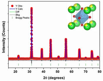
The refined powder XRD pattern for the BSTO samples is shown in Figure 1. Rietveld refinement for the diffraction data were carried by using GSAS-II program (Toby and Von Dreele, Reference Toby and Von Dreele2013). The initial structural parameters for refinement were taken from previous reports (Kim et al., Reference Kim, Jung and Ryu2004). The background was modeled using a Chebyshev polynomial. The fixed positional and isotropic displacement parameters were constrained for Ba and Sr to be the same and Ti, Zr, and Mn to be the same. Site occupancies were set and held fixed to the values determined by weights of the various starting materials mixed together and calcined given that no impurity phases evolved, even when sintered at higher temperatures. The powder data fitted well for Pm-3m space group and the fractional coordinates are shown in Table I. The results of the refinement (lattice parameter and R factors) are shown in Table II. The crystal structure of BSTO has been elucidated by Vesta software and shown as the inset in Figure 1 (Momma and Izumi, Reference Momma and Izumi2013). The experimental data were also indexed by Powder X software (Dong, Reference Dong1999) and the results of the refined parameters are shown in Tables II and III. K-alpha-2 stripping has been done before peak indexing in Powder X software. The various metal–oxygen bond lengths from the refined data are given in Table IV.
Rietveld refinement of powder XRD data of BSTO polycrystalline sample. The inset shows the BSTO crystal structure.

Fractional coordinates for BSTO samples.

Crystal data from GSAS program and Powder X software.

Powder diffraction data of BSTO polycrystalline material.

Selected bond lengths for BSTO samples.

IV. CONCLUSION
Polycrystalline samples of BSTO are prepared by a conventional solid-state reaction. Powder XRD patterns for the prepared sample confirmed the single-phase formation of the compound without any impurities. The prepared sample crystallized in a cubic structure (Pm-3m space group) with lattice parameters a = b = c = 4.0253 Ǻ, α = β = γ = 90°. Rietveld refinement for the XRD pattern was done by GSAS program and the peak indexing was carried out by Powder X software.
V. DEPOSITED DATA
The Crystallographic Information Framework (CIF) file was deposited with the ICDD. The data can be requested at pdj@icdd.com.