Key findings
-
• Albendazole, the reference drug, is inadequate for treating trichinellosis.
-
• Citrus paradisi peel extract demonstrated therapeutic and synergistic effects in trichinellosis management.
-
• Administration of the drugs early during the enteral phase exhibited the best efficacy.
-
• The combination therapy group had the lowest mean muscle larvae count, improved muscle integrity, and the highest reduction in NF-κB expression.
Introduction
Trichinellosis, a zoonotic parasitic disease, is caused by nematodes of the genus Trichinella. The parasite affects animals and humans and has a global distribution. In humans, Trichinella spiralis (T. spiralis) is a pathogenic one affecting nearly 11 million individuals worldwide (Caron et al. Reference Caron, Bory, Pluot, Nheb, Chan, Prum, Lim, Sim, Sengdoeurn, Sovann, Khieu, Vallée and Yera2020; Wakid et al. Reference Wakid, El Kholy, Alsulami and El-Wakil2024).
Human infection is transmitted by consuming raw or undercooked meat from pigs or wild pigs harboring infective larvae. This leads to intestinal and muscular phases characterized by severe inflammation and tissue damage (Mohammed et al. Reference Mohammed, Heikal, Ellessy, Aboushousha and Ghareeb2023; Nada et al. Reference Nada, Mohammad, Moad, El-Shafey, Ghandour and Ibrahim2018).
In the early infection stage, the infection can present with gastrointestinal complaints, including vomiting, diarrhea, and abdominal pain. Conversely, during the infection muscle phase, typically commencing one week post-infection, the larvae migration to skeletal muscles may result in facial edema, rash, fever, and myalgia (Gottstein et al. Reference Gottstein, Pozio and Nöckler2009).
Standard treatment with benzimidazole derivatives, including albendazole (ABZ) and mebendazole, effectively targets the intestinal stages of T. spiralis infection. However, these medications show only modest effectiveness against muscle-encysted larvae; some have high resistance levels and poor absorption. Due to safety concerns, their use is also prohibited in children younger than three years old and pregnant women (Yadav Reference Yadav and Temjenmongla2012).
These limitations highlight the urgent need to develop novel, effective, and safe therapeutic options for trichinellosis. In this regard, medicinal plants have drawn interest as a viable substitute because they are less expensive, less toxic, and less likely to cause negative side effects than synthetic medications. (Rahman et al. 2019; Salama et al. Reference Salama, Mostafa, Abd El-Aal, Moawad, Hammad, Adel and Mostafa2022).
The genus Citrus (C.) belongs to the Rutaceae family. This genus is famous for its beneficial fruits, which are a vital source of vitamins and important nutrients (Mohammed et al. Reference Mohammed, Ibrahim, Abdel-Aziz and Ghareeb2024). Phytochemically, C. paradisi (grapefruit) has high concentrations of phytochemicals such as flavonoids, polyphenols, carotenoids, and vitamins. Notably, the peel contains flavonoid glycosides like naringin and hesperidin, which exhibit antioxidant, antimutagenic, analgesic, and anti-inflammatory properties. The peel extract also demonstrates significant potential in inhibiting bacteria and fungi and neutralizing free radicals (Abed et al. Reference Abed, AlAubaidee, Azzawi and Alabbasy2022; Ortuño et al. Reference Ortuño, Báidez, Gómez, Arcas, Porras, García-Lidón and Del Río2006).
C. paradisi has demonstrated anti-parasitic activities in many previous studies. It has an anthelminthic impact on Schistosoma haematobium (Mahmoud et al. Reference Mahmoud, Ebeid and Nosseir2006). Besides, antiprotozoal effects were detected against cryptosporidiosis (Alsulami and El-Wakil Reference Alsulami and El-Wakil2024), giardiasis (De Almeida et al. Reference De Almeida, Bezagio, Colli, Romera, Ferrari and Gomes2022), malaria (Ogunjinmi et al. Reference Ogunjinmi, Adegbola, Ajayi and Orike2024), and Eimeria infection (Pérez-Fonseca et al. Reference Pérez-Fonseca, Gutiérrez, Sumano, Salem, Ortega-Cerrilla, Villa-Mancera and Alcala-Canto2022).
Hence, the present study aimed to assess the efficacy of C. paradisi peel extract as a therapeutic agent against T. spiralis or synergistic to the reference drug, ABZ, in experimentally infected mice.
Materials and methods
Plant material
C. paradisi fruits were purchased from a local Market in Cairo, Egypt. The plant was kindly identified by Dr Therese Labib, Department of Flora and Taxonomy, El-Orman Garden, Giza, Egypt. A voucher specimen number (C.p/f/2023) of the plant was deposited in the herbarium of the Medicinal Chemistry Department, Theodor Bilharz Research Institute, Giza, Egypt.
Preparation of C. paradisi extract
The peels were removed, collected, and dried in the shade away from direct sunlight. At room temperature, the dry powdered peels (550 g) were extracted via maceration using methanol (4 x 1.5 L). After the peels were taken off and gathered, they were left to dry in the shade, away from the sun. Using methanol (4 x 1.5 L) at room temperature, the dry powdered peels (550 g) were extracted by maceration. The combined extracts were concentrated at 40°C using a rotatory evaporator (Buchi R-300, Switzerland). For later use in biological and phytochemical studies, the resulting methanolic extract (103 g) was stored at room temperature in a secure container (Mohammed et al. Reference Mohammed, Ibrahim, Abdel-Aziz and Ghareeb2024).
High-Performance Liquid Chromatography (HPLC) analysis
Chromatographic separation was performed using an Agilent 1260 series HPLC system with a Zorbax Eclipse Plus C8 column (4.6 × 250 mm, 5 μm). The mobile phase consisted of water (A) and 0.05% trifluoroacetic acid in acetonitrile (B) at a flow rate of 0.9 ml/min. The mobile phase was programmed consecutively in a linear gradient as follows: 0 min (82% A); 0–1 min (82% A); 1–11 min (75% A); 11–18 min (60% A); 18–22 min (82% A); 22–24 min (82% A).
Detection was carried out at 280 nm using a multi-wavelength detector. Samples were injected at a volume of 5 μL, and the column temperature was kept at 40°C (Abdel-Wareth et al. Reference Abdel-Wareth and Ghareeb2018; Nasr et al. Reference Nasr, Ghareeb, Mohamed, Elwan, Abdel-Aziz and Abdel-Aziz2018).
Animals
This study utilized 66 male Swiss albino mice (5–6 weeks old, 25 ± 5 g body weight) bred under pathogen-free conditions at the Theodor Bilharz Research Institute (TBRI) animal facility in Giza, Egypt. All animals were maintained with ad libitum access to standard pellet feed and water throughout the experimental period.
Experimental grouping
The mice were randomly allocated into 5 groups as follows:
-
Group I: healthy group (the control negative group) (6 mice)
-
Group II: Infected, untreated group (the control positive group) (6 mice)
-
Group III: Infected and treated with ABZ (Sigma Aldrich, Egypt) administered orally through an esophageal tube, with a daily dosage of 50 mg/kg (Attia et al. Reference Attia, Mahmoud, Farrag, Makboul, Mohamed and Ibraheim2015) (18 mice)
-
Group IV: Infected and treated with C. paradisi extract administered using an esophageal tube orally, with a daily dosage of 100 mg/kg (De Almeida et al. Reference De Almeida, Bezagio, Colli, Romera, Ferrari and Gomes2022) (18 mice)
-
Group V: Infected and received a combination of ABZ and C. paradisi extract (18 mice).
The study groups from Group III–V were further subdivided into three subgroups: Subgroup A: infected and treated during the enteral phase [2–6 days post-infection (dpi)], Subgroup B: infected and treated during the invasive phase (migratory phase) [10–14 dpi] and Subgroup C: infected and treated during the encapsulated phase [30–34 dpi]. Each subgroup contained 6 mice. All mice were sacrificed at 35 dpi to determine the effect of the administered drug at different infection intervals on the outcomes of trichinellosis infection.
The parasite and infection
T. spiralis utilized in this study was supplied by the Medical Parasitology Department, TBRI. It was kept by consecutive passages on mice. Briefly, infected mice were sacrificed 30 dpi, skinned, and eviscerated. Unnecessary parts (tail, teeth, and ears) were removed, and the remaining body was placed in a glass beaker with artificial gastric juice (1% pepsin and 1% concentrated HCl in distilled water), incubated at 37°C overnight for larval isolation. A copper sieve (50 mesh/inch) removed coarse particles, and a finer sieve (200 mesh/inch) collected excysted larvae, which were washed and allowed to settle. The supernatant was removed, and larvae in the sediment were counted using a McMaster counting chamber. The hemocytometer was loaded with 100 μl of the sediment and placed on the microscope, and the entire chamber counting gird was viewed at 4X objective. The gird contains 4 large corner squares with a surface area of 1 mm2, each consisting of 16 small squares. The depth of the chamber is 0.1 mm. Larvae were counted in each of these corner sets of 16 squares under the 10X objective. Larvae on the right and bottom lines were counted, while larvae on the left and top lines were not counted to avoid overlap and false counting. The average larvae count was calculated as follows: Larva count in 1 ml = (total number of larvae / 4) x 10,000 (Shalaby et al. Reference Shalaby, Moghazy, Shalaby and Nasr2010).
All mice except group I were infected orally using an esophageal tube with 200 to 300 larvae (Abou Rayia et al. Reference Abou Rayia, Saad, Wang, Gong, Zhang and Zhang2017).
Parasitological examination
On the 35th day post-infection, mice were sacrificed and the skeletal muscles were taken and digested using the pepsin digestion method to detect and count encysted larvae of T. spiralis (Basyoni et al. Reference Basyoni2013; Muñoz-Carrillo et al. Reference Muñoz-Carrillo, Maldonado-Tapia, López-Luna, Muñoz Escobedo, Flores de la Torre and Moreno García2018). The number of encysted muscle larvae was expressed according to Denham (Reference Denham1965) per gram of muscle. The reduction percentage was calculated as follows: RP% = A - C/A x 100, where A is the mean muscle larvae count from the control positive group, and B is the mean muscle larvae count from the treated groups (El-wakil et al. Reference El-Wakil, Shaker, Aboushousha, Abdel-Hameed and Osman2023).
Histopathological examination
The procedures and protocols adopted for sample fixation and staining adhered to the established standards of Suvarna et al. (Reference Suvarna, Layton, Bancroft, Suvarna, Layton and Bancroft2019). The skeletal muscle tissue samples were dissected, cleaned, and then preserved for 48 hours in 10% neutral buffered formalin. After being processed in successively higher grades of ethanol and cleaned in xylene, the samples were embedded in Paraplast Plus media for tissue embedding. To demonstrate muscle bundles, a rotatory microtome cut serial sections that were 5 μm thick and adhered to glass slides. Hematoxylin and Eosin, a general staining technique for microscopic inspections, were applied to them, followed by a blind examination by a qualified pathologist.
Immunohistochemical (IHC) examination
Immunohistochemical analysis was performed to ascertain the levels of the nuclear factor kappa-light-chain-enhancer of activated B cells (NF-κB) expression, indicating inflammation (El-Sayed et al. Reference El-Sayad, El-Wakil, Moharam, Abd El-Latif, Ghareeb and Elhadad2023). The manufacturer’s instructions were followed to prepare tissue sections that were 5 microns thick and embedded in paraffin. Sections of deparaffinized tissue that had been recovered were exposed to 0–3% H2O2 for 20 minutes. Next, the muscle samples were incubated with an antibody against NF-κB p65 (GTX54672, GeneTex Inc.) (1:100) overnight at 4°C. Phosphate-buffered saline (PBS) was used to wash the tissue sections. After 20 minutes of incubation with the secondary antibody Horseradish Peroxidase (HRP) [Envision kit (DAKO)], the sample was rinsed out and incubated with diaminobenzidine (DAB) for 15 minutes. After that, a PBS wash, hematoxylin counterstaining, xylene drying, and coverslipping were done for microscopic inspection. At least 6 non-overlapping random fields were selected and scanned from muscle samples to determine the Optical density, in immunohistochemically stained sections, of muscular tissue immunoexpression levels of NF-κB. All light microscopic examinations and data were obtained using the Leica application module for histological analysis attached to a Full HD microscopic imaging system (Leica Microsystems GmbH, Germany).
Statistical analysis
Data analysis was done using IBM SPSS Statistics (Version 26) and Microsoft Excel 2016. Mean ± standard deviation (SD) was used to report normally distributed continuous variables. Group comparisons for normally distributed data were performed using Student’s t-test, while multigroup analyses utilized ANOVA, followed by a post hoc test, Tukey’s HSD. A P-value < 0.05 was deemed statistically significant, and P < 0.01 was considered highly significant.
Results
Plant extract chemical components
Table 1 and Figures 1 and 2 show that HPLC analysis revealed that the tested extract contains nine phenolic acids; their concentrations per gm in the plant were gallic acid (Conc.: 787.93μg/g), chlorogenic acid (Conc.: 717.62μg/g), caffeic acid (Conc.: 60.53μg/g), coumaric acid (Conc.: 65.10μg/g), syringic acid (Conc.: 248.71μg/g), ellagic acid (Conc.: 208.18μg/g), ferulic acid (Conc.: 7438.10μg/g), rosmarinic acid (Conc.: 1149.46μg/g), and cinnamic acid (Conc.: 24.49μg/g). The flavonoidal compounds were detected as catechin (Conc.: 3461.39μg/g), rutin (Conc.: 467.47μg/g), naringenin (Conc.: 118362.61μg/g), daidzein (Conc.: 75.38μg/g), quercetin (Conc.: 331.47μg/g), kaempferol (Conc.: 464.68μg/g) and hesperetin (Conc.: 64.36μg/g). Moreover, phenolic acid derivatives were identified as methyl gallate (Conc.: 230.97μg/g). It is worth noting that in the extract, naringenin, with a retention time of 10.42 min and an area percentage of 83.04%, was the predominant flavonoid compound, with a concentration of 118,362.61 μg/g, while ferulic acid (Rt = 9.52 min, area % = 8.37%) was the most abundant phenolic acid at 7,438.10 μg/g. In contrast, caffeic acid (Rt = 5.89 min, area % = 0.05%) showed the lowest detected concentration (60.53 μg/g), and vanillin (Rt = 9.17 min) was not detected.
Areas under peaks and concentrations of the identified phenolic compounds in the C. paradisi methanolic extract compared to eighteen standard (STD) phenolic compounds

Retention Time (Rt, min): the time at which a compound elutes from the HPLC column
Area %: relative abundance of each detected compound
Concentration (μg/ml): the calculated amount of the compound in the sample
Concentration (μg/g): amount of compound per gram of the plant material
High-Performance Liquid Chromatography (HPLC) chromatogram of eighteen standard phenolic compounds.
X axis: time in minutes. Y axis: mAU: milli-absorbance units, indicates the absorption of analytes by the detector, helps in determining the concentration of the analyte.

High-Performance Liquid Chromatography (HPLC) chromatogram of C. paradisi methanolic extract.

Effect of different therapeutic approaches on muscle-encysted larval counts
In the current study, the therapeutic agents were administered to the different experimental groups at different time intervals after infection to investigate the effect of these therapeutic agents when given during the enteral (Subgroup A), migratory (Subgroup B), or encapsulated phases (Subgroup C) of infection on the muscle-encysted larval counts at the end of the experiment (35 dpi).
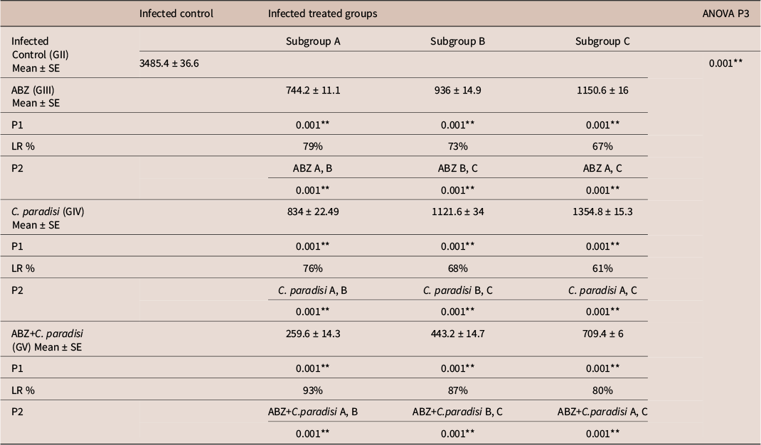
The positive control group (GII) exhibited a mean larval count of 3485.4 ± 36.6 per gram of muscle. All treated groups displayed a highly significant difference (p < 0.001) when compared with the positive control group (GII) (Table 2).
The mean larval count and larval reduction percentage (LR%) among the different study groups

-
• T. spiralis encysted larvae count per gram of muscle is represented as Mean ± SE
-
• P1: comparison between treated mice and control group using Student’s test
-
• P2: comparison between pairs of groups using post hoc test (Tukey’s)
-
• P3: comparing between groups depending on the One Way ANOVA test (compare more than two arithmetic means)
-
• *Significantly different at P < 0.05. ** Highly significantly different at P < 0.01.
When comparing the different therapeutic regimens on the mean larval count, the subgroup A mice showed the lowest larval count, with the highest reduction percentages, followed by the subgroup B mice, and then the subgroup C mice with a highly significant difference (p < 0.001) between them (Table 2).
Regarding drug administration in subgroup A, the ABZ-treated group (GIII) demonstrated a mean encysted larvae count of 744.2 ± 11.1, the C. paradisi extract-treated group (GIV) showed a mean encysted larvae count of 834 ± 22.49, and the combination of ABZ and C. paradisi-treated group (GV) yielded the highest efficacy with the least mean count value of 259.6 ± 14.3, with reduction percentages of 79%, 76%, and 93%, respectively (Table 2).
When the drugs were administered in subgroup B, the ABZ-treated group (GIII) demonstrated a mean encysted larvae count of 936 ± 14.9, the C. paradisi extract-treated group (GIV) showed a mean encysted larvae count of 1121.6 ± 34, and the combination of ABZ and C. paradisi-treated group (GV) yielded the highest efficacy with the least mean count value of 443.2 ± 14.7, with reduction percentages of 73%, 68%, and 87%, respectively (Table 2).
The administration of the therapeutic agents in subgroup C showed the highest larval count, with the lowest reduction percentages as per Table 2.
Histopathological examination
Regarding histopathological examination, skeletal muscle sections from the healthy group (GI) showed intact skeletal muscle bundles and intermuscular spaces without abnormal alterations or infiltrates (Figure 3A). In contrast, the positive control group (GII) revealed necrosis, severe inflammation, and dense encysted larvae (Figure 3B). In treated groups (Figure 3C–K), especially those receiving combination therapy (Figure 3I–K), significant reductions in inflammatory cell infiltrates and improved muscle architecture were observed.
Histopathological photomicrographs of skeletal muscle sections. (a) The healthy group shows intact skeletal muscle bundles. (b) The infected positive control group shows densely packed encysted larvae (at least 12 Larvae/ microscopic field) (red arrowhead). (c) ABZ (Subgroup A) shows a significant decrease of encysted larvae (red arrowhead) (2 to 3 larvae/ microscopic field). (d) ABZ (Subgroup B) shows figures similar to those of the previous group, with persistent severe inflammatory cell infiltrates. (e) ABZ (Subgroup C) shows persistent records of encysted larvae (red arrowhead) (at least 6 larvae/ microscopic field). (f) C. paradisi (Subgroup A) shows moderately higher records of encysted larvae. (g) C. paradisi (Subgroup B) shows persistent moderately higher records of encysted larvae (red arrowhead) (at least 10 larvae/ microscopic field). (h) C. paradisi (Subgroup C) shows similar records to the previous group, with moderate records of encysted larvae. (i) ABZ+C. paradisi (Subgroup A) shows no figures of encysted larvae. (j) ABZ+C. paradisi (Subgroup B) shows minimal encysted larvae with few ruptured larval capsules (magnification X100). (k) ABZ+C. paradisi (Subgroup C) showing figures similar to the previous group’s.

The combined ABZ and C. paradisi extract administration in subgroup A showed the best protective efficacy without figures of encysted larvae in almost all tissue samples, with multiple focal inflammatory cell infiltrates and minimal necrotic and degenerated muscle fibers (Figure 3I). Groups that received the combination of drugs in subgroup B or subgroup C had similar results of minimal figures of encysted larvae and intense reactive inflammatory cells infiltrate with minimal necrotic and degenerated muscle fibers (Figure 3J–K).
Immunohistochemical examination
The NF-κB expression in mice’s skeletal muscle sections is depicted in Table 3 and Figure 4. The healthy non-infected group (GI) showed low expression of NF-κB with a mean ± SE of 0.17 ± 0.01(Figure 4A). The positive control infected non-treated group (GII) showed an increase in NF-κB levels, with 0.8 ± 0.02 (Figure 4B). Treated groups showed reductions of NF-κB expression. The group administered ABZ in subgroup A showed 0.32 ± 0.02, 0.36 ± 0.01 in subgroup B, and 0.43 ± 0.03 in subgroup C (Figure 4C–E). While the group that received C. paradisi extract showed 0.5 ± 0.02 in subgroup A, 0.58 ± 0.03 in subgroup B, and 0.67 ± 0.02 in subgroup C, which were the highest values among all the groups (Figure 4 F–H).
Optical density of muscular tissue IHC expression of NF-κB in different study groups

** Initial P-value < 0.01 is highly significant
Muscular tissue IHC expression of P NF-κB (a) The healthy group shows low expression of NF-κB. (b) The positive control group shows an increase in NF-κB levels. (c–e) The ABZ-treated groups demonstrate reductions in NF-κB expression. (f–h) The C. paradisi-treated groups show notable reductions in NF-κB expression. (i–k) ABZ+C. Paradisi-treated groups demonstrated the best reduction in NF-κB expression.

The combined ABZ and C. paradisi extract administration showed a mean ± SE of 0.21 ± 0.01 in subgroup A, 0.29 ± 0.01 in subgroup B, and 0.31 ± 0.01 in subgroup C (Figure 4 I–K). The combination of ABZ and C. paradisi extract showed the most reduction in mean ± SE and a statistically significant difference (p < 0.001) compared to the positive control group.
Discussion
Trichinellosis, caused by Trichinella spiralis, remains a global concern, particularly in regions with limited food safety and healthcare measures (Bai et al. Reference Bai, Hu, Liu, Tang and Liu2017). Current treatments, such as Albendazole (ABZ), are associated with significant adverse effects, including encephalitis and epilepsy, and demonstrate limited efficacy against encysted larvae (Pozio et al. Reference Pozio and Bruschi2001; Yadav Reference Yadav and Temjenmongla2012).
This underscores the urgent need for safer and more effective anthelmintic agents. Recently, there has been a lot of interest in the use of medicinal plants to treat a variety of illnesses, including parasitic infections, because of their minimal adverse effects and better tolerability compared to synthetic drugs (Alsulami and El-Wakil Reference Alsulami and El-Wakil2024; Rahman et al. Reference Rahman, Jahan and Mim2019).
Citrus fruits, particularly C. paradisi, stand out for their nutritional and medicinal value. Being rich in biologically active compounds, including phenolic compounds and flavonoids, citrus fruits exhibit potent antioxidant properties, making them a promising alternative in pharmacological research (Elizondo-Luévano et al. Reference Elizondo-Luévano, González-Aguilar and Alvarez-Parrilla2021; Rahman et al. Reference Rahman, Jahan and Mim2019).
C. paradisi extract is rich in flavonoids, such as naringin and hesperidin, which exhibit diverse biological activities such as antioxidant, antimutagenic, analgesic, and anti-inflammatory effects. Additionally, the peel extract of C. paradisi demonstrates significant potential in inhibiting bacterial strains and fungal growth, as well as scavenging free radicals (Abed et al. Reference Abed, AlAubaidee, Azzawi and Alabbasy2022; De Almeida et al. Reference De Almeida, Bezagio, Colli, Romera, Ferrari and Gomes2022; Ortuño et al. Reference Ortuño, Báidez, Gómez, Arcas, Porras, García-Lidón and Del Río2006).
Therefore, this study assessed the efficacy of C. paradisi extract against T. spiralis in experimentally infected mice.
Adham et al. (Reference Adham2015) reported that C. paradisi fruit extract contains hesperidin at a concentration of 204.2 μg / gram peel. On the other side, Sudanese C. paradisi peel extract contains high amounts of naringenin (28.09%) (Alrasheid et al. Reference Alrasheid, Mohamed, Mohieldin, Eldein, Yassin, Widdatallh, Alnour, Eltilib, Ahmed and Ayoub2019). Also, prior research has demonstrated that Ultra High-Performance Liquid Chromatography (UHPLC) profiling of C. paradisi extract led to the identification of narirutin (92 mg/100 g FW), poncirin (11.2 mg/100 g FW), neohesperidin6 (15.6 mg/100 g FW), poncirin (11.2 mg/100 g FW), taxifolin (1.5 mg/100 g FW), and gallic acid (2.7 mg/100 g FW) (Ahmed et al. Reference Ahmed, Azmat, Qayyum, Bibi, Ali, Ahmed and Ramadan2023).
The present study revealed a significant decrease in the number of encysted larvae in muscle found in all treated groups (throughout enteral, migratory, and encapsulated phases) compared with the untreated infected group on the 35th dpi.
The mean count value of muscle larvae for the group that received the combined treatment of ABZ and C. paradisi extract was (259.6 ± 14.3), (443.2 ± 14.7), and (709.4 ± 6) in subgroups A, B, and C, respectively. It was found that muscle larvae recovered from the group that received the combined treatment of ABZ and C. paradisi extract (GV) had the least mean muscle larva count as compared with the control positive group and as compared with other groups treated with either ABZ or C. paradisi only with the highest percentage of reduction of 93%, 87% and 80% in subgroups A, B, and C, respectively.
These findings align with previous studies demonstrating the anti-parasitic potential of citrus extracts attributed to their high flavonoid content, including naringin and hesperidin (De Almeida et al. Reference De Almeida, Bezagio, Colli, Romera, Ferrari and Gomes2022; Elom et al. Reference Elom, Uche, Ukwah, Usanga, Okpara-Elom, Kalu and Ibe2021).
Flavonoids exhibit various bioactivities, such as lowering intestinal inflammation, modulating immune responses, and inhibiting parasite adhesion. For instance, Kallaf et al. (Reference Khallaf, Othman, Mahmoud, Attia and Tahawy2018) reported the therapeutic effects of Citrus sinensis against T. spiralis. They concluded that the effectiveness might be attributed to their content of polymethoxylated flavonoids: pentamethoxyflavone, heptamethoxyflavone, and hexamethoxyflavon.
Additionally, Elom et al. (Reference Elom, Uche, Ukwah, Usanga, Okpara-Elom, Kalu and Ibe2021) and De Almeida et al. (Reference De Almeida, Bezagio, Colli, Romera, Ferrari and Gomes2022) reported the anti-parasitic effects of citrus extracts against Plasmodium spp and Giardia duodenalis (G. duodenalis), respectively, linking efficacy to their immunomodulatory and anti-inflammatory properties. Moreover, the anti-cryptosporidiosis effects of different citrus species were reported in previous studies. Hafez et al. (Reference Hafez and Hamed2021) reported that C. maxima significantly decreased C. parvum oocysts numbers compared to the infected non-treated group and the group treated with the standard drug nitazoxanide. Abd El Wahab et al. (Reference Abd El Wahab, Shaapan, El-Naggar, Ahmed, Owis and Ali2022) studied the therapeutic efficacy of C. sinensis peel extract on Cryptosporidium-infected mice. Two doses of C. sinensis extract, a high dose (30 mg/kg) and a low dose (15 mg/kg), were investigated compared to nitazoxanide (NTZ). Parasitological results showed a statistically significant reduction in the percentage of oocyst shedding in fecal samples, more in the high dose than the low dose C. sinensis extract-treated mice.
Regarding the histopathology, a microscopic examination of skeletal muscles was carried out and revealed that the infected non-treated group (GII) sections in muscles at 35th dpi showed high numbers of densely packed encysted larvae with necrotic and degenerated muscle fibers and sequestrated with severe mononuclear inflammatory cells infiltrates. The C. paradisi combined with ABZ-treated mice in subgroup A showed the best results with no encysted larvae in almost all tissue samples and multiple foci of intermuscular mixed inflammatory cell infiltrates with minimal figures of necrotic and degenerated muscle fibers. In ABZ-treated mice in subgroup A, muscle sections revealed a moderate protective effect with persistent moderate higher records of encysted larvae, and reactive inflammatory cell infiltrates were shown among many records of necrotic and degenerated muscle fibers.
De Almeida et al. (Reference De Almeida, Bezagio, Colli, Romera, Ferrari and Gomes2022) found that C. paradisi had a therapeutic effect on G. duodenalis-infected mice, with treated groups showing no trophozoites in the intestinal mucosa, compared to ABZ and metronidazole-treated groups (2 trophozoites/field) and the positive control group (50+ trophozoites/field). Hafez et al. (Reference Hafez and Hamed2021) showed that Citrus maxima extract reduced Cryptosporidium parvum (C. parvum) oocysts in immunocompromised mice, with mild inflammation and less villous damage, likely due to its flavonoid and phenolic content. Abd El Wahab et al. (Reference Abd El Wahab, Shaapan, El-Naggar, Ahmed, Owis and Ali2022) also reported that Citrus treatment in C. parvum-infected mice maintained normal villi appearance and minimal inflammation due to the flavonoids and phenolic compounds in Citrus that inhibit Cryptosporidium colonization.
An essential function of NF-κB is to control the immune system’s reaction to inflammation and infection. In the case of trichinellosis, it is known that during infection, host immune cells activate NF-κB. The transcription of pro-inflammatory cytokines, including TNF-α, IL-1β, and IL-6, is triggered by this activation and is crucial for starting and maintaining the immune response against the parasite. However, severe or prolonged NF-κB activation can lead to persistent inflammation and tissue injury, especially in muscular tissues where the larvae encyst (Liu et al. Reference Liu, Meng, Le Li, Xu, Lin, Dong and Wang2023).
In the current study, NF-κB expression was done to evaluate the extent of inflammatory signaling in the host during infection and assess how the treatment modulates this pathway. A reduction in NF-κB activity after treatment would indicate a potential anti-inflammatory effect and suggest mitigating tissue pathology caused by the parasite-induced immune response.
Regarding the immunohistochemical studies, NF-κB was crucial for the host’s defense against T. spiralis. The present study shows that the healthy control group showed low expression of NF-κB, having a mean ± SE of 0.17 ± 0.01, while the positive control group showed elevated NF-κB levels with 0.8 ± 0.02. A significant decrease in NF-κB levels was observed across all treated groups, with the most notable reduction in mean ± SE recorded in the groups receiving the combined treatment of ABZ and C. paradisi with 0.21 ± 0.01 in subgroup A, 0.29 ± 0.01 mice in subgroup B, and 0.31 ± 0.01 in subgroup C.
Furthermore, NF-κB levels correlate with the larval burden detected in this study, suggesting that NF-κB downregulation is probably linked to the clearance of encysted larvae.
This is in accordance with Symeonidou et al. (Reference Symeonidou, Pappa, Kourelis, Anogeianaki, Frydas, Karagouni and Hatzistilianou2010), who conducted a microarray analysis of NF-κB signaling in mice infected with T. spiralis, showing changes in gene expression, indicating that NF-κB activity is modulated during infection, potentially affecting the parasite’s survival and the host’s immune response. Similarly, Xu et al. (Reference Xu, Yu, Wu, Liu and Lu2019) deduced that infection with T. spiralis leads to the activation of NF-κB and the secretion of pro-inflammatory cytokines. Furthermore, they reported that the NF-κB signaling pathway contributes substantially to the host’s immune response to T. spiralis infections, where elevated NF-κB activity has been observed in macrophages during such infections, suggesting a correlation between increased NF-κB activation and heightened T. spiralis presence.
These observations align with Liu et al. (Reference Liu, Zhang, Joo and Sun2017), who demonstrated that T. spiralis cathepsin L (rTsCatL2) induces macrophage polarization via the NF-κB pathway. This polarization enhances antibody-dependent cell-mediated cytotoxicity (ADCC) against newborn larvae, indicating that increased NF-κB activity contributes to the host’s defense mechanisms against the parasite. Stabrauskiene et al. (Reference Stabrauskiene, Marksa, Ivanauskas, Viskelis, Viskelis and Bernatoniene2023) also suggested that naringin and neohesperidin from C. paradisi could inhibit NF-KB activation, reducing pro-inflammatory cytokines like IL-33, TNF-, IL-1, and IL-6.
Similarly, Desmiaty et al. (Reference Desmiaty, Sandhiutami, Mulatsari, Maziyah, Rahmadhani, Algifari and Jantuna2024) proposed that ABZ acts by inhibiting NF-κB, which modulates the expression of NOD-like receptor family pyrin domain-containing protein 3 (NLRP3), caspase-1, and pro-IL-1β. Consequently, combined therapy with ABZ and C. paradisi led to a significant decrease in NF-κB levels, which was explained by the possibility of their synergistic effect.
Conclusions
The findings of the present study suggest that C. paradisi extract could represent a safe and natural therapeutic option that could be utilized in the treatment of T. spiralis infections either alone or in combination with the standard therapy through reduction of muscle larvae count, improved muscle integrity, and reduction in NF-κB expression. Additional studies are required to clarify the underlying pathways through which C. paradisi affects trichinellosis and its possible synergistic interaction with albendazole.
Financial support
This research did not receive any specific funding from public, commercial, or not-for-profit organizations.
Competing interests
The authors declare no competing interests.
Ethical standard
The authors assert that all procedures contributing to this work were conducted in compliance with the ethical standards of the October 6th University Research Ethical Committee under approval number (PME-Me-2306002). The October 6th University Research Ethical Committee is organized and operates in accordance with the guidelines of the International Council for Harmonisation (ICH), the Islamic Organization for Medical Sciences (IOMS), the United States Office for Human Research Protections (OHRP), and the United States Code of Federal Regulations, under Federal-wide Assurance Number FWA 00017585