The correct regulation of insulin secretion in response to fluctuating blood glucose concentrations is of vital importance. Too much or too little insulin can have very profound physiological consequences, as demonstrated by the various forms of diabetes (subnormal insulin secretion) and hyperinsulinism (excess insulin secretion). Insulin is a peptide hormone secreted from the pancreatic β-cell when blood glucose levels rise above the normal range (∼5 mm). How the pancreatic β-cell is able to sense the increase in blood glucose and subsequently secrete the appropriate amount of insulin has been the subject of intense investigation (for a comprehensive review see Ref. Reference MacDonald, Joseph and Rorsman1).
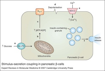
Briefly, elevation of blood glucose level leads to an increase in the amount of glucose taken up into the pancreatic β-cell via the GLUT2 glucose transporter. This allows an increase in the rate of glucose metabolism, resulting in an increase in the amount of ATP relative to ADP within the cell. This rise in the cytoplasmic [ATP]/[ADP] ratio leads to the closure of ATP-sensitive potassium (KATP) channels, resulting in membrane depolarisation (Refs Reference Nichols2, Reference Ashcroft3). Membrane depolarisation leads to the opening of L-type voltage-gated calcium channels, and the resultant influx of calcium stimulates the fusion of insulin-containing vesicles with the plasma membrane (Fig. 1). Thus, KATP channels play a central role in regulating insulin secretion in response to fluctuations in blood glucose levels by linking the metabolic status of the cell (represented by [ATP]/[ADP]) to the electrical excitability of the cell membrane. In summary, active KATP channels suppress insulin secretion whereas inactive channels promote insulin secretion. It should be noted that, in the majority of species, insulin secretion is biphasic (Ref. Reference Straub and Sharp4). The first phase of secretion is characterised by a rapid release of insulin that peaks ∼3–4 min following a stimulus with elevated glucose and then declines rapidly to a nadir at ∼8 min. Following the nadir, insulin secretion increases steadily, reaching a plateau over the next 20–25 min (second-phase insulin secretion). It is generally accepted that first-phase insulin secretion is regulated primarily by the KATP-channel-dependent mechanism (see below), whereas second-phase insulin secretion is regulated by KATP-channel-independent mechanisms (Refs Reference Taguchi5, Reference Aizawa6).
Stimulus–secretion coupling in pancreatic β-cells. Schematic of events that couple glucose metabolism to insulin secretion. (a) An increase in blood glucose levels results in an increase in the rate of glucose uptake into the pancreatic β-cell via GLUT2 glucose transporters. (b) The increased intracellular glucose level leads to an increase in the rate of metabolism, resulting in an increase in the ratio of intracellular ATP and ADP concentrations ([ATP]/[ADP]). (c) This in turn inhibits ATP-sensitive potassium (KATP) channels, which (d) allows the plasma membrane to depolarise more easily. (e) This membrane depolarisation causes the opening of voltage-dependent calcium channels (VDCCs), allowing an influx of Ca2+ into the cell. (f) This influx of Ca2+ stimulates the translocation and fusion of insulin-containing dense-core secretory granules with the plasma membrane, leading to insulin secretion.

The remarkable ability of the pancreatic β-cell to sense the amount of glucose in its environment and to secrete insulin accordingly therefore relies on the maintenance of an appropriate level of KATP channel activity. The net K+ ion current flowing through these channels at any given time, and hence the magnitude of change in membrane potential, depends on both the number of channels at the plasma membrane (N) and the probability with which the channels are able to open (P o). Disruption of either of these parameters will therefore be expected to interfere with normal insulin secretion and lead to a disease state. Indeed, two distinct types of inherited genetic mutations have been reported: one that alters the functional properties of the channel and another that affects the cell-surface channel density. Mutations that lead to a reduction in KATP channel function (loss-of-function mutations), or prevent cell-surface expression (trafficking mutations), lead to congenital hyperinsulinism. This is a rare genetic disorder where the affected patient secretes excess insulin despite low blood glucose levels (Ref. Reference Dunne7). The majority of loss-of-function mutations appear to reduce the ability of the channel to open in response to a reduced [ATP]/[ADP] ratio (Refs Reference Dunne7, Reference Nichols8, Reference Shyng9). As a consequence, KATP channels in these patients remain closed even when blood glucose levels fall to dangerously low levels, leading to uncontrolled insulin secretion. The trafficking mutations impair the ability of channels to reach the cell surface (Refs Reference Partridge, Beech and Sivaprasadarao10, Reference Cartier11, Reference Taschenberger12), as a result of which few or no channels would be present in the membrane to suppress membrane depolarisation and insulin secretion. Transgenic mice expressing reduced levels of β-cell KATP channels (∼60% of the normal) reiterate the hyperinsulinaemic phenotype (Refs Reference Remedi13, Reference Remedi14).
By contrast, mutations that enhance the function of KATP channels (gain-of-function mutations), or increase the channel numbers at the cell surface (trafficking mutations), could present the opposite condition – namely diabetes mellitus. Such mutations have been reported in patients with permanent neonatal diabetes mellitus (PNDM) (Ref. Reference Gloyn15), transient neonatal diabetes mellitus (TNDM) (Ref. Reference Gloyn16) and maturity-onset diabetes of the young (MODY) (Ref. Reference Huopio17). In the majority of cases, these mutations appear to reduce the ability of the channel to close in response to increased [ATP]/[ADP] ratio (Refs Reference Ashcroft3, Reference Gloyn15, Reference Ashcroft18). Although some mutations cause only small changes in P o, the effect on insulin secretion can be significant because tiny changes in K+ current amplitudes are thought to be enough to suppress firing and, consequently, insulin secretion (Ref. Reference Ashcroft and Rorsman19). Thus, failure of KATP channels to close even when blood glucose levels are abnormally high results in reduced insulin secretion. In two cases of PNDM, mutations increase the channel numbers at the cell surface by inhibiting their removal from the cell membrane (see later). A link between mutations in Kir6.2 and the more common form of diabetes, namely type 2 diabetes mellitus, has been reported (Refs Reference Riedel20, Reference Riedel, Steckley and Light21), but the mechanisms by which these mutations reduce insulin secretion are poorly understood.
KATP channels: structure
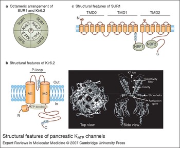
The pancreatic KATP channel is composed of four copies each of two separate proteins: Kir6.2 and the sulphonylurea receptor (SUR1) (Fig. 2) (Ref. Reference Aguilar-Bryan and Bryan22). Kir6.2 is a member of the inwardly rectifying potassium (Kir) channel family and forms the highly selective potassium ion (K+)-conducting pore of the fully formed channel (Refs Reference Inagaki23, Reference Tucker24). Kir6.2 is composed of two membrane-spanning helices (M1 and M2) either side of an extracellularly facing re-entrant P-loop that contains the conserved peptide sequence GFG responsible for selective transport of K+, termed the K+ selectivity filter (Fig. 2b) (Ref. Reference MacKinnon25). The three-dimensional structure of the channel is not known, but the structure of the pore-forming Kir6.2 could be modelled on the basis of the crystal structures of the bacterial Kir channel homologue Kirbac1.1 (Ref. Reference Kuo26) and the intracellular domains of Kir3.1 (Ref. Reference Nishida and MacKinnon27). The inner M2 helices are tilted with respect to the membrane and are slightly kinked such that they can support the structure of the selectivity filter at the extracellular end of the pore, but also form a ‘bundle-crossing’ at the cytoplasmic face of the pore. This bundle crossing serves as the ‘activation gate’ of the channel. Between the bundle crossing and the selectivity filter is a water-filled cavity. In the closed state of the channel, the opening of the activation gate is too narrow to allow any ions to pass through, but when the gate opens fully in response to a stimulus (decreased [ATP]/[ADP] ratio), ions (predominantly cations) enter the water-filled cavity. The selectivity filter, however, only allows K+ ions to pass from the cavity to the extracellular face of the pore. The small ‘slide helix’, which precedes the first transmembrane domain (M1), is thought to play a key role in controlling the closing and opening of the activation gate – a process termed ‘gating’ (Ref. Reference Antcliff28).
Structural features of pancreatic KATP channels. (a) ATP-sensitive potassium (KATP) channels are made up of two types of separate subunit – Kir6.2 and SUR1 – that assemble to form an octamer of four subunits each: four subunits of Kir6.2 form the inner pore domain with a central K+-conducting pore (P) and are surrounded by four subunits of regulatory SUR1. (b, left) Kir6.2 is composed of two membrane-spanning helices (M1 and M2) either side of the re-entrant P-loop, which contains the ‘GFG’ motif characteristic of K+-selective channels. The intracellular C-terminus contains several signals that govern trafficking of the channel, including the ‘RKR’ motif for retention in the endoplasmic reticulum (ER). A functionally important inhibitory ATP-binding site is formed by elements of both the N- and C-termini. (b, right) A top view of the three-dimensional structural model of the pore domain shows the four Kir6.2 subunits surrounding a central pore domain; a side view shows two diagonally placed subunits. Key features are indicated with arrows. The coordinates of the Kir6.2 model were provided by Prof. M.S.P Sansom, Oxford University, UK. (c) SUR1 comprises 17 transmembrane helices arranged into three transmembrane domains (TMD0–2). Two nucleotide-binding folds – NBF1 and NBF2 – are located intracellularly; these bind and hydrolyse nucleotides (ATP and Mg2+–ADP) during the channel-gating process. SUR1 also contains an ‘RKR’ ER-retention motif in its intracellular domains. Two glycosylation sites (Ψ) in the extracellular domains of SUR1 are important in regulating forward trafficking of the channel complex.

SUR1 is a member of the ATP-binding cassette (ABC) protein family, to which the cystic fibrosis transmembrane regulator belongs (Refs Reference Aguilar-Bryan29, Reference Tusnady30). SUR1 is located at the periphery of the channel complex (Fig. 2a) and has a regulatory role, providing the majority of channel sensitivity to nucleotides (Refs Reference Nichols2, Reference Ashcroft3). It contains 17 transmembrane helices and has an extracellular N-terminus and an intracellular C-terminus (Refs Reference Tusnady30, Reference Conti31). The transmembrane portion of SUR1 is organised into three distinct domains: TMD0, TMD1 and TMD2 (Fig. 2c). TMD0 is absent in the majority of other members of the ABC protein family. In KATP channels, this domain appears to interface with Kir6.2, with a role in both gating (Refs Reference Babenko and Bryan32, Reference Fang, Csanady and Chan33) and trafficking (Ref. Reference Chan, Zhang and Logothetis34). Although an electron microscopic image of the channel complex has recently been published, the resolution is not high enough to discern the molecular interface between Kir6.2 and SUR1 (Ref. Reference Mikhailov35). The cytoplasmic portion of SUR1 contains two nucleotide-binding folds (NBF1 and 2) (Fig. 2c). Binding and hydrolysis of nucleotides at the two NBF domains regulates channel gating (Refs Reference Ueda, Inagaki and Seino36, Reference Gribble37, Reference Matsuo, Kimura and Ueda38) (see below). SUR1 also contains distinct binding sites for the potassium channel opener drug diazoxide, and the inhibitory sulphonylurea drugs tolbutamide and glibenclamide (Refs Reference Babenko, Gonzalez and Bryan39, Reference Proks40, Reference Ashcroft and Gribble41). Both types of drug are currently in clinically use: diazoxide is used to suppress insulin secretion in patients with congenital hyperinsulinism whereas the sulphonylurea drugs are used to stimulate insulin secretion in patients with type 2 diabetes.
KATP channels: channel regulation
As mentioned above, one important property of the channel underpinning its role in the regulation of insulin secretion is its ability to be regulated by the [ATP]/[ADP] ratio. In its free state, ATP inhibits KATP channels by binding to an intracellular site present in the proximal C-terminus of Kir6.2 (Refs Reference Tucker24, Reference Antcliff28). In complex with Mg2+, however, ATP and ADP activate the channels; by binding to the NBFs of SUR1, Mg2+–nucleotides relieve the inhibition caused by free ATP. Precisely how binding of Mg2+–nucleotides to SUR1 reverses the ATP inhibition at Kir6.2 is not entirely clear. According to a current model, binding and hydrolysis of Mg2+–ATP (or possibly binding of Mg2+–ADP) by NBF2 is required to activate the channel (Refs Reference Nichols2, Reference Ueda42). Binding of Mg2+–ATP to NBF1, also necessary for channel activation, is thought to be stabilised by the high-affinity binding of Mg2+–nucleotides to NBF2 (Ref. Reference Ueda42). The conformational changes resulting from the binding/hydrolysis of nucleotides at SUR1 are likely transduced to the activation gates of the pore domain via the slide helix of Kir6.2 (Refs Reference Antcliff28, Reference Lin43).
KATP channels can also be activated by phosphatidylinositol phosphates, such as phosphatidylinositol (Reference Straub and Sharp4,Reference Taguchi5)-bisphosphate [PtdIns(Reference Straub and Sharp4,Reference Taguchi5)P 2] and long-chain fatty acyl CoA (LC-CoA). The binding site for PtdIns(Reference Straub and Sharp4,Reference Taguchi5)P 2 is located on the C-terminus of Kir6.2 and overlaps with that for ATP. Accordingly, PtdIns(Reference Straub and Sharp4,Reference Taguchi5)P 2 antagonises the inhibitory effect of ATP on Kir6.2 and stabilises the open state of the channel (Refs Reference Nichols2, Reference Ribalet44). Application of LC-CoA stimulates KATP channel activity also by reducing the sensitivity of the channel to ATP (Refs Reference Gribble45, Reference Branstrom46). The stimulatory effect of LC-CoA on KATP channels might have clinical implications since obesity-linked type 2 diabetes is often accompanied by elevated levels of free fatty acids and LC-CoAs within the pancreatic β-cell. Indeed, mutations of Kir6.2 known to be associated with the onset of type 2 diabetes have been shown to hypersensitise channels to activation by LC-CoA (Ref. Reference Riedel20).
Molecular cell biology of KATP channels
As mentioned above, studies of genetic mutations provide compelling evidence for a relationship between the number of KATP channels at the cell surface and the pathophysiology of insulin secretion (Refs Reference Nichols2, Reference Ashcroft3, Reference Dunne7, Reference Partridge, Beech and Sivaprasadarao10, Reference Cartier11, Reference Mankouri47, Reference Seino and Miki48). However, the precise mechanisms by which these mutations alter the cell-surface density of the channels are largely unclear. This is because we know very little about how KATP channels traffic to, and out of, the plasma membrane, and how these opposing traffic pathways balance each other to regulate channel numbers at the cell surface. Below, we review our current understanding of the biosynthetic pathway, which controls the synthesis and delivery of channels to the cell surface, and the endocytic trafficking pathway, which removes the channels from cell membrane for subsequent degradation, storage and recycling.
KATP channel biosynthesis and forward trafficking
Kir6.2 and SUR1 are encoded by the KCNJ11 and ABCC8 genes, respectively. The genes are located together on chromosome 11 (gene locus 11p15. 1) and are under the control of a common promoter and regulated by the same transcription factors, including the Foxa2 transcription factor (Ref. Reference Lantz49). This allows expression of both genes to be intrinsically linked. It has been demonstrated that the expression of KATP channels may be regulated at both the transcriptional and translational level by fluctuations in glucose levels. An acute reduction of ambient glucose levels leads to a rapid increase in the rate of synthesis of both Kir6.2 and SUR1 via an increase in the rate of translation (Ref. Reference Smith50). By contrast, longer-term exposure to elevated glucose levels has been shown to down-regulate the rate of transcription (Ref. Reference Moritz51). These changes in control of KATP channel expression may partially underlie the loss of pancreatic β-cell differentiation seen in type 2 diabetes (Ref. Reference Jonas52).
Following synthesis, the subunits of KATP assemble and mature before delivery to the plasma membrane (Fig. 3). The quality-control system at the endoplasmic reticulum (ER), and perhaps also at the Golgi complex, ensures that only fully assembled channels reach the cell surface. This quality control is achieved through the use of an ER retention/retrieval signal, consisting of the sequence ‘RKR’, found in the cytoplasmic domains of both Kir6.2 and SUR1 (Fig. 2) (Ref. Reference Zerangue53). The precise mechanisms that override the function of these retention/retrieval signals are not entirely clear. During channel assembly these signals are thought to be masked, allowing exit from the ER. Any subunits that fail to assemble contain an exposed RKR signal that is thought to be recognised by COP1 (coat protein complex) (Ref. Reference Michelsen, Yuan and Schwappach54), leading to retention in the ER. Unassembled subunits that escape the ER quality-control system and enter the Golgi (or the ER–Golgi intermediate compartment) are thought to be retrieved to the ER via COP1-coated vesicles, again by binding to COP1. However, whether RKR serves as a true retention or retrieval signal has yet to be ascertained. Recent studies suggest that epsilon and zeta isoforms of 14-3-3 play a key role in forward trafficking (Refs Reference Yuan, Michelsen and Schwappach55, Reference Heusser56). These regulatory proteins are thought to bind the RKR motifs of the fully assembled channel subunits and, by inactivating the RKR signal, promote trafficking to the plasma membrane. Since both 14-3-3 and COPI compete for the RKR motif, the equilibrium between the two competing reactions likely determines forward trafficking of KATP channels. In addition to the RKR-based ER-retention motif, several other motifs are thought to control the biosynthetic export of KATP channels. These include a dileucine-based motif (LL-X7-F; where X is any amino acid) (Ref. Reference Sharma57) and two N-linked glycosylation sites all found in SUR1, which are thought to be important in regulating various stages of ER export and transit through the Golgi (Ref. Reference Conti, Radeke and Vandenberg58). Channel subunits that are not incorporated into fully assembled channels are marked for removal by polyubiquitination followed by degradation via the ER-associated degradation (ERAD) and proteasome pathway (Ref. Reference Yan59).
The biosynthetic pathway of KATP channels. Following synthesis in the endoplasmic reticulum (ER) the newly synthesised subunits of ATP-sensitive potassium (KATP) channels (Kir6.2, orange shapes; SUR1, light-blue shapes) assemble to form fully functioning octameric channels, thereby masking the ‘RKR’ ER-retention motifs (red spots), allowing export from the ER. Subunits that fail to assemble will be polyubiquitinated (pink diamonds), thereby marking the protein for degradation by the proteasome/ERAD pathway. Upon leaving the ER, the channels are transported in vesicles (possibly coated with the coat protein complex COPII) to the Golgi complex by a process that involves the binding of 14-3-3 proteins. Once at the Golgi complex, the proteins are glycosylated at two specific sites in the SUR1 subunit, and exit the Golgi and traffic to the cell surface.

Endocytic trafficking of KATP channels
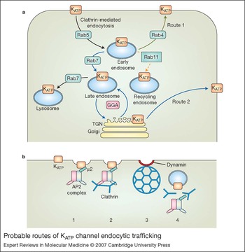
Following biosynthetic delivery, many membrane proteins remain at the cell surface for only a finite period before they are removed from the membrane by a process known as endocytosis. Endocytosed proteins can be either recycled back to the cell surface, or directed to intracellular lysosomes where they are degraded. Thus, while biosynthetic and endocytic recycling processes recruit proteins to the cell membrane, endocytosis removes them. Relative to the biosynthetic delivery process, however, very little is known about the endocytic trafficking of KATP channels. Yet, a better understanding of the endocytic mechanisms is important because, unlike the biosynthetic delivery mechanisms, endocytic sorting mechanisms have the ability to rapidly adjust the density of a given membrane protein at the cell surface. For this reason, many cell signalling events appear to deploy endocytic, rather than biosynthetic, mechanisms to adjust the cell-surface density of target membrane proteins. Examples include several G-protein-coupled receptors (GPCRs), ligand-gated ion channels and transporters, where endocytosis terminates the effect of extracellular signals (Refs Reference Marchese60, Reference Nong, Huang and Salter61). Although there are very little published data, endocytic sorting of KATP channels likely plays a similar signalling role, in the regulation of insulin secretion. Figure 4a provides an overview of the known and predicted endocytic trafficking pathways for KATP channels.
Probable routes of KATP channel endocytic trafficking. (a) ATP-sensitive potassium (KATP) channels are internalised from the cell surface by clathrin-mediated endocytosis and first enter the sorting early endosomes. From there, they could either recycle back to the plasma membrane directly (route 1) or via the late endosome and trans-Golgi network (TGN) route (route 2). The channels appear to avoid the conventional route via the endosomal recycling compartments (broken arrow). A proportion of the channels may enter the lysosomal compartment for subsequent degradation. The routes depicted are predicted based on the studies by Hu et al. (Ref. Reference Hu62) and Smith et al. (Ref. Reference Smith70). Rab proteins that mediate the individual traffic steps are indicated (Ref. Reference Zerial and McBride93). GGA (Golgi-localising γ-adaptin ear homology ARF-binding protein) proteins control traffic between the TGN and endosomes (Ref. Reference Bonifacino and Traub65). (b) Steps involved in clathrin-mediated endocytosis. Briefly, the µ2 subunit (turquoise) of the adaptor protein complex 2 (AP2) binds to a specific motif located in the cytoplasmic domains of Kir6.2 (step 1). This is followed by the recruitment of clathrin coat proteins and formation of clathrin-coated buds (step 2). The dynamin GTPase pinches the bud off the membrane, releasing the vesicle into the cytoplasm (step 3), where the coat and AP2 complexes dissociate (step 4) leaving the uncoated vesicle to fuse with the sorting early endosomes. From the early endosomes, the channel can be sorted to various destinations within the cell, as depicted in part (a).

Recent studies have demonstrated that endocytosis of KATP channels occurs by clathrin-mediated endocytosis (Refs Reference Mankouri47, Reference Hu62), a mechanism commonly used by most membrane proteins (Refs Reference Conner and Schmid63, Reference Kennedy and Ehlers64). During clathrin-mediated endocytosis, specific peptide motifs often containing tyrosine or leucine residues are recognised and bound by cytoplasmic multiprotein complexes, termed adaptor protein (AP2) complexes (Refs Reference Bonifacino and Traub65, Reference McMahon and Mills66). Two functional trafficking motifs have been reported in the C-terminus of Kir6.2: a dileucine (LL) motif (Ref. Reference Hu62) and a tyrosine-based motif (YXXΦ, where Y is a tyrosine, X is any amino acid, and Φ is a hydrophobic residue) (Ref. Reference Mankouri47). Although both signals might contribute to endocytosis of KATP channels, studies by Mankouri et al. (Ref. Reference Mankouri47) suggest that the tyrosine-based motif plays a dominant role. Despite its size and large C-terminal domains, SUR1 does not appear to contain endocytic signals (Ref. Reference Mankouri47). Biochemical and dominant-negative approaches demonstrate that KATP channels are recognised by the µ2 subunit of the AP2 complex (Ref. Reference Mankouri47). Channels bound by the AP2 complexes recruit clathrin to the membrane, which then begins to polymerise into a mesh, causing a portion of the membrane to invaginate. Once the region of membrane has almost formed a complete sphere, a large GTPase protein – dynamin – cleaves the newly formed vesicle from the membrane and releases it into the cytoplasm (Fig. 4b).
The process of endocytosis can be constitutive, as is the case with the low-density lipoprotein receptor and the transferrin receptor (Refs Reference Conner and Schmid63, Reference Kennedy and Ehlers64, Reference Bonifacino and Traub65), or could be dependent on a cellular signal, as with certain GPCRs (Ref. Reference Drake, Shenoy and Lefkowitz67). Two studies – one by Hu et al. (Ref. Reference Hu62) and the other by Mankouri et al. (Ref. Reference Mankouri47) – report that KATP channels can undergo endocytosis constitutively. However, according to Hu et al., activation of protein kinase C (PKC) is essential for robust endocytosis, whereas Mankouri et al. did not note significant differences in the extent of endocytosis with and without PKC activation. The reason for this discrepancy is not clear. However, an understanding of the role of PKC in endocytic sorting of KATP channels is important because PKC plays a crucial role in the regulation of insulin secretion (Ref. Reference Nesher68).
Following the release of clathrin-coated vesicles into the cytoplasm, the clathrin coat and adaptor proteins dissociate, allowing the vesicles to fuse with early endosomes (Refs Reference Conner and Schmid63, Reference Kennedy and Ehlers64, Reference Bonifacino and Traub65). Early endosomes are sorting stations where internalised proteins are sorted to specific destinations of the cell, including recycling back into the plasma membrane (Fig. 4; for a comprehensive discussion refer to Ref. Reference Pfeffer69). Few studies have addressed endocytic sorting of KATP channels. Internalised channels are able to recycle back to the plasma membrane (Ref. Reference Smith70), but this recycling does not appear to occur via the conventional recycling endosomal compartments because endocytosed channels showed limited colocalisation with co-internalised transferrin, a protein that is internalised by clathrin-mediated endocytosis and recycles to the plasma membrane largely via the recycling endosomal compartments (Ref. Reference Hu62). Internalised channels showed significant colocalisation with the cation-independent mannose-6-phopshate receptor, a late-endosomal marker (Ref. Reference Smith70). Furthermore, internalised channels showed significant overlapping with co-internalised wheat-germ agglutinin, which prominently labels the trans-Golgi network (Refs Reference Hu62, Reference Smith70). These data suggest that as the early endosomes mature, channels may enter the late endosomes and traffic to the trans-Golgi network, where they may join the biosynthetic secretory pathway and return to the plasma membrane. Such recycling routes are used by some proteins, such as furin (Refs Reference Rohn71, Reference Schapiro72). Consistent with this proposition is the fact that the C-terminus of Kir6.2 contains a signal known as the acidic cluster dileucine (DXXLL) motif, which has been shown to mediate vesicular traffic between the trans-Golgi network and the late endosomes (Refs Reference Bonifacino and Traub65, Reference Ghosh and Kornfeld73). Further studies are required to confirm this recycling route for KATP channels.
A small proportion of the internalised KATP channels also showed colocalisation with Rab7 and Lamp1 proteins (Refs Reference Hu62, Reference Smith70), which mark the late-endosomal and lysosomal compartments (Refs Reference Bonifacino and Traub65, Reference Zerial and McBride74); this suggests that endocytosed KATP channels are directed to elements of the lysosomal system for subsequent degradation. However, it is currently unclear to what extent endocytosed channels are targeted to lysosomes, to what extent they are recycled back to the cell surface, and if the sorting decisions are subjected to regulation by cellular signals. Activity-dependent and cell-signal-regulated trafficking has been reported for a number of other cell-surface channels (e.g. AMPA receptors) (Ref. Reference Kennedy and Ehlers64), transporters (e.g. GLUT4) (Ref. Reference Dugani and Klip75) and receptors (Refs Reference Drake, Shenoy and Lefkowitz76, Reference Gaborik and Hunyady77).
Another possible cellular destination for endocytosed channels is insulin-secretory vesicles, which have been shown to be associated with both Kir6.2 and SUR1 (Refs Reference Geng78, Reference Varadi79). It was suggested that these vesicles serve as sorting stations for delivery of channels to the cell surface (Ref. Reference Varadi79). It remains to be seen if channels associated with the secretory vesicles are recruited from the endocytosed pool of channels or have come from the biosynthetic pathway. Some reports place some form of KATP channel in the mitochondria, which have been shown to possess an ATP-dependent K+ conductance (Ref. Reference Jiang80). However, the molecular identity of these so-called mitoKATP channels remains controversial (Refs Reference Seino and Miki48, Reference Minners, McLeod and Sack81). Moreover, recent immunocytochemical and cell fractionation studies failed to confirm the presence of Kir6.2 and SUR1 in mitochondria (Ref. Reference Varadi79).
KATP channels in neonatal diabetes mellitus
Neonatal diabetes mellitus is a rare condition affecting ∼1 in 400 000 neonates and is defined as insulin-requiring hyperglycaemia within the first 3 months of life (Ref. Reference Shield82). In approximately half of all cases the condition is transient (TNDM), becoming resolved within the first 3 months, whereas in the remaining patients diabetes is permanent (PNDM). Both PNDM and TNDM are associated with mutations of KCNJ11 and ABCC8. To date, ∼40 mutations of either Kir6.2 or SUR1 have been identified in sufferers of either PNDM or TNDM (Table 1). The majority of these mutations affect functional properties of the channels.
Functional consequences of mutations of KCNJ11 (Kir6.2) and ABCC8 (SUR1) associated with permanent and transient neonatal diabetes mellitus

aAlso show increased activation by Mg2+–ATP.
bPatients show signs of developmental delay (DEND syndrome).
Abbreviations: AP, adaptor protein; COP, coat protein complex; ER, endoplasmic reticulum; GPCR, G-protein-coupled receptor; KATP channel, ATP-sensitive potassium channel; Kir, inwardly rectifying potassium channel; LC-CoA, long-chain fatty acyl CoA; MODY, maturity-onset diabetes of the young; NBF, nucleotide-binding fold; PKC, protein kinase C; PNDM, permanent neonatal diabetes mellitus; PtdIns(4,5)P 2, phosphatidylinositol (4,5)-bisphosphate; SUR1, sulphonylurea receptor; TGN, trans-Golgi network; TNDM, transient neonatal diabetes mellitus.
Neonatal diabetes mellitus and functional defects
The first proven link between PNDM and KATP channel dysfunction was reported in 2004 when Gloyn et al. (Ref. Reference Gloyn15) identified six novel heterozygous missense mutations in the KCNJ11 gene isolated from ten patients. Four of these patients had the same mutation, a substitution of the arginine at position 201 for a histidine (R201H), which was shown to reduce the ability of ATP to inhibit channel activity. Several other PNDM- and TNDM-causing mutations have since been discovered in KCNJ11 (see Table 1 for further details). A significant proportion of these mutations reduce the ATP sensitivity of the channel by disrupting the ATP-binding site in Kir6.2 (Ref. Reference Ashcroft83). Other PNDM-causing mutations, however, do not affect ATP binding directly; instead, they increase the intrinsic P o (the fraction of time the channel remains open in the absence of ATP) of the channel (Refs Reference Proks84, Reference Proks85). Since the affinity of the open state for ATP is less than that of the closed state, mutant channels are less well inhibited by ATP. Many of the mutations affecting the intrinsic P o appear to reside close to the cytoplasm–membrane interface in the slide helix (e.g. Q52R, V59G) (Ref. Reference Proks84) or close to the bundle crossing (I296L) (Ref. Reference Proks85) – both regions thought to be intimately involved in channel gating (see above). The third most common mechanism leading to KATP channel dysfunction in PNDM results from an increase in the ability of Mg2+–ADP to activate channels. These mutations are primarily found to affect SUR1 and are commonly located in or near the NBFs of the channel (Ref. Reference Babenko86). An important outcome of these studies, from a therapeutic point of view, is the realisation that sulphonylurea therapy can be used in place of insulin therapy to treat patients with PNDM (Refs Reference Stanik87, Reference Zung88).
Neonatal diabetes mellitus and trafficking defects
Functional consequences of two PNDM mutations – Y330C and F333I – located in the predicted ATP-binding pocket of Kir6.2 have recently been reported (Ref. Reference Tammaro89). F333I caused impaired ATP binding/transduction, whereas Y330C impaired ATP binding/transduction as well as causing an increase in the intrinsic P o of the channel (Ref. Reference Tammaro89). In addition to these effects, both Y330C and F333I disrupt a trafficking motif responsible for clathrin-mediated endocytosis of the channel (Ref. Reference Mankouri47) (see above). Disruption of the 330YSKF333 motif in Kir6.2 led to a twofold increase in the number of channels at the cell surface (Ref. Reference Mankouri47). Such an increase in channel density in β-cells would be expected to increase the overall KATP current and thereby suppress insulin secretion, because the overall KATP current is determined not only by the P o of the channel but also by the channel density at the cell surface. These data point towards an important role for regulated trafficking of KATP channels in determining insulin secretion and association of trafficking defects with neonatal diabetes.
Congenital hyperinsulinism and trafficking disorders
Congenital hyperinsulinism is a rare genetic disorder affecting 1 in 50 000 in general populations, but its incidence is high (1 in 2500) in certain Finnish and Saudi Arabian populations (Refs Reference Ashcroft3, Reference Dunne7, Reference Glaser90). More than 100 mutations associated with this disease have been reported in KCNJ11 and ABCC8. Children affected by this disease secrete insulin continuously despite severe hypoglycaemia. Not surprisingly, mutations associated with congenital hyperinsulinism have an effect opposite to those observed with mutations causing neonatal diabetes mellitus: some mutations increase the sensitivity of the channel to the [ATP]/[ADP] ratio (less sensitive to activation by Mg2+–ADP) (Refs Reference Shyng9, Reference Nichols8); others impair the biosynthetic delivery of these channels to the plasma membrane (Refs Reference Partridge, Beech and Sivaprasadarao10, Reference Cartier11, Reference Taschenberger12). However, so far there are no reports of mutations affecting the endocytic pathway contributing to decreased cell-surface density. The impairment of biosynthetic delivery of mutant KATP channels occurs as a result of retention in the ER (Refs Reference Cartier11, Reference Yan, Casey and Shyng91) or the trans-Golgi network (Ref. Reference Partridge, Beech and Sivaprasadarao10). ER retention is likely to result from misfolding of the channel subunits. Folding defects caused by some mutations could be rescued by treating the cells expressing the mutant channels with sulphonylurea drugs, but not by diazoxide (Ref. Reference Yan, Casey and Shyng91). Sulphonylurea drugs, however, are unsuitable for the treatment of congenital hyperinsulinism, because they also block KATP channels. Compounds that lack the inhibitory effect and, preferably, have an ability to activate the channel would be useful. Pharmacological chaperones (ligands with pharmacological selectivity) offer promise as a new class of therapeutic agents and have been reported for several medically important proteins including the cystic fibrosis transmembrane regulator (for a review see Ref. Reference Loo and Clarke92).
Concluding remarks
Recent advances in our understanding of the molecular basis of neonatal diabetes mellitus have revealed multiple mechanisms by which mutations in the KATP channel genes lead to the disease state. Regardless of the mechanism, as one would expect, all mutations cause an increase in channel activity. Some mutations affect the functional properties of the channel, whereas others affect trafficking. Functional effects thus far reported include an increase in the intrinsic P o of the channel, or a decrease in the sensitivity of the channel to inhibition by ATP, or an increase in the ability of channel to be activated by Mg2+–ADP. Trafficking mutations could increase the channel density by a variety of mechanisms. They could increase the biosynthetic delivery of channels to the cell surface, reduce their removal from the cell membrane by endocytosis, enhance recycling of endocytosed channels back to the cell surface, or reduce channel degradation. These possibilities have not been fully investigated, except in one study where mutations impaired the ability of the channel to undergo endocytosis, thereby increasing the channel density at the cell surface (Ref. Reference Mankouri47). Future studies may reveal further mutations affecting trafficking of the channel.
Unlike neonatal diabetes mellitus, type 2 diabetes is a widespread disease affecting more than 150 million people worldwide; its incidence is predicted to increase to 300 million by 2025. Although mutations (polymorphisms) in both KCNJ11 (e.g. E23K) and ABCC8 have been shown to be associated with enhanced susceptibility to type 2 diabetes, the underlying mechanisms are far from clear. Functional studies of several mutations have thus far failed to provide unambiguous results. This raises the possibility that some such mutations might affect trafficking of the channels. In order for this possibility to be properly addressed, however, a comprehensive understanding of the routes via which KATP channels traffic, the signals that control the traffic and the associated mechanisms is required. Some progress has been made, as outlined in this review, but given the complexity of trafficking of membrane proteins, significant progress has yet to be achieved with KATP channels. Once we have an improved knowledge of the channel biosynthesis, sorting and trafficking, investigations into the mechanisms by which genetic mutations might affect trafficking could commence. Such studies have the potential to reveal opportunities for new therapeutic strategies to treat a range of insulin-secretory disorders, enabled by an ability to control the number of channels at the cell surface.
Acknowledgements and funding
We acknowledge the Medical Research Council, UK, for financial support. A.J.S. was supported by the Emma and Leslie studentship. We thank Prof. M.S.P. Sansom, Oxford University, UK, for providing the coordinates of the Kir6.2 model.