Case report
Our patient is a 4-year-old male with truncus arteriosus type I who underwent repair with a ventricular septal defect patch closure and placement of a right ventricle to pulmonary artery conduit shortly after birth.
Postoperative course was complicated by extracorporeal mechanical oxygenation with subsequent need for trans-catheter creation of atrial level communication to facilitate with recovery. A cardiac catheterisation also showed right pulmonary stenosis requiring stent placement. Further complications included cannulation of the right internal jugular vein for extra corporeal mechanical oxygenation in the setting of subsequent biventricular failure, development of occlusive thrombi in the right internal jugular vein, right femoral and iliac veins, and left femoral vein and inferior caval vein. He ultimately recovered and was discharged home.
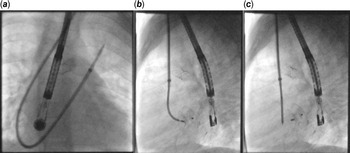
By 4 years of age, he had recurrence of right pulmonary artery stenosis and right heart dilation from the atrial septal defect requiring repeat catheterisation. The extensive history of venous occlusion necessitated the use of the left internal jugular vein for access to perform the procedure. A 6 Fr sheath was placed in the left internal jugular vein and a 0.035’’ J wire was advanced through the left innominate vein and the superior caval vein into the right atrium. The 6 Fr sheath was exchanged for a 7 Fr long sheath for stent placement in the right pulmonary artery. Attention was then turned to the atrial septal defect, which measured 9 × 10 mm with adequate atrial rims and a total septal length measuring 33 mm by trans-esophageal echocardiography. A 13 mm AmpatzerTM Septal Occluder device (Boston Scientific, Marlborough, MA) was chosen for closure. Due to the need to deliver the device from the left internal jugular vein, we planned to cannulate a pulmonary vein to have a secure wire position before advancing the delivery system. A combination of a 5-Fr Cobra glide catheter (Terumo Medical, Somerset, NJ) and a 0.035’ Angle glide wire was utilised to cross the atrial septum and cannulate the left-upper pulmonary vein. Then, a 0.035" AmplatzerTM Super Stiff Guidewire (Boston Scientific, Marlborough, MA) was placed in the left-upper pulmonary vein. The AmplatzerTM TrevisioTM 45-degree delivery system (Boston Scientific, Marlborough, MA) was advanced across the atrial septum into the left-upper pulmonary vein. The device was then screwed to the AmplatzerTM TrevisioTM delivery cable (Boston Scientific, Marlborough, MA) and then loaded into the delivery system and advanced to the tip. Under fluoroscopic and echocardiographic guidance, the entire system was pulled back into the left atrium. The left disc was deployed, and the entire system was pulled back until the disc caught on the septum. At that point, the right disc was deployed (Fig 1). Once the device was adequately placed but prior to its release, transoesophageal echocardiography showed that the device was on either side of the septum with no residual shunting. The patient tolerated the procedure well with no complications. Of note, releasing the device was easy, and no significant arrhythmias or haemodynamic instabilities were encountered during the process.
( a ) Antero-posterior image with the delivery sheath crossing the atrial septal defect and positioned in the left-upper pulmonary vein. ( b ) Lateral image showing the deployed device still connected to the TrevisioTM cable. Note the degree of flexibility of the distal most part of the cable. (c) Lateral image showing the device position immediately after release, note the minimal change in device position before and after release.

Transthoracic echocardiography the next day confirmed good positioning and no residual shunt. His last follow-up 6 months post defect closure continued to show a stable clinical status with no residual shunt and appropriate device position.
Discussion
Traditionally, atrial septal defect closure is performed through femoral venous access. Multiple reports have detailed alternative access sites for such procedures including transhepaticReference Sadiq1 and right internal jugular venous access.Reference Oliveira, Moura, Almeida, Ribeiro and Nascimento2,Reference Butera, Lovin and Basile3 Here, we report a case of atrial septal defect closure using the left internal jugular vein in a patient with situs solitus. To our knowledge, this is the first report that describes such an approach in situs solitus anatomy.
Femoral venous access was not possible in our case due to occlusion of the inferior caval vein. Similarly, the right internal jugular vein, which would have provided an easier access, was occluded. Therefore, we elected to use the left internal jugular vein with hepatic access being reserved as a backup. While this proved to be a successful access point, it is worth noting that left internal jugular access does create some difficulty with obtaining adequate transoesophageal echocardiographic images as they are typically both done from the same side. The combination of Cobra glide catheter and glide wire enabled us to cross the atrial septal defect and access the left-upper pulmonary vein. Having a stiff wire deep inside the vein enabled us to advance the delivery system. Next, the TrevisioTM delivery cable played a major role in maintaining the device position and not pulling through the atrial septal defect. The distal most part of the cable, which is made from a Nitinol core surrounded by a loosely wound stainless-steel coil, allows for increased flexibility. This provided the ease of device positioning and allowed for echocardiographic evaluation while still connected. Indeed, the TrevisioTM delivery system was recently developed to decrease the tension placed on the device and the intracardiac structures during device release.Reference Randall, Morgan and Zablah4 This characteristic was leveraged in our case to maintain appropriate device position during echocardiography and device release, despite the tortuous path from access site to atrial septum. We believe this characteristic can also lend itself for use in atrial septal defect closure in smaller size patients and ones with a deficient retro-aortic rim, where the stiffness of the standard cable makes device closure difficult. Potential uses can even be extended to ventricular septal defect closure, where the flexible tip can decrease strain on surrounding cardiac structures during device positioning and release. The decreased strain can also provide a more faithful prediction of device position and interaction with surrounding structures after release.
Conclusion
Although the left internal jugular vein is not a convenient access site for atrial septal defect closure due to the tortuous path that the catheter and delivery system need to take, it is feasible and safe. We report a case of atrial septal defect closure in a patient with truncus arteriosus and situs solitus using the left internal jugular vein. The combination of a secure wire position and the use of TrevisioTM cable were key in successful closure despite the difficult course. The usefulness of TrevisioTM cable in difficult anatomy and small patients’ needs to be confirmed by further studies.
Acknowledgements
None.
Financial support
This research received no specific grant from any funding agency, commercial, or not-for-profit sectors.
Conflict of interest
None.
Ethical standards
No specific ethical approval from Institutional Reviews Boards are necessary for this type of publication. The authors assure that all patient data provided in this case report are anonymised.