Introduction
The success of in vitro embryo production depends mainly on the selection of good quality oocytes (Ashry & Smith, Reference Ashry and Smith2015). Morphological evaluation of the cumulus–oocyte complex is a widely used method for selection of oocytes for in vitro embryo production, not only in the dromedary camel but in almost all other species. Brilliant cresyl blue (BCB) staining is a non-invasive method for screening for oocyte quality that has been used in different animal species (Roca et al., Reference Roca, Martinez, Vazquez and Lucas1998; Rodriguez-Gonzalez et al., Reference Rodriguez-Gonzalez, Lopez-Bejar, Velilla and Paramio2002; Bhojwani et al., Reference Bhojwani, Alm, Torner, Kanitz and Poehland2007; Manjunatha et al., Reference Manjunatha, Gupta, Devaraj, Ravindra and Nandi2007; Wu et al., Reference Wu, Liu, Zhou, Lan, Han, Miao and Tan2007a; Ishizaki et al., Reference Ishizaki, Watanabe, Bhuiyan and Fukui2009; Rodrigues et al., Reference Rodrigues, Rodriguez, Silva, Cavalcante, Feltrin and Rodrigues2009; Catalá et al., Reference Catalá, Izquierdo, Uzbekova, Morato, Roura, Romaguera, Papillier and Paramio2011; Wang et al., Reference Wang, Lin, Huang, Wang, Zhao and Chen2012; Silva et al., Reference Silva, Rodriguez, Galuppo, Arruda and Rodrigues2013; Pereira et al., Reference Pereira, Lorenzo, Carneiro, Bilodeau-Goeseels, Kastelic, Esteller-Vico, Lopez-Bejar and Liu2014). Recent studies on human oocytes have also been reported (Alcoba et al., Reference Alcoba, Conzatti, Ferreira, Pimentel, Kussler, Capp, von Eye Corleta and Brum2016, Reference Alcoba, Schneider, Arruda, Martiny, Capp, von Eye Corleta and Brum2017). BCB staining depends on measuring glucose-6-phosphate dehydrogenase (G6PDH) activity. G6PDH is an enzyme synthesized by the growing oocytes (Mangia & Epstein, Reference Mangia and Epstein1975; Pujol et al., Reference Pujol, Lopez-Bejar and Paramio2004). G6PDH activity is reduced once the oocytes have finished the growth phase and possibly have acquired developmental competence (Wassarman, Reference Wassarman and Neill1988; Tian et al., Reference Tian, Braunstein, Pang, Stuhlmeier, Xi, Tian and Stanton1998). Therefore, oocytes that have finished their growth phase have low levels of G6PDH and exhibit a cytoplasm with a blue color (BCB+) while growing or less competent oocytes have a high levels of G6PDH, and are able to reduce the blue color of the dye, and display colorless cytoplasm (BCB−) (Ericsson et al., Reference Ericsson, Boice, Funahashi and Day1993; Tian et al., Reference Tian, Braunstein, Pang, Stuhlmeier, Xi, Tian and Stanton1998). Previous studies in different animal species have demonstrated that screening of oocytes using BCB staining improved the development potential of the embryos derived from (BCB+) oocytes. Indices of early embryonic development, including cleavage, and development to morula and blastocyst, were significantly higher in BCB+ derived embryos (Alm et al., Reference Alm, Torner, Lohrke, Viergutz, Ghoneim and Kanitz2005; Bhojwani et al., Reference Bhojwani, Alm, Torner, Kanitz and Poehland2007; Manjunatha et al., Reference Manjunatha, Gupta, Devaraj, Ravindra and Nandi2007; Wang et al., Reference Wang, Lin, Huang, Wang, Zhao and Chen2012). The molecular identity of oocytes screened with BCB staining is completely different in many aspects related to oocyte quality including gene expression (El Shourbagy et al., Reference El Shourbagy, Spikings, Freitas and St John2006).
Using BCB staining for screening camel oocytes has not been reported previously to the best of our knowledge. Therefore, the objectives of the current studies were to investigate the developmental competence of dromedary camel oocytes selected by BCB staining. Effects of BCB staining on oocyte maturation, fertilization and early embryonic development were elucidated. The differential expression of select candidate genes that are important for folliculogenesis and oocyte development [growth differentiation factor 9 (GDF9) and bone morphogenetic protein 15 (BMP15)], plus cell cycle regulators [Cyclin B1 and cyclin-dependent kinase1 (CDK1)], maternal effect genes required for embryo development [zygote arrest factor 1 (Zar1) and Maternal Antigen that embryos require (Mater)] or genes that have transcription factor activity [OCT4 and Signal transducer and activator of transcription 3 (STAT3)], was also investigated.
Materials and Methods
Materials
Unless stated otherwise, chemicals and reagents were purchased from Sigma–Aldrich (S.A., Egypt).
Ovary collection and oocyte retrieval
Dromedary camel oocytes were retrieved from ovaries collected from local slaughterhouse (Cairo, Egypt). Oocytes retrieval and selection were performed as described elsewhere (Fathi et al., Reference Fathi, Seida, Sobhy, Darwish, Badr and Moawad2014). Briefly, the ovaries were transported to the laboratory within 2–3 h after slaughter in a thermos flask filled with prewarmed (30°C) sterile normal saline solution (NSS, 0.9% NaCl). Cumulus–oocyte complexes (COCs) were aspirated from 2 to 8 mm follicles using a 20-gauge needle attached to a 20-ml syringe (Moawad et al., Reference Moawad, Darwish, Badr and El-Wishy2011). Morphologically good quality oocytes were selected under a stereomicroscope.
Brilliant cresyl blue staining
BCB staining was carried out as previously described (Ashry et al., Reference Ashry, Lee, Mondal, Datta, Folger, Rajput, Zhang, Hemeida and Smith2015). Briefly, BCB stain was diluted in Dulbecco's phosphate buffered saline containing 0.4% BSA (mDPBS). Morphologically good quality oocytes were stained with BCB at 38.5°C in a 5% CO2 humidified air atmosphere. The control group was kept in mDPBS under the same conditions. After staining, COCs were washed twice with mDPBS and classified into BCB+, with a blue cytoplasm or BCB−, with a colorless cytoplasm. Firstly, we tested the effect of three different BCB concentrations (13, 26 and 39 µM) on oocyte maturation to select the appropriate staining protocol for camel oocytes. Based on the results of first experiment, 26 µM of BCB was used in the subsequent experiments.
In vitro embryo production
Dromedary camel oocyte recovery, in vitro maturation (IVM), in vitro fertilization IVF (IVF) and embryo culture were performed as described previously (Moawad et al., Reference Moawad, Darwish, Badr and El-Wishy2011; Fathi et al., Reference Fathi, Seida, Sobhy, Darwish, Badr and Moawad2014). Briefly, after BCB staining, selected COCs were matured in TCM-199 with Earle's salts (supplemented with 10 µg/ml FSH, 10% FCS, 50 µg/ml sodium pyruvate, 2.6 mg/ml sodium bi-carbonate, and 50 µg/ml gentamycin). COCs were cultured in 100-µl drops of maturation medium (10–15 oocytes/drop) covered with mineral oil at 39°C in 5% CO2 in humidified air for 30 h. For assessment of nuclear maturation, cumulus cells were stripped off, then denuded oocytes were transferred to 1% sodium citrate solution for 3 min. Oocytes were fixed in ethanol:acetic acid (3:1) for 24 h, then stained with 1% orcein and examined under a phase contrast microscope in which MII stage oocytes were considered mature. In vitro fertilization was carried out using dromedary camel epidydimal spermatozoa. Epididymal spermatozoa were collected by flushing the body and cauda epididymis and separated by swim up technique in the presence of 5 mM caffeine as a capacitating agent (Fathi et al., Reference Fathi, Seida, Sobhy, Darwish, Badr and Moawad2014). Matured COCs were co-incubated with spermatozoa (2 × 10⁶ motile spermatozoa/ml) in 50-µl drops of fertilization medium (10 oocytes/group) at 39°C in 5% CO2 humidified air for 18–20 h. After fertilization, cumulus cells were stripped by repeated pipetting. Fertilization was then determined by fixation and staining of presumptive zygotes using aceto-orcein staining as detailed above. Normal fertilization was evidenced by the presence of spermatozoon, sperm head, or male and/or female pronuclei in the ooplasm. The presence of more than one spermatozoon or two pronuclei in the ooplasm was considered to demonstrate polyspermic fertilization.
Eighteen hours post insemination, presumptive zygotes were washed and cultured in modified KSOMaa (Khatir et al., Reference Khatir, Anouassi and Tibary2007), 5 zygotes/50 µl drop under mineral oil at 39°C in 5% CO2 humidified air atmosphere until day 7 (day 0 = day of insemination). The number of cleaved embryos (2–8 cells) was determined at 48 hpi. The development to morula and blastocyst stages was evaluated at day 4 and day 7 post insemination respectively.
RNA isolation and real-time PCR
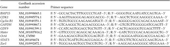
After exposure to BCB staining, cumulus cells were stripped off by repeated pipetting and GV stage oocytes from BCB+, BCB− and control groups (n = 20 oocytes/group, n = 4 replicates) were snap frozen for further use in RNA isolation. RNA extraction, cDNA synthesis and real-time PCR analysis of gene expression were carried out as described before (Bettegowda et al., Reference Bettegowda, Patel, Lee, Park, Salem, Yao, Ireland and Smith2008; Ashry et al., Reference Ashry, Lee, Mondal, Datta, Folger, Rajput, Zhang, Hemeida and Smith2015). Briefly, an RNeasy® Micro Kit (Qiagen, Germantown, MD, USA) was used for total RNA extraction and cDNA synthesis was done using an iScript cDNA synthesis kit (BioRad, Hercules, CA, USA) according to the manufacturer's protocols. Quantification of relative transcript abundance of each gene was done by the 2−ΔΔCt method as described elsewhere (Livak & Schmittgen, Reference Livak and Schmittgen2001). Transcript abundance of the studied genes was normalized relative to the abundance of GAPDH as an endogenous control and expressed as fold change relative to the control group. PCR primers were designed using the NCBI Primer designing tool (https://www.ncbi.nlm.nih.gov/tools/primer-blast/). Primers sequences and GenBank accession numbers are presented in Table 1.
Primers used for RT-PCR analysis

F: forward primer, R: reverse primer.
Statistical analysis
Differences in gene expression were analyzed by one-way analysis of variance (ANOVA) using SAS (SAS Institute Inc., NC, USA). For other experiments, percentage data were arcsine transformed before analysis with ANOVA. Differences amongst treatment means were detected using Fisher's protected least significant difference test (LSD).
Results
Effect of BCB staining on oocyte selection efficiency and in vitro maturation
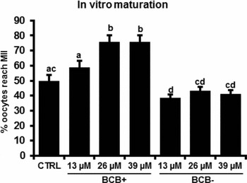
To determine the appropriate BCB concentration for staining of camel oocytes we tested three different BCB concentrations (13, 26 and 39 µM). As depicted in Fig. 1, staining of dromedary camel oocytes with 26 or 39 µM of BCB for 90 min resulted in a significant increase in the maturation rate of BCB+ oocytes compared with controls and the 13 µM BCB concentration. No significant difference in the maturation rate was observed between 26 and 39 µM concentrations. Based on the results of this experiment the 26 µM concentration was used for subsequent experiments.
Effects of different BCB concentrations on camel oocyte maturation. Cumulus–oocyte complexes (COCs) from control, BCB+ and BCB− groups (n = 20–30 oocytes/group; n = 4 replicates) were subjected to in vitro maturation for 30 h, then nuclear maturation status was determined by phase contrast microscopy. Data are shown as mean ± standard error of the mean (SEM). a,b,c,dValues with different superscripts across treatments indicate significant differences (P < 0.05).

Effects of BCB on in vitro maturation and fertilization of dromedary camel oocytes
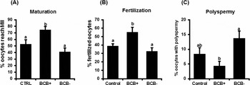
In terms of in vitro maturation and fertilization, screening of camel oocytes with BCB staining before maturation resulted in significant increase in the maturation and fertilization rates and reduced the incidence of polyspermy in the BCB+ group. BCB+ oocytes showed a significantly higher maturation rate compared with BCB− and the control oocytes, with maturation rates of 74.48, 41, and 52.88% for BCB+, BCB− and control respectively (Fig 2 A). In vitro fertilization rate was also increased in the BCB+ group with a low incidence of polyspermy (Fig. 2 B, C). the fertilization rate was 55.33, 32.5 and 38.86%, while the polyspermy was 4.29, 8.4 and 13.75% for BCB+, BCB− and control groups respectively. Suggesting that screening of camel oocytes with BCB staining prior to in vitro maturation would select the more competent oocytes that have a higher potential to resume meiosis and able to be normally fertilized.
Effects of BCB staining on camel oocytes maturation and in vitro fertilization. After BCB staining, BCB+, BCB− and Control oocytes (n = 4 replicates, 20–25 oocytes/group) were subjected to in vitro maturation, and in vitro fertilization. The ratio of oocytes reaching MII at 30 h (A), normally fertilized oocytes (B) and polyspermic fertilization (C) were recorded. Data are shown as mean ± standard error of the mean (SEM). a,bValues with different superscripts across treatments indicate significant differences (P < 0.05).

Effects of BCB staining on the development of early dromedary camel embryos in vitro
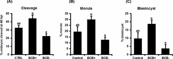
To further investigate the influence of BCB staining on camel oocyte developmental potential, in vitro produced presumptive zygotes of BCB+, BCB− and control groups were cultured in vitro up to day 7. Indices of early embryonic development; cleavage, development to morula and blastocyst stages were significantly improved in BCB+ groups compared with the BCB− and control groups. Cleavage rate at 48 hpi was 44.29, 22.5 and 32.84% for BCB+, BCB− and control groups respectively (Fig. 3 A). the percentage of embryos that reached morula stage at day 5 was 29.8, 12.5 and 19.4% for BCB+, BCB− and control groups respectively (Fig. 3 B), while blastocyst development rate at day 7 was 18.7, 3.75 and 9.77 for BCB+, BCB− and control groups respectively. Collectively, results suggested that screening of camel oocytes with BCB staining would select more competent oocytes for in vitro embryo production.
Effects of BCB staining on camel early embryonic development in vitro. Presumptive zygotes of BCB+, BCB− and control groups (n = 4 replicates; 20–25 zygotes) were cultured in KSOM medium until day 7. Indices of early embryonic development were recorded: (A) cleavage at 48 hpi; (B) day 5 morula; and (C) day 7 blastocyst. Data are shown as mean ± standard error of the mean (SEM). a,bValues with different superscripts across treatments indicate significant differences (P < 0.05).

Expression of select candidate genes in dromedary camel oocytes screened with BCB staining
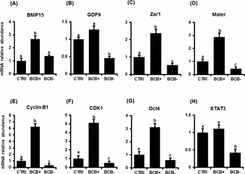
To investigate the effect of BCB staining on the molecular level, we analyzed the transcripts abundance of select transcripts in GV stage camel oocytes screened with BCB staining. Transcripts were selected based on the biological function in the oocytes and/or early embryos. Expression of BMP15 was >2-fold higher (P < 0.05) in BCB+ relative to BCB− and control groups, whereas no significant difference was observed between BCB− and control groups (Fig. 4 A). Although, mRNA abundance for GDF9 was higher in the BCB+ group compared with BCB− oocytes, mRNA abundance did not differ between BCB+ and control groups (Fig. 4 A, B). Transcript abundance of Zar1 and Mater was significantly higher (P < 0.05) in the BCB+ group versus BCB− and control groups. Both transcripts were significantly lower in the BCB− group relative to controls (Fig. 4 C, D). Expression of cyclin B1 was >6-fold and 3-fold higher (P < 0.05) in the BCB+ group compared with the BCB− and control groups respectively (Fig. 4 E). CDK1 was upregulated in the BCB+ group with >5-fold and 2-fold increase compared with the BCB− and control groups respectively (Fig. 4 F). Oct4 was overexpressed in the BCB+ and control groups relative to the BCB− group, expression of Oct4 was also higher in the BCB+ group compared with controls (Fig. 4 G). No significant difference was observed for STAT3 expression between the BCB+ and control groups, whereas both groups showed higher expression relative to the BCB− group (Fig. 4 H). Collectively, results indicated that the BCB+ dromedary camel oocytes were enriched with several transcripts that regulate crucial functions during oocyte and follicular development.
Expression of some candidate genes in camel oocytes screened with BCB staining. After BCB staining, GV stages oocytes (n = 4 replicates; 20 oocytes/group) were stripped off cumulus cells and used for RT-PCR analysis for (A) BMP15, (B) GDF9, (C) Zar1, (D) Mater, (E) Cyclin B1, (F) CDK1, (G) Oct4 and (H) STAT3. Data were normalized relative to the expression of GAPDH as endogenous control and expressed as fold change relative to control. Data are shown as mean ± standard error of the mean (SEM). a,b,cValues with different superscripts across treatments indicate significant differences (P < 0.05)

Discussion
Although BCB staining has been widely used for screening of oocytes in several animal species, its use in camels has not been reported previously. The results of the current studies revealed that the BCB test would serve as a reliable method for selecting more competent camel oocytes for in vitro embryo production. BCB+ oocytes showed higher maturation, fertilization and embryonic development rates compared with BCB− and control oocytes selected solely based on morphological criteria. The transcript level of several developmentally important transcripts was higher in BCB+ GV stages oocytes.
Previous studies have demonstrated that BCB+ oocytes are more competent and yield more blastocysts compared with BCB− oocytes. It has been shown that the BCB test allowed the selection of larger oocytes with higher in vitro maturation and fertilization rates and an increased percentage of normally fertilized oocytes (Wang et al., Reference Wang, Lin, Huang, Wang, Zhao and Chen2012). The potential of IVF embryos derived from BCB+ oocytes to develop to morula and blastocyst stages was significantly higher compared with oocytes selected solely by morphological criteria (Rodriguez-Gonzalez et al., Reference Rodriguez-Gonzalez, Lopez-Bejar, Velilla and Paramio2002; Alm et al., Reference Alm, Torner, Lohrke, Viergutz, Ghoneim and Kanitz2005; Manjunatha et al., Reference Manjunatha, Gupta, Devaraj, Ravindra and Nandi2007; Silva et al., Reference Silva, Rodriguez, Galuppo, Arruda and Rodrigues2013). In the present study, BCB+ dromedary camel oocytes showed higher maturation, fertilization rates compared with BCB− and control counterparts. Indices of early embryonic development: cleavage, development to morula and blastocyst stages, were also higher in the BCB+ group. Previous studies have demonstrated that BCB selects larger oocytes with more mitochondria and maturation-promoting factor (MPF) activity (Catalá et al., Reference Catalá, Izquierdo, Uzbekova, Morato, Roura, Romaguera, Papillier and Paramio2011). Conversely, less competent (BCB–) oocytes undergo fertilization failure or developmental arrest due to delayed mitochondrial DNA (mtDNA) replication that resulted from delayed onset of expression of their nuclear-encoded replication factors. The oocyte challenges to rescue this failure during the late stages of maturation. Subsequently, oocyte competence in terms of mtDNA replication and composition is not fully synchronized and oocytes fail to behave normally (May-Panloup et al., Reference May-Panloup, Vignon, Chretien, Heyman, Tamassia, Malthiery and Reynier2005; Spikings et al., Reference Spikings, Alderson and St John2007; Opiela et al., Reference Opiela, Lipinski, Slomski and Katska-Ksiazkiewicz2010; Catalá et al., Reference Catalá, Roura, Izquierdo, Hammammi, Uzbekova and Paramio2013).
Growth differentiation factor 9 (GDF9) and bone morphogenetic protein 15 (BMB15) are known to play an important role during folliculogenesis and oocyte development (Chang et al., Reference Chang, Brown and Matzuk2002; McNatty et al., Reference McNatty, Juengel, Reader, Lun, Myllymaa, Lawrence, Western, Meerasahib, Mottershead, Groome, Ritvos and Laitinen2005). Increased expression of GDF9 and BMP15 is positively correlated to oocyte and embryo developmental competence (Wu et al., Reference Wu, Tang, Cai, Lu, Xu, Zhu, Luo and Huang2007b; Li et al., Reference Li, Li, Ou, Zhang, Ren, Wei, Zhang and Yang2014; Ashry et al., Reference Ashry, Lee, Mondal, Datta, Folger, Rajput, Zhang, Hemeida and Smith2015).
Results of present studies have demonstrated the higher expression of GDF9 and BMP15 in good quality oocytes selected by BCB screening (BCB+) compared with control oocytes selected exclusively based on morphological criteria and with less competent oocytes (BCB−). ZAR1 and Mater are maternal effect transcripts that accumulate during oogenesis (Uzbekova et al., Reference Uzbekova, Roy-Sabau, Dalbies-Tran, Perreau, Papillier, Mompart, Thelie, Pennetier, Cognie, Cadoret, Royere, Monget and Mermillod2006). Previous studies have reported the higher expression of both transcripts in BCB+ ovine oocytes (Wang et al., Reference Wang, Lin, Huang, Wang, Zhao and Chen2012).
Cyclin B1 and CDK1 are implicated in the regulation of the cell cycle and resumption of meiosis in oocytes. (Castedo et al., Reference Castedo, Perfettini, Roumier and Kroemer2002). Oct4 is a transcription factor known to regulate cell fate decision in early embryos (Wu & Scholer, Reference Wu and Scholer2014). STAT3 is also essential for early embryonic development (Teng et al., Reference Teng, Diao, Ma, Xu and Yang2004). Previous studies (El-Sayed, Reference El-Sayed2013; El-Sayed & Ghanem, Reference El-Sayed and Ghanem2015) have demonstrated the higher expression of cyclin B1 and Oct4 in morphologically good quality dromedary camel oocytes.
Results of the present study demonstrated higher expression of cyclin B1, CDK1, Oct4 and STAT3 in BCB+ oocytes. Enrichment of BCB+ oocytes with the aforementioned transcripts that support several functions during oocyte maturation and/or early embryonic development explains, to some extent, the high developmental competence of BCB+ oocytes.
In conclusion, the present study provided novel information about the use of BCB staining for selection of good quality oocytes to improve the in vitro embryo production in the dromedary camel. Good quality camel oocytes were enriched with several transcripts that play a crucial role in folliculogenesis and oocyte development (GDF9 and BMP5), regulate the cell cycle (Cyclin B1, STAT3), are maternal effect genes required for embryo development (ZAR1 and Mater) or have transcription factor activity (OCT4).