Introduction
Rhinosporidiosis is a common disease in tropical countries and usually involves the nose, nasopharynx and ocular conjunctiva.Reference Thomas, Gopinath and Betts1 Involvement of the trachea and bronchial tree is very rare, and diagnosis by bronchoscopic biopsy is dangerous due to the risk of bleeding.Reference Rekha, Thomas, Pappachan, Venugopal, Jayakumar and Sukumaran2 Recurrence after surgical resection is quite common in nasal lesions; however, the exact risk of recurrence in tracheo-bronchial lesions is not known due to the small number of cases reported.Reference Rekha, Thomas, Pappachan, Venugopal, Jayakumar and Sukumaran2
We describe an elderly man, a known case of nasopharyngeal rhinosporidiosis, who developed tracheal extension of the infection. This rare pattern of involvement is presented, along with a brief review of the literature.
Case report
A 73-year-old man was admitted to the casualty department in severe respiratory distress. There was a history of sudden onset of breathlessness.
On examination, there was severe respiratory embarrassment with engorgement of neck veins, a pulse rate of 150 beats/min and a respiratory rate of 40 breaths/min.
An attempt to pass an endotracheal tube failed, and an emergency tracheostomy was performed to relieve the patient's respiratory distress. At tracheostomy, a fleshy mass protruded from the wound. This was removed piecemeal and sent for histopathological examination, and the wound was closed after placing a tracheostomy tube.
It was then revealed that the patient was a known case of rhinosporidiosis of the nose and nasopharynx for the last 17 years. He had undergone surgery, with local excision of fungal masses, seven times during this period. There was no history of similar illness in the family.
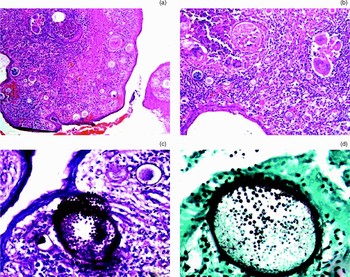
Histopathological examination of the fleshy mass from the tracheostomy wound showed a respiratory epithelial lining with a subepithelial granulomatous response. Numerous sporangia of Rhinosporidium seeberi were seen in the epithelium and subepithelium. The sporangia showed the characteristic pink-staining, thick, hyaline cyst walls. Many sporangia contained spores, while others were empty and a few had ruptured in the subepithelium, evoking a granulomatous response. The spores were better visualised on staining with periodic acid–Schiff and silver methanamine stains (Figure 1). A final diagnosis of tracheal involvement by rhinosporidiosis was made.
Photomicrographs of the tracheal mass showing: (a) respiratory epithelium with subepithelial sporangia, some containing spores (H&E; ×40); (b) granulomatous reaction surrounding ruptured sporangia (H&E; ×100); and spores clearly demonstrated by (c) period acid–Schiff and (d) silver methanamine stains (×200).

After initial stabilisation, complete excision of the tracheal mass was carried out under general anaesthesia.
No recurrence was observed over six months of follow up.
Discussion
Rhinosporidiosis, caused by the aquatic parasite Rhinosporidium seeberi, is a common infection in the tropics, with the highest prevalence in southern India and Sri Lanka.Reference Fredricks, Jolley, Lepp, Kosek and Relman3 The condition commonly involves the nose, nasopharynx, lacrimal sac, larynx and conjunctiva, in that order of frequency.Reference Thomas, Gopinath and Betts1 Involvement of the tracheo-bronchial tree is very rare, having been reported in an occasional case.Reference Thomas, Gopinath and Betts1, Reference Rekha, Thomas, Pappachan, Venugopal, Jayakumar and Sukumaran2, Reference Shah and Ingle4 The infection is transmitted to humans by direct contact with spores via dust, infected clothing or swimming in stagnant water contaminated with spores.Reference Rekha, Thomas, Pappachan, Venugopal, Jayakumar and Sukumaran2
Extension of the nasal or nasopharyngeal infection to the trachea, as in the present case, may be the result of: implantation of spores during intubation for surgical resection; inhalation of discharged spores; or trickling of infected saliva into the trachea.Reference Rekha, Thomas, Pappachan, Venugopal, Jayakumar and Sukumaran2, Reference Grewal and Rangam5 Tracheal masses pose many diagnostic and therapeutic challenges. Diagnosis by bronchoscopic biopsy is fraught with the danger of bleeding from these highly vascular masses.Reference Rekha, Thomas, Pappachan, Venugopal, Jayakumar and Sukumaran2 Computed tomography (CT) scanning is preferred for diagnosis in these cases, since it provides details of the extent of the lesion. Another emergent diagnostic modality which is useful in this regard is virtual bronchoscopy; especially in cases in which rigid bronchoscopy is dangerous or might not indicate the full extent of the lesion.Reference Rekha, Thomas, Pappachan, Venugopal, Jayakumar and Sukumaran2 Virtual bronchoscopy involves a three-dimensional reconstruction of the tracheo-bronchial tree using a double-detector helical CT scan and a virtual endoscopy software program. This technique is useful in detecting bronchial obstructions and endoluminal lesions, as well as for assessing the tracheo-bronchial tree beyond stenoses.Reference De Wever, Vandecaveye, Lanciotti and Verschakelen6 Since the patient in our report presented in severe respiratory distress, an emergency tracheostomy had to be performed, which revealed the tracheal mass.
To date, treatment of rhinosporidiosis is an unresolved problem. Diathermy excision is the first line of treatment in most cases. However, recurrences are common, even at more than one site.Reference Shah and Ingle4 There is a 10 per cent chance of recurrence after excision of nasal rhinosporidiosis; however, the incidence of recurrence of tracheo-bronchial lesions is unknown.Reference Rekha, Thomas, Pappachan, Venugopal, Jayakumar and Sukumaran2 Other therapeutic options include laser cauterisation through a bronchoscope,Reference Shah and Ingle4 combined rigid bronchoscopy and tracheostomy,Reference Rekha, Thomas, Pappachan, Venugopal, Jayakumar and Sukumaran2 or lobectomy if local excision is deemed impossible.Reference Thomas, Gopinath and Betts1 Dapsone has been tried in some cases, with partial relief.Reference Job, Venkateswaran and Mathan7 In the present case, partial excision was carried out at tracheostomy. After stabilisation of the patient, complete excision was performed under general anaesthesia.
Conclusion
Tracheo-bronchial involvement by rhinosporidiosis is an extremely rare occurrence. Such lesions pose great diagnostic and therapeutic problems, and should be kept in mind in known cases presenting with respiratory distress.