Introduction
The semicircular canals, acting as the primary sensors of angular acceleration within the human vestibular system, form the crucial anatomical framework for the vestibulo-ocular and vestibulo-spinal reflexes.Reference Mohamed, Ibraheem and Gad1–Reference Khan and Chang3 Precise characterisation of semicircular canal geometry is vital for understanding vestibular biomechanics and elucidating the structural underpinnings of pathologies like benign paroxysmal positional vertigo (BPPV), superior canal dehiscence syndrome, Ménière’s disease and congenital inner-ear malformations.Reference Della Santina, Potyagaylo, Migliaccio, Minor and Carey4–Reference El Khoury, Braga, Dumoncel, Nancy, Esclassan and Vaysse7 Over the past decades, the foundational understanding of semicircular canal morphology has progressed rapidly from early ex vivo cadaveric and micro-computed tomography (CT) evaluationsReference Cardenas-Serna and Jeffery8 to high-resolution in vivo modalities, including high-resolution temporal bone CT, cone-beam CT and three-dimensional (3D) T2-weighted magnetic resonance imaging (MRI) sequences.Reference Huang, Liu, Tang, Xu, Xie and Yang9–Reference Casselman, Offeciers, De Foer, Govaerts, Kuhweide and Somers14
These imaging advancements have catalysed a major paradigm shift from manual tracing toward standardised 3D co-ordinate frameworksReference Della Santina, Potyagaylo, Migliaccio, Minor and Carey4–Reference Zhou, Mao, Li, Li and Yang11, Reference Joshi, Navlekar, Kishore, Reddy and Kumar15–Reference Yu, Feltrin, Bathla, Raj, Agarwal and Lee20 and, more recently, fully automated deep-learning segmentation pipelines.Reference Vaidyanathan, Van Der Lubbe, Leijenaar, Van Hoof, Zerka and Miraglio21–Reference Stebani, Blaimer, Zabler, Neun, Pelt and Rak30 Nonetheless, broad clinical translation remains constrained by heterogeneous imaging protocols, unstandardised geometric indices, a lack of large-scale multi-centre validation and a persistent separation between structural image data and functional neurotological outcomes.Reference Della Santina, Potyagaylo, Migliaccio, Minor and Carey4–Reference Lane, Lindell, Witte, Delone and Driscoll12, Reference Feraco, Piccinini and Gagliardo16–Reference Yu, Feltrin, Bathla, Raj, Agarwal and Lee20, Reference Stebani, Blaimer, Zabler, Neun, Pelt and Rak30–Reference Seiwerth, Dlugaiczyk, Schmäl, Rahne, Kösling and Plontke52
To bridge these critical gaps, this review provides a comprehensive and critical synthesis that contextualises the entire technological trajectory of human semicircular canal measurement. We systematically evaluated the transition from classical manual morphometry to state-of-the-art artificial intelligence (AI)-driven workflows, critically compared mainstream clinical imaging techniques and analysed the clinical evidence linking structural variations to vestibular disorders. Ultimately, this work aimed to outline a standardised, forward-looking roadmap for integrating automated morphometric analysis into routine clinical workflows.
Anatomy and physiology of semicircular canals
The inner ear consists of a bony labyrinth excavated within the petrous temporal bone, enclosing a corresponding membranous labyrinth. Each of the three orthogonal canals—anterior, posterior and lateral—contains a membranous duct filled with endolymph, with sensory receptors (cristae ampullares) housed within their ampullated ends.Reference Mohamed, Ibraheem and Gad1–Reference Khan and Chang3 The non-ampullary limbs of the vertical canals join to form the common crus, while the lateral canal opens independently into the vestibule.Reference Khan and Chang3 Adopting the pioneering framework by Della Santina,Reference Della Santina, Potyagaylo, Migliaccio, Minor and Carey4 standardised 3D multi-planar reconstructions have established that while inter-canal angles remain relatively orthogonal to optimise angular motion encoding, significant population-level variations exist in canal orientation, size and curvature across age groups, sexes and individuals.Reference Della Santina, Potyagaylo, Migliaccio, Minor and Carey4–Reference Lee, Shin, Kim, Yoo, Song and Koh6 Furthermore, comparative morphometric analyses indicate that these geometric parameters are closely constrained by primate phylogeny, craniofacial architecture and ontogenetic development.Reference El Khoury, Braga, Dumoncel, Nancy, Esclassan and Vaysse7, Reference Cardenas-Serna and Jeffery8
Physiologically, head rotation induces inertial endolymph flow that deflects the cupula and modulates vestibular afferent firing.Reference Mohamed, Ibraheem and Gad1–Reference Khan and Chang3 This intrinsic ‘anatomy–hydrodynamics–symptom’ pathway dictates that subtle spatial or positional shifts in the semicircular canals can disrupt normal fluid mechanics to drive distinct clinical syndromes.Reference Della Santina, Potyagaylo, Migliaccio, Minor and Carey4–Reference Huang, Liu, Tang, Xu, Xie and Yang9 Pathological alterations to this system—such as the intra-luminal displacement of otoconia in BPPV or the focal osseous defect creating a ‘third mobile window’ in superior canal dehiscence syndrome—fundamentally violate normal vestibular physiology. Subtle variations in canal orientation and dimensions can therefore act as anatomical determinants of disease susceptibility and dictate subsequent surgical boundaries,Reference Huang, Liu, Tang, Xu, Xie and Yang9 highlighting the profound clinical requirement for precise geometric tracking.
Imaging techniques for the semicircular canals
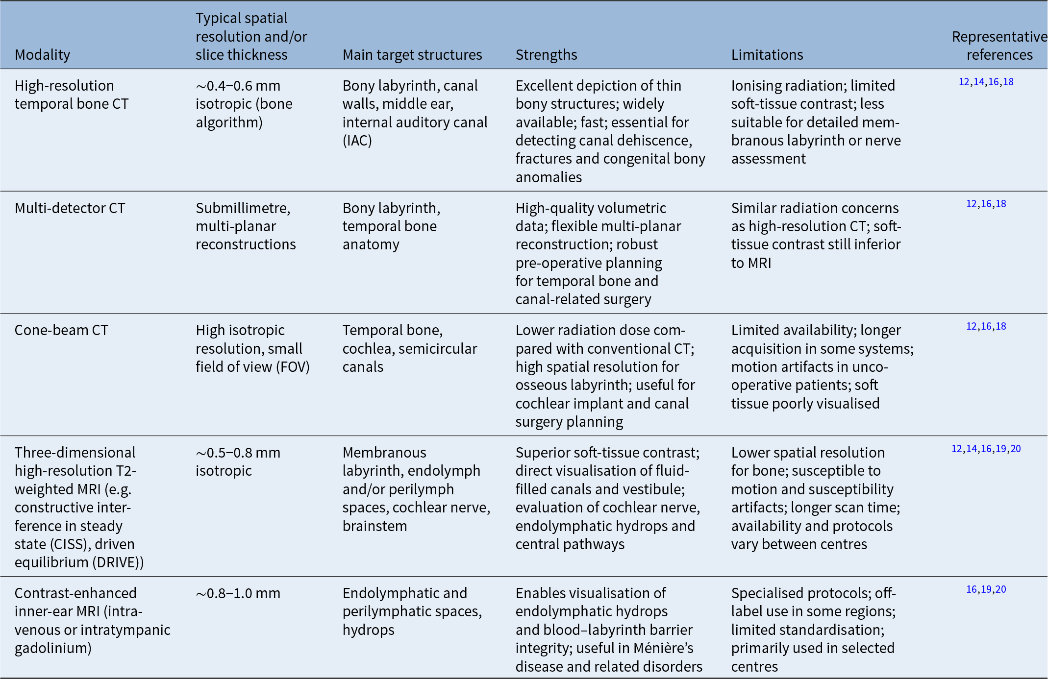
Cross-sectional imaging enables in vivo visualisation of the labyrinth. The primary characteristics of current modalities are summarised in Table 1.Reference Brotto, Ariano, Sozzi, Cenedese, Muraro and Sorrentino32–Reference Jackler, Luxford and House34, Reference Ward, Van De Berg, Van Rompaey, Bisdorff, Hullar and Welgampola53–Reference Xie, Wang, Jiang, Xie, Zhang and Liu56
Comparison of imaging modalities for semicircular canal assessment

Table 1 Long description
The table compares high-resolution temporal bone CT, multidetector CT, cone-beam CT, 3D high-resolution T2-weighted MRI and contrast-enhanced inner-ear MRI. CT-based methods primarily assess the bony labyrinth and canal walls with high spatial resolution and are useful for dehiscence, congenital bony anomalies and surgical planning, but involve radiation and limited soft-tissue contrast. MRI-based methods assess the membranous labyrinth, endolymphatic or perilymphatic spaces, cochlear nerve and brainstem without ionizing radiation, but have lower bone resolution, longer acquisition time and greater protocol variability. Representative references are listed for each modality.
CT = computed tomography; MRI = magnetic resonance imaging.
Computed tomography (high-resolution computed tomography, multi-detector computed tomography and cone-beam computed tomography)
High-resolution CT and cone-beam CT constitute the reference standard for osseous labyrinth evaluation, utilising submillimetre multi-planar reformations to accurately depict canal morphology, bony dehiscence and congenital malformations.Reference Lane, Lindell, Witte, Delone and Driscoll12–Reference Yu, Feltrin, Bathla, Raj, Agarwal and Lee20 While high-resolution CT remains indispensable for intricate surgical planning in superior canal dehiscence syndrome and cochlear implantation, cone-beam CT offers an excellent lower-radiation alternative with high isotropic resolution for pre-operative assessment. These reproducible radiological datasets serve as the primary structural inputs for downstream 3D morphometric frameworks and deep-learning segmentation pipelines.Reference Vaidyanathan, Van Der Lubbe, Leijenaar, Van Hoof, Zerka and Miraglio21–Reference Stebani, Blaimer, Zabler, Neun, Pelt and Rak30
Magnetic resonance imaging and inner-ear magnetic resonance hydrography
Three-dimensional high-resolution T2-weighted MRI (e.g. constructive interference in steady state (CISS), driven equilibrium (DRIVE) and advanced contrast-enhanced magnetic resonance hydrography provide an exceptional membranous-level aperspective of the inner ear, capturing the soft-tissue labyrinth, vestibulocochlear nerve and endolymphatic hydrops in Ménière’s disease.Reference Benson, Carlson and Lane13, Reference Joshi, Navlekar, Kishore, Reddy and Kumar15 This soft-tissue characterisation fundamentally complements CT-based osseous assessments.Reference Feraco, Piccinini and Gagliardo16, Reference Yu, Feltrin, Bathla, Raj, Agarwal and Lee20 Together, these complementary radiological modalities establish a synergistic, multi-modal imaging foundation that powers downstream 3D quantitative morphometry and automated AI analysis pipelines.
Intramodality comparison registration for longitudinal follow up
In longitudinal clinical follow up, patients must undergo repeated imaging over months or years. This is essential for monitoring the progressive erosion of the osseous labyrinth, tracking the potential expansion of bony clefts in superior canal dehiscence syndrome or evaluating post-operative bone healing and structural stability following semicircular canal-plugging surgical procedures.Reference Leng, Diehn, Lane, Koeller, Witte and Carter57 However, variations in patient head positioning, scan angles and slice coverage across different imaging sessions introduce spatial discrepancies that make direct parameter comparison unreliable. To resolve these positioning shifts and ensure accurate longitudinal evaluation, intramodality comparison registration (e.g. CT-to-CT or MRI-to-MRI alignment) must be utilised.Reference Liu, Durongbhan, Davey and Stok58
Intramodality registration operates under the rigid-body assumption, applying translation and rotation parameters to align follow-up scans to the baseline co-ordinate space, as the dense otic capsule is immune to soft-tissue deformation.Reference Liu, Durongbhan, Davey and Stok58 The alignment process is optimised using intensity-based similarity metrics, such as mutual information or normalised mutual information. To preserve the fidelity of fine inner-ear structures and avoid blurring from standard linear interpolation, sinc-based interpolation is preferred for image resampling, achieving sub-voxel registration accuracy.Reference Hajnal, Saeed, Soar, Oatridge, Young and Bydder59 CT-to-CT registration can facilitate longitudinal comparison of osseous structures across serial examinations, provided that differences in acquisition and reconstruction parameters are appropriately considered.Reference Liu, Durongbhan, Davey and Stok58 For MRI-to-MRI registration, aligning sequential 3D T2-weighted images enables precise subtraction maps that reveal microstructural deviations in the fluid-filled membranous ducts,Reference Cho, Song, Cho and Chung60 ensuring highly reproducible longitudinal morphometric assessments.
Methods for measuring semicircular canal size and spatial configuration
Canal height, width, radius of curvature and inter-canal angles
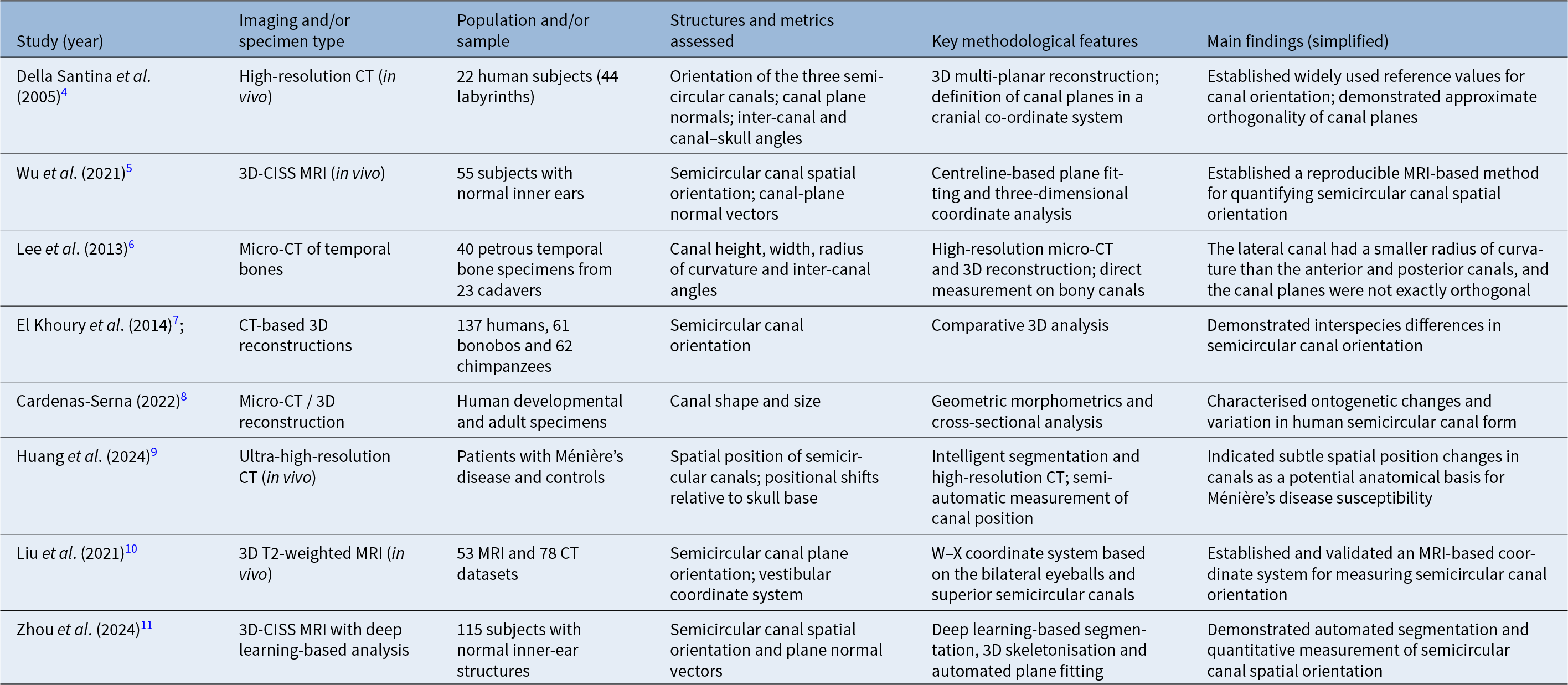
Quantitative assessment of semicircular canal geometry has evolved from direct measurements on anatomical specimens to sophisticated 3D analyses on in vivo CT and MRI, and more recently to fully automated pipelines based on deep learning. Current approaches differ in spatial resolution, structure of interest (bony vs membranous labyrinth) and suitability for large clinical cohorts. Representative morphometric studies are summarised in Table 2.
Summary of landmark morphometric studies on human semicircular canals

Table 2 Long description
The table summarises representative semicircular canal morphometric studies using CT, micro-CT, MRI and artificial intelligence-assisted imaging analysis. The studies evaluate features including canal orientation, plane normal vectors, inter-canal angles, canal dimensions, radius of curvature, spatial position and automated quantitative measurements. They include anatomical, developmental, comparative and clinical imaging studies and collectively demonstrate the evolution from manual and three-dimensional morphometric analysis to standardised coordinate systems and automated image-analysis approaches.
CT = computed tomography; 3D = three-dimensional; MRI = magnetic resonance imaging; CISS = constructive interference in steady state
Anatomical specimens and micro-computed tomography
Historically, the quantitative assessment of semicircular canal geometry relied on cadaveric specimens and micro-CT imaging to establish foundational morphometric baselines, such as canal arc length, spatial orientation and the characteristically smaller radius of curvature observed in the lateral canal.Reference Della Santina, Potyagaylo, Migliaccio, Minor and Carey4, Reference Lee, Shin, Kim, Yoo, Song and Koh6–Reference Cardenas-Serna and Jeffery8
Beyond supplying initial geometric data, these ex vivo studies formed the anatomical bedrock for early biomechanical models of endolymph dynamics and served as a vital ‘gold standard’ for validating subsequent in vivo measurements. However, specimen-based morphometry is inherently constrained by restricted sample sizes, post-mortem tissue degradation, selection biases and a fundamental inability to capture dynamic structure–function relationships in living subjects. These systematic limitations have directly driven the technological transition toward the high-throughput, in vivo imaging-based measurement frameworks detailed below.
Computed tomography-based in vivo three-dimensional measurements
In vivo semicircular canal morphometry is now mainly performed on high-resolution CT or cone-beam CT datasets to reconstruct 3D configurations and define spatial orientations. Following the foundational methodology introduced by Della Santina et al., 3D multi-planar reconstructions are widely utilised to fit canal planes, compute normal vectors and quantify inter-canal and canal-cranial angles within standardised cranial co-ordinate systems.Reference Della Santina, Potyagaylo, Migliaccio, Minor and Carey4, Reference Wu, Lin, Zheng, Zhou, Liu and Yang5
This framework was subsequently adapted by Wu et al., who used three-dimensional CISS MRI in 55 subjects to reconstruct the semicircular canals, fit canal planes from their centre lines and quantify spatial attitude using plane normal vectors.Reference Wu, Lin, Zheng, Zhou, Liu and Yang5 Clinically, as highlighted by Casselman, Joshi and colleagues, these standardised measurements are indispensable for surgical risk mitigation and pre-operative planning, offering critical geometric insights into canal proximity to adjacent structures, such as the facial nerve, during neurotological interventions and cochlear implantation.Reference Casselman, Offeciers, De Foer, Govaerts, Kuhweide and Somers14, Reference Joshi, Navlekar, Kishore, Reddy and Kumar15, Reference Hiremath, Biswas, Mndebele, Schramm, Ertl-Wagner and Blaser18–Reference Yu, Feltrin, Bathla, Raj, Agarwal and Lee20
Consequently, CT-based morphometry is highly effective for delineating osseous canal configurations at the cohort level and guiding surgical decision-making. However, a major lingering challenge is that these measurements remain highly sensitive to institutional variations in CT acquisition parameters, reconstruction kernels and downstream post-processing algorithms, emphasising the critical need for cross-centre protocol standardisation.
Magnetic resonance imaging-based in vivo measurements
Three-dimensional high-resolution T2-weighted MRI directly visualises the membranous semicircular ducts and endolymphatic spaces, capturing morphometric dimensions closely tied to endolymph hydrodynamics and duct compliance. Liu et al.Reference Liu, Chen, Xie, Liang, Zhang and Liao10 proposed the W–X co-ordinate system for vestibular MRI, using the centres of the bilateral eyeballs and the vertices of the superior semicircular canals as anatomical references to quantify semicircular canal orientation.
Magnetic resonance imaging-based morphometry is particularly relevant in disorders in which membranous morphology or fluid distribution is altered, such as endolymphatic hydrops in Ménière’s disease. Furthermore, MRI avoids ionising radiation and allows simultaneous assessment of the cochlear nerve and central vestibular pathways, facilitating structure–function correlation across the entire vestibular system.Reference Huang, Liu, Tang, Xu, Xie and Yang9, Reference Casselman, Offeciers, De Foer, Govaerts, Kuhweide and Somers14, Reference Feraco, Piccinini and Gagliardo16 Limitations include lower spatial resolution for osseous structures compared with CT, and greater susceptibility to motion and susceptibility artifacts.
Taken together, CT- and MRI-based measurements provide complementary views of semicircular canal geometry: CT emphasises the osseous canal walls and their relationship to adjacent temporal bone structures, whereas MRI emphasises the membranous duct and fluid spaces. Harmonisation of measurement definitions and co-ordinate systems across modalities is crucial for integrating these datasets.
Deep learning-based automatic segmentation and quantitative analysis
Traditional semicircular canal measurements require extensive manual contouring or semi-manual landmark placement, which is time-consuming and subject to inter- and intra-observer variability. In recent years, deep learning-based segmentation has transformed inner-ear morphometry by enabling automated, observer-independent delineation of the bony labyrinth and fluid spaces.
On MRI, Vaidyanathan et al. used a 3D U-Net architecture to segment the entire inner-ear fluid space, achieving high Dice similarity coefficients and robust cross-centre performance.Reference Vaidyanathan, Van Der Lubbe, Leijenaar, Van Hoof, Zerka and Miraglio21 Ahmadi et al. introduced IE-Vnet, a deep-learning model specifically optimised for segmentation of the inner ear’s total fluid volume on MRI, which outperformed traditional atlas-based methods and operated with substantially reduced computation time.Reference Ahmadi, Frei, Vivar, Dieterich and Kirsch22
On CT, Hussain et al. built an auto-context convolutional neural network that automatically segments the cochlea, vestibule and semicircular canals, providing a basis for quantitative analysis of bony labyrinth morphology.Reference Hussain, Lalande, Girum, Guigou and Bozorg Grayeli23 Li et al. proposed Labyrinth-Net, which uses a multi-model inconsistency constraint to improve robustness when segmenting complex labyrinthine structures on temporal bone CT.Reference Li, Zhu, Yin, Wang, Zhuo and Zhou24 Wang, Ke and colleagues extended these approaches to fully automated segmentation of adult and paediatric temporal bone CT, demonstrating high accuracy and substantial reductions in manual workload.Reference Wang, Lv, Wang, Ma, Du and Fan25, Reference Ke, Lv, Ma, Du, Xiong and Wang26 Lim et al. trained and externally validated a general U-Net model for inner-ear segmentation across multiple centres, reporting average Dice coefficients above 0.8 and highlighting clinical scalability.Reference Lim, Abily, Ben Salem, Gaillandre, Attye and Ognard27 Li et al. also introduced a 3D deep-supervised densely connected network tailored for small organ segmentation in the temporal bone.Reference Li, Gong, Yin, Zhang, Wang and Zhuo28
A recent systematic review by Li et al. concluded that most state-of-the-art temporal bone segmentation methods are based on U-Net-like architectures and are evolving toward integrated frameworks that combine segmentation, landmark detection and quantitative analysis.Reference Li, Wijewickrema, Margeta, Kamraoui, Hussain and Gerard29 In this direction, Stebani et al. proposed a joint segmentation and landmark detection framework that outputs voxel-wise inner-ear segmentations and geometric landmarks, enabling fully automated computation of semicircular canal curvature, arc length and plane orientation from CT or micro-CT data.Reference Stebani, Blaimer, Zabler, Neun, Pelt and Rak30 A scoping review on deep learning for otologic imaging further supports the feasibility of accurately segmenting small, complex structures such as the semicircular canals.Reference Ross, Tanna, Lilaonitkul and Mehta61
Collectively, these advances indicate that deep learning-based pipelines can transform semicircular canal morphometry from a labour-intensive research tool into a high-throughput component of clinical workflows and large-scale imaging studies. By providing standardised, reproducible measurements of canal geometry, such methods pave the way for multi-centre normative databases and sophisticated structure–function analyses. The overall workflow is illustrated in Figure 1.
Deep learning-based pipeline for automated semicircular canal segmentation and quantitative morphometry. Volumetric high-resolution computed tomography (CT) or three-dimensional (3D) T2-weighted magnetic resonance imaging (MRI) of the temporal bone is first acquired. After pre-processing (cropping to the inner-ear region and intensity normalisation), a 3D convolutional neural network (CNN)—typically a U-Net–based architecture such as 3D U-Net, IE-VNet, or Labyrinth-Net—is applied to segment the inner ear. Post-processing steps (canal isolation and landmark detection) then enable extraction of semicircular canal metrics including volume, arc length, curvature and plane orientation, which can be integrated with vestibular function tests and clinical outcomes for structure–function analysis.

Figure 1 Long description
The workflow starts with volumetric high-resolution CT or 3D T2-weighted MRI acquisition of the temporal bone. Images are preprocessed by cropping to the inner-ear region and intensity normalization. A three-dimensional convolutional neural network, typically a U-Net-based model such as 3D U-Net, IE-VNet or Labyrinth-Net, segments the inner ear. Post-processing isolates the semicircular canals and detects landmarks. Quantitative metrics including volume, arc length, curvature and plane orientation are then extracted and integrated with vestibular function tests and clinical outcomes for structure-function analysis.
Clinical limitations and technical bottlenecks of artificial intelligence models
Despite the outstanding high-throughput capability of deep-learning algorithms on standardised benchmarks, several critical technical and clinical roadblocks continue to hinder their reliable translation into real-world neurotological workflows.Reference Wang, Lv, Wang, Ma, Du and Fan25 These challenges are driven by a complex interplay of spatial, algorithmic and pathology-induced factors that mandate careful methodological scrutiny.
First, severe spatial resolution constraints and inherent partial volume effects present a foundational challenge in inner-ear image analysis.Reference Kim, Kang, Lee, Lee and Nam62 Because the bony and membranous semicircular canals are exceptionally narrow microstructures, they typically span a mere 2 to 3 voxels on clinical CT or MRI scans utilising standard slice thicknesses of 0.5–1.0 mm. When the fine boundaries of these thin pathways blend with the surrounding high-density otic capsule or adjacent soft tissues, pronounced voxel-level intensity blurring frequently confounds segmentation networks. This spatial degradation triggers catastrophic topological errors, including false boundary delineations, artificial lumen fusions or fragmented disconnections along the canal arc, which heavily compromise downstream hydrodynamic modelling.Reference Kim, Kang, Lee, Lee and Nam62
Beyond resolution constraints, deep-learning architectures remain notoriously hypersensitive to domain shifts across heterogeneous imaging protocols.Reference Zemmoura, Burkhardt and Herbet63 When segmentation models optimised on homogeneous, single-centre datasets are deployed across different scanner vendors, divergent magnetic field strengths or varied CT reconstruction kernels, the resulting shifts in grey-level histograms and signal-to-noise ratios typically cause a drastic decline in performance.Reference Ahmadi, Frei, Vivar, Dieterich and Kirsch22 This lack of cross-vendor generalisability emphasises that without extensive multi-centre domain adaptation or transfer learning, models remain overly fragile when encountering the scanning variations typical of real-world clinical environments.
Furthermore, a profound clinical bottleneck arises from algorithmic performance failures under severe pathological conditions.Reference Wang, Lv, Wang, Ma, Du and Fan25 The vast majority of current AI tools are trained on anatomically normal templates, rendering their underlying structural assumptions vulnerable when encountering structural anomalies. In patients with congenital inner-ear malformations, such as Mondini dysplasia, these pre-configured geometric priors are completely violated. Similarly, in conditions involving intraluminal obliteration—such as fibrous tissue encapsulation or neo-ossification in labyrinthitis ossificans and advanced otosclerosis—the loss of normal MRI fluid signals and corresponding alterations in CT bone density drive massive segmentation errors. Quantitative evidence demonstrates that in such pathological ears, the Dice similarity coefficient for the labyrinth plummets from a robust 0.910 down to 0.775, while the Dice similarity coefficient for the adjacent facial nerve drops to an entirely unusable 0.506.Reference Wang, Lv, Wang, Ma, Du and Fan25
Finally, the development of truly robust networks is severely throttled by statistical fragility in low-data regimens, largely because of the profound paucity of annotated, high-quality pathological datasets. Within these restricted data domains, marginal variations in training and testing splits can completely reshuffle different models’ performance rankings.Reference Wang, Lv, Wang, Ma, Du and Fan25 This instability strongly indicates that many highly publicised ‘leaderboard’ superiorities on public benchmarks are frequently mere statistical artifacts of noise rather than reflections of genuine clinical generalisability, underlining the critical need for rigorous external validation before clinical deployment.
Morphological variation of the semicircular canals and clinical implications
Normal variation and congenital inner-ear malformations
Micro-CT and in vivo CT and/or MRI studies demonstrate that the three semicircular canals exhibit appreciable inter-individual variability in dimensions, curvature and spatial orientation while maintaining good bilateral symmetry of corresponding canals.Reference Della Santina, Potyagaylo, Migliaccio, Minor and Carey4–Reference Cardenas-Serna and Jeffery8 This physiological variation underpins the definition of normative ranges and may contribute to disease susceptibility in certain vestibular disorders.Reference Wu, Lin, Zheng, Zhou, Liu and Yang5, Reference Huang, Liu, Tang, Xu, Xie and Yang9, Reference Liu, Chen, Xie, Liang, Zhang and Liao10
Congenital inner-ear malformations are a major cause of severe-to-profound sensorineural hearing loss in children, and canal malformations—such as hypoplastic, dysplastic or absent canals—are frequent components of these anomalies. Brotto et al. reported that about half of children with inner-ear malformations have vestibular dysfunction or delayed balance development, emphasising the importance of semicircular canal and vestibular assessment when planning cochlear implantation or other interventions.Reference Brotto, Ariano, Sozzi, Cenedese, Muraro and Sorrentino32
From a structural perspective, Jackler et al. proposed a classic embryogenesis-based classification that distinguishes common cavity, incomplete partition types and other subtypes according to the timing of developmental arrest.Reference Jackler, Luxford and House34 This foundational framework was subsequently updated by Sennaroğlu and Bajin to adapt to modern management paradigms and guide cochlear implantation in severely malformed cochleovestibular systems.Reference Sennaroğlu and Bajin35, Reference Sennaroglu36 Recent radiological reviews by Bhagat, O’Brien and colleagues refined imaging criteria for individual malformation subtypes and proposed algorithmic CT and/or MRI assessment workflows, which are highly relevant for pre-operative evaluation of canal anomalies.Reference Bhagat, Kumar, Garg, Prakash, Meher and Arya38, Reference O’Brien, D’arco, Onofrj and Koch39
Histopathological studies by Monsanto et al. highlighted potential pitfalls of cochlear implantation in malformed inner ears, including perilymphatic gusher, aberrant cochlear or vestibular wall defects, and atypical cochlea–vestibule–canal relationships.Reference Monsanto, Sennaroglu, Uchiyama, Sancak, Paparella and Cureoglu45 Systematic reviews by Farhood, Shah and Sennaroğlu and co-workers showed that, despite these challenges, many patients with inner-ear malformations can achieve favourable speech perception outcomes when implantation strategies are carefully tailored to the specific anatomy and vestibular status.Reference Sennaroglu36, Reference Farhood, Nguyen, Miller, Holcomb, Meyer and Rizk46, Reference Shah, Walters, Langlie, Davies, Finberg and Tuset47 These data underscore that detailed imaging-based characterisation of semicircular canal morphology is integral to risk stratification, surgical planning and prognostication in congenital inner-ear malformations.
Semicircular canal morphology and benign paroxysmal positional vertigo
Benign paroxysmal positional vertigo, the most common peripheral vestibular disorder, is widely attributed to otoconia dislodged from the utricle that enter the semicircular canals and move with head position, abnormally stimulating the crista ampullaris. While the standardised clinical practice guidelines by Bhattacharyya et al.Reference Bhattacharyya, Gubbels, Schwartz, Edlow, El-Kashlan and Fife40 and comprehensive pathophysiological updates by SwainReference Swain42 provide robust clinical frameworks for canal-specific diagnosis and canalith repositioning manoeuvres, recent neurotological interest has increasingly focused on ‘structural susceptibility’. This paradigm explores whether inherent, individual semicircular canal geometry influences otoconial deposition and migration dynamics. In this context, emerging editorial perspectives by Bhandari and colleaguesReference Bhandari, Sharma, Zuma, Ramos and Martin-Sanz49 emphasise the need for refined morphometric phenotyping of canal involvement, especially because population-level evaluations by Si et al.Reference Si, Ling, Li, Li, Shen and Yang48 demonstrated that multi-canal BPPV variants present with significantly higher recurrence rates and more complex nystagmus patterns than single-canal pathologies, pointing toward a distinct anatomical or metabolic predisposition.
Direct morphometric evidence remains limited. Yılmazer et al. used CT to compare semicircular canal arc lengths between BPPV patients and controls, and found significantly longer anterior and posterior canals in BPPV, proposing ‘elongated canals’ as a potential imaging risk marker.Reference Yılmazer, Bircan, Erk, Kış, Göker and Hanci64 Ping et al. described a short-arm variant of posterior canal BPPV in which otoconia are located in the short ampullary limb rather than the long non-ampullary limb, and showed that its clinical course and optimal repositioning strategy differ from the classic long-arm variant.Reference Ping, Yi-Fei, Shu-Zhi, Yan-Yan and Xiao-Kai65 For lateral canal BPPV, Maia and co-workers systematically reviewed management strategies across pathophysiological forms (canalithiasis vs cupulolithiasis; geotropic vs apogeotropic nystagmus), highlighting the importance of canal orientation and ampullary position when selecting maneuvers.Reference Maia, Ramos, Cal, Brock, Mangabeira Albernaz and Strupp50
Overall, available data linking semicircular canal morphology to BPPV susceptibility come from small, single-centre studies with heterogeneous imaging protocols and measurement methods, and reported associations are not fully consistent. Larger, multi-centre case–control studies using standardised CT and/or MRI morphometry are needed to determine whether specific geometric features—such as increased arc length, altered curvature or canal orientation—truly increase BPPV risk or affect the likelihood of multi-canal involvement and response to treatment.
Superior canal dehiscence syndrome and canal plugging and/or occlusion surgery
Superior canal dehiscence syndrome is characterised by a focal bony defect in the roof of the superior semicircular canal, which creates a third mobile window in the inner ear and leads to sound- and pressure-induced vestibular symptoms and auditory manifestations such as autophony and conductive hyperacusis. The Bárány Society consensus document by Ward et al. established diagnostic criteria requiring characteristic symptoms, objective vestibular or audiological signs (e.g. low-threshold vestibular evoked myogenic potentials, abnormal video head impulse test or characteristic nystagmus) and high-resolution imaging evidence of superior canal dehiscence on CT or MRI.Reference Ward, Van De Berg, Van Rompaey, Bisdorff, Hullar and Welgampola53 Suzuki et al. emphasised that CT evaluation should include oblique reformations parallel and perpendicular to the superior canal to reduce partial-volume artifacts and avoid overcalling thin but intact bone as true dehiscence.Reference Suzuki, Ota, Takanami, Yoshino and Masuda54
Semicircular canal occlusion, or plugging, was first introduced for refractory BPPV and has since been applied to selected cases of Ménière’s disease and superior canal dehiscence syndrome. Zhang et al. described triple semicircular canal plugging as a novel surgical modality for intractable Ménière’s disease and reported satisfactory vertigo control with partial hearing preservation in most patients.Reference Zhang, Fan, Han, Lv, Li and Wang55 Xie et al. evaluated triple semicircular canal occlusion combined with endolymphatic sac decompression and showed durable vertigo control with acceptable audiovestibular outcomes.Reference Xie, Wang, Jiang, Xie, Zhang and Liu56 For superior canal dehiscence syndrome, Seiwerth et al. reported on transmastoid two-point superior canal plugging with preservation of the vestibulo-ocular reflex, showing that carefully planned canal isolation can effectively control symptoms while minimising long-term vestibular dysfunction.Reference Seiwerth, Dlugaiczyk, Schmäl, Rahne, Kösling and Plontke52
These studies support canal plugging or occlusion as a viable option for carefully selected patients; however, optimal indications, configuration of canal occlusion and long-term outcomes remain under investigation.Reference Tian, Yin, Zhang, Zhang, Zeng and Li51–Reference Ward, Van De Berg, Van Rompaey, Bisdorff, Hullar and Welgampola53, Reference Zhang, Fan, Han, Lv, Li and Wang55, Reference Xie, Wang, Jiang, Xie, Zhang and Liu56 Precise pre-operative imaging assessment of superior canal morphology—including the size and location of dehiscence, canal curvature and relationship to adjacent structures—is therefore essential for surgical planning and for balancing vertigo control against the risk of iatrogenic vestibular hypofunction.
Future directions: standardised measurement and artificial intelligence-assisted analysis
Remaining challenges
Despite substantial advances in anatomical characterisation, imaging technology and automatic segmentation, several important challenges continue to limit the translational impact of semicircular canal morphometry.
Primarily, marked heterogeneity in multi-centre imaging and post-processing protocols—spanning divergent CT and/or MRI acquisition parameters, reconstruction kernels and reference co-ordinate systems—precludes direct cross-cohort comparisons. This is further compounded by a profound lack of standardisation in geometric indices; the absence of consensus regarding landmarks and centreline fitting strategies for core metrics (such as ‘arc length’, ‘height’ or ‘canal plane’) introduces methodological artifacts that heavily obscure true biological variations and complicate robust meta-analyses.
Furthermore, the field is severely limited by a paucity of large-scale, multi-centre normative databases. Most available morphometric datasets originate from modest, single-centre retrospective cohorts, leaving the compounding influences of age, sex, ethnicity and craniofacial morphology incompletely characterised. Crucially, the symbiotic integration of structural morphometry with objective vestibular function tests and patient-reported symptom severity remains sparse. Bridging this persistent structure–function gap is a critical prerequisite for establishing the definitive clinical significance and diagnostic validity of quantitative semicircular canal variations in neurotological practice.
Research priorities
The advent of automated, precise semicircular canal morphometry opens up exciting new frontiers. To realise the full clinical potential of these technologies, the research community should focus on the following strategic priorities.
First, building large-scale, standardised multi-centre databases. The single greatest limitation currently is the lack of large, diverse datasets. We advocate for a global initiative to create a federated database of high-resolution temporal bone images, coupled with standardised clinical and functional vestibular data. This resource would be indispensable for training robust, generalisable AI models and establishing normative morphometric references across different demographics.
Second, integrating structure and function. The next leap forward will come from quantitatively linking semicircular canal morphology to physiological function. Future studies must be designed to correlate geometric parameters (e.g. radius of curvature, canal non-planarity) with objective functional measures (e.g. video head impulse test (vHIT) gains and vestibular evoked myogenic potential (VEMP) responses). This will enable the development of biomechanical models that can predict an individual’s susceptibility to conditions like BPPV or canal dehiscence.
Third, clinical workflow integration and prospective validation. For these tools to impact patient care, they must be seamlessly integrated into the clinical workflow. This requires the development of user-friendly, US Food and Drug Administration (FDA)-cleared software that can provide radiologists and otologists with rapid, reproducible morphometric reports. Crucially, prospective clinical trials are needed to validate the diagnostic accuracy and prognostic value of these AI-derived imaging biomarkers in predicting disease onset, progression and treatment outcomes.
Finally, exploring new morphometric biomarkers. Beyond radius and orientation, AI can uncover subtle morphological signatures of disease that are invisible to the human eye. Future research should leverage advanced techniques like radiomics and geometric deep learning to identify novel imaging phenotypes associated with vestibular migraine, Ménière’s disease and age-related vestibular loss, paving the way for a new era of precision otology.
Conclusions
The semicircular canals are indispensable components of the vestibular system, and their detailed anatomy and imaging characteristics are closely linked to a broad spectrum of vestibular disorders. Advances in high-resolution CT and MRI, together with standardised co-ordinate systems, have greatly improved in vivo quantification of canal geometry. Deep learning-based segmentation and 3D morphometric analysis now make it possible to obtain automated, reproducible measurements of canal volume, curvature and spatial orientation across large imaging datasets.
Emerging evidence suggests that semicircular canal morphology may influence disease susceptibility and treatment response in BPPV, superior canal dehiscence syndrome, Ménière’s disease and congenital inner-ear malformations. However, existing studies are limited by heterogeneous methods, small sample sizes and incomplete integration of structural and functional data. Future efforts should therefore focus on harmonising imaging and measurement protocols, constructing multi-centre normative databases and combining morphometric data with vestibular function tests and clinical outcomes.
By uniting rigorous anatomical and imaging-based measurement with artificial intelligence and advanced visualisation technologies, semicircular canal metrics have the potential to become key components of precision diagnosis, surgical planning and individualised management in clinical vestibulology. In the longer term, standardised, AI-assisted semicircular canal morphometry may also provide a framework for incorporating vestibular anatomy into broader models of balance control and spatial orientation.
• Variations in semicircular canal geometry are increasingly recognised as critical factors in the pathogenesis and clinical manifestation of disorders such as benign paroxysmal positional vertigo[Q2], superior canal dehiscence syndrome and Ménière’s disease
• Current clinical assessment relies on high-resolution computed tomography and three-dimensional T2-weighted magnetic resonance imaging to visualise the bony and membranous labyrinths
• This review systematically synthesised the technological evolution of semicircular canal morphometry, transitioning from foundational cadaveric studies to state-of-the-art artificial intelligence-driven automated analysis pipelines
• The emergence of deep-learning architectures, such as U-Net and Labyrinth-Net, which enable reproducible, high-throughput segmentation and automated landmark detection, is detailed
• Recent clinical evidence linking specific structural markers, such as canal arc length and orientation, to disease susceptibility and surgical treatment outcomes is consolidated
• A forward-looking roadmap is proposed, emphasising the necessity of multi-centre data standardisation and the integration of structural and functional data to advance precision otology
Data availability statement
Data sharing is not applicable to this article as no new data were created or analysed in this study.
Competing interests
None declared.
Acknowledgements
In accordance with ethical publication standards, we disclose that ChatGPT was used for grammar checks and no AI tools were used for data generation or interpretation. We have ensured that the final content remains accurate and compliant with journal guidelines.