Introduction
Toxocara canis and T. cati are the most common parasite species of dogs and cats in the world. Their prevalence is strictly associated with the host's age, being the highest in puppies and kittens up to 24 weeks old; however, the infection is also frequent in stray animals (O'Lorcain, Reference O'Lorcain1994; Saeki et al., Reference Saeki, Yokoi and Yamamoto1997; Kulišić et al., Reference Kulišić, Pavlović, Milutinović and Aleksić-Bakrać1998). An increasing number of household dogs and cats has been noted in recent years in many European countries. Regular deworming of pets is not very common and that, together with the long survival of Toxocara spp. eggs in the outer environment, results in accumulation of eggs in the soil in public and private areas. The cases of human toxocarosis recognized each year reflect the contamination of the environment with T. canis and T. cati eggs (Giacometti et al., Reference Giacometti, Cirioni, Fortuna, Osimani, Antonicelli, del Prete, Riva, D'Errico, Petrelli and Scalise2000; Gawor & Borecka, Reference Gawor and Borecka2004; Muradian et al., Reference Muradian, Gennari, Glickman and Pinheiro2005). Eggs of both species are very much alike. Light and scanning microscopic observations are insufficient for their differentiation. Subspherical eggs are of similar size. Their respective surface structures are not reliable features as 16% of T. canis and 29% of T. cati eggs showed surface pitting uncharacteristic of their species (Uga et al., Reference Uga, Matsuo, Kimura, Rai, Koshino and Igarashi2000).
The elaborated molecular methods for discrimination of both species were based on polymerase chain reaction (PCR) analysis of genomic DNA extracted from adult worms, or embryonated eggs collected from the uteri of female worms (Jacobs et al., Reference Jacobs, Zhu, Gasser and Chilton1997; Wu et al., Reference Wu, Nagano, Xu and Takahashi1997). The described methods of DNA isolation were inadequate in routine examinations when numerous samples need to be tested. As soil contains natural inhibitors of the PCR reaction, attempts must be made to avoid their effects. Recently, this issue has been addressed, but the methods used are unsuitable for routine diagnostics due to the time consuming and laborious extraction of gDNA for genetic analysis (Borecka, Reference Borecka2004; Fogt-Wyrwas et al., Reference Fogt-Wyrwas, Jarosz and Mizgajska-Wiktor2007). We have described a PCR-restriction fragment length polymorphism (RFLP) technique for the differentiation of Toxocara eggs isolated from soil (Borecka, Reference Borecka2004); however, the method of obtaining gDNA from eggs was complicated and unsatisfactory.
The present study was undertaken to develop a modification of gDNA extraction for the PCR method used for the detection and differentiation of Toxocara spp. eggs in soil or sediments.
Materials and methods
Thirty-six 100–150 g sand samples were taken from sandpits in housing estates in Warsaw, Poland. From each sample 10 g was examined by means of the flotation technique to evaluate contamination with ascarid eggs, as the use of only positive samples in further PCR procedures was considered more suitable, i.e. less time and cost consuming. Toxocara spp. eggs were extracted using the modified Dada method (Dada, Reference Dada1979). Dried samples were sieved through a 0.5 mm mesh; 10 g samples were transferred to volumetric flasks containing 50 ml of 0.1% Tween 80 and vortexed for 30 min. The suspensions were transferred to 60 ml centrifuge tubes and then centrifuged at 1500 rpm for 10 min. After that the supernatant free of eggs was decanted. A saturated flotation solution (ZnSO4, 1.52 specific gravity) was added to the tubes containing the remaining sediment and the tubes were vortexed again and later centrifuged at 1500 rpm for 10 min. The solution was then added to form a meniscus and a coverslip was overlaid. After 15 min the coverslip was transferred to a glass slide which was examined at 100 × magnification and Toxocara spp. eggs were counted. Aliquots of sand (2.5 g) from samples found to be positive were taken for further molecular examination.
Direct isolation of genomic DNA from sand samples
The isolation was performed using GeneMATRIX tissue kit (Eurx, Poland). The kit protocol was modified by the authors for use with sand samples. For each 2.5 g sample of sand, 1 ml of buffer Lyse-T, 280 μl of buffer RN and 80 μl of proteinase K solution were added. The samples were mixed by vortexing and were incubated at 56°C until complete lysis was obtained (overnight incubation – a minimum of 16 h). In the next step, incubated samples were centrifuged for 1 min at 1200 rpm; 440 μl of supernatants were mixed with 350 μl of SOL-T buffer and incubated for 10 min at 56°C. After the incubation, 300 μl of 96% ethanol was added and then samples were centrifuged for 1 min at 1200 rpm; 600 μl of the obtained supernatants were transferred to spin-columns, placed in collection tubes and centrifuged for 1 min at 1200 rpm. The filters with bound gDNA were washed twice using 600 μl and 300 μl of Wash-TX buffer. In the next step, the spin-columns were transferred to new collections of tubes and 20 μl of the elution buffer heated to 70°C was added to elute the bound DNA. Samples were incubated for 3 min at room temperature and then centrifuged for 1 min at 1200 rpm. The extracted DNA was used directly for PCR or frozen at − 20°C.
PCR method
PCR amplification of internal transcribed spacer 2 (ITS-2) was described by Jacobs et al. (Reference Jacobs, Zhu, Gasser and Chilton1997), who extracted genomic DNA either from adult worms collected from dogs, foxes and cats or embryonated eggs recovered from the uteri of female worms. The region spanning ITS-2 was amplified from the gDNA (10–15 ng) by PCR using oligonucleotide species-specific primers:
(1) Tcan1 (5′-AGTATGATGGGCGCGCCAAT-3′) and NC2 (5′-TAGTTTCTTTTCCTCCGCT-3′) for T. canis; and
(2) Tcat1 (5′-GGAGAAGTAAACTC-3′) and NC2 for T. cati.
The Tcan1–NC2 primer set amplified the PCR product of c. 380 bp from T. canis, and the Tcat1–NC2 set amplified the product of 370 bp from T. cati.
PCR reactions (50 μl) were performed in 10 mm Tris–HCl, pH 8.4; 50 mm KCl; 3.0 mm MgCl2; 250 μm of each of dATP, dCTP, dGTP, dTTP; 100 pmol of each primer with 2 U Taq polymerase, under the following conditions: 94°C, 30 s (denaturation); 55°C, 30 s (annealing); 72°C, 30 s (extension) for 30 cycles (Thermocycler, Biometra, Germany). A sample of 1 μl (usually equal to 10–15 ng) of genomic DNA was added to each PCR reaction. Samples without DNA were included in each amplification run to exclude ‘carry-over’ contamination. The obtained fragments were separated on 2.5% agarose gels containing ethidium bromide. The pUC 1444-80 marker (produced by BTL, Poland) was used to estimate the size of the obtained fragments. Gels were transilluminated in UV light and photographed.
Results
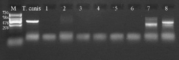
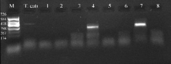
Contamination of 8 (22.2%) out of the 36 examined sand samples from the sandpits with Toxocara spp. eggs was confirmed by means of the flotation technique. In each positive sample 1–5 Toxocara eggs were recovered. No attempt was made to differentiate T. canis and T. cati using microscopy as extracted eggs were damaged. For molecular examinations, 2.5 g of sand was taken from the samples which contained the parasite eggs. The use of proteinase K for DNA extraction directly from the sand excluded the necessity of previous isolation of eggs by means of the flotation method. The PCR technique enabled us to distinguish the two ascarid species present in the examined samples. gDNA of T. canis was found in two samples (nos 1 and 3), gDNA of T. cati in three samples (nos 4, 5, 7) and gDNA of both species was detected in three samples (nos 2, 6, 8). The expected PCR products of c. 380 bp from T. canis and c. 370 bp from T. cati of some examined samples (nos 4, 6, 7, 8) were visible on agarose gels (figs 1 and 2).
Product of amplification of gDNA extracted from sand samples using primers specific for T. canis (Tcan1–NC2): lane M, pUC marker (1444-80 bp); lane T. canis, product of PCR for standard gDNA isolated from adult T. canis worms; lane 1, no DNA control; lanes 2–6, negative results of PCR reaction for non-contaminated samples; lanes 7 and 8, product of PCR reaction for DNA extracted from contaminated sand samples (nos 6 and 8).

Product of amplification of gDNA extracted from sand samples using primers specific for T. cati (Tcat1–NC2): line M, pUC marker (1444-80 bp); lane T. cati, product of PCR for standard gDNA isolated from adult T. cati worms; lanes 1, 2, 3, 5, 6, negative results of PCR reaction for non-contaminated samples; lanes 4 and 7, products of PCR reaction for DNA extracted from contaminated sand samples (nos 4 and 7); lane 8, no DNA control.

Discussion
In this study a modification of gDNA extraction for the PCR technique was developed to be used in routine examination of soil, sand or sediments for the presence of Toxocara spp. eggs. In previous investigations we used a PCR-linked RFLP method for the detection and differentiation of Toxocara eggs from soil samples (Borecka, Reference Borecka2004), but this method was more expensive and time consuming when compared to the one presented here. In the former study the most serious problem was connected with the extraction of embryos from eggs (Borecka, Reference Borecka2004). Freezing in liquid nitrogen ( − 70°C), boiling for 10 min, and incubation with proteinase K (10 mg/ml) at 65°C for 2 h did not lead to the disintegration of eggshells. For this reason embryonic material was removed from the eggs using a needle. In the present study the modified protocol of a commercial kit (Eurx, Poland) was used. According to the experiment procedure, incubating of the sample was performed overnight (16 h) until complete lysis was achieved. The use of proteinase K resulted from our experience and data obtained by other authors. Halos et al. (Reference Halos, Jamal, Vial, Maillard, Suau, Le Menach, Boulouis and Vayssier-Taussat2004), in their genetic studies on ticks, showed that proteinase K was efficient for the destruction of the chitinous exoskeleton of those arachnids. According to the kit protocol (GeneMATRIX tissue kit) proteinase K activity is amplified by buffer, aiding tissue and cell disintegration. The extended period of incubation implemented in the present study led to damage of eggs. DNA extraction was confirmed by specific products of amplification of the ITS-2 region of Toxocara.
The application of DNA extraction directly from soil enables the examination of many samples in a short time. In our former study, genomic material was removed from Toxocara eggs, suspended in distilled water on a slide, by using a needle, which was the most difficult step of the examination (Borecka, Reference Borecka2004). In an experiment carried out by Fogt-Wyrwas et al. (Reference Fogt-Wyrwas, Jarosz and Mizgajska-Wiktor2007), who used PCR for the differentiation of T. canis and T. cati eggs isolated from soil, the samples were naturally contaminated with 1–2 eggs and were also prepared to contain 10 and 430 eggs/40 g of soil. The large number of eggs in the soil is unusual in material collected from public places. Even in shelters for dogs and cats some authors detected up to 10 eggs/50 g sample (Dubna et al., Reference Dubna, Langrova, Jankovska, Vadlejch, Pekar, Napravnik and Fechtner2007). The artificial inoculation of samples with enormous numbers of eggs seems to be inappropriate when the PCR method is applied for environmental examination, especially when considering the very high sensitivity of this reaction. In our investigation, samples were collected from sandpits supposed to be contaminated with Toxocara eggs. Following the confirmation that 10 g samples contained such eggs, by the flotation method, 2.5 g of sand from the sample was taken for molecular testing. First, gDNA was released using proteinase K, thereafter further PCR processing was performed. We examined samples with low levels of contamination, as 1–5 eggs were found per 10 g sand sample. The elaborated method of gDNA extraction makes it possible to examine small samples. On the one hand, the use of a small amount of soil shortens the preparation time and reduces the concentrations of inhibitory substances but, on the other hand, there is a smaller chance of recovering the parasite DNA. Nevertheless, using this technique numerous samples can be tested, providing an opportunity for detailed screening of a location. The direct digestion of soil samples by proteinase K removes the need for flotation and the subsequent manipulation of eggs.
To date, the direct digestion of soil to search for ascarid gDNA has not been performed. Extraction of DNA from embryos released from the eggshells of Toxocara species was an indispensable step of the procedure in previous studies (Borecka, Reference Borecka2004; Fogt-Wyrwas et al., Reference Fogt-Wyrwas, Jarosz and Mizgajska-Wiktor2007) and this method was relatively laborious. Fogt-Wyrwas et al. (Reference Fogt-Wyrwas, Jarosz and Mizgajska-Wiktor2007) recommended crushing the eggs, by pressing a cover slip on a microscope slide, as the only method to get the embryonic material. The weak point in this technique, apart from isolation of the eggs, is that embryos are polluted by the flotation solution and subsequently have to be rinsed with large amounts of water. Another difficulty is the presence of organic and inorganic compounds in soil, which inhibit the amplification of nucleic acids by PCR. They can interfere with the reaction at several levels, leading to different degrees of attenuation and even to complete inhibition (Moreira, Reference Moreira1998). The use of enzymatic lysis (proteinase K), bead beating or sonication can inactivate these inhibitors (Yeates et al., Reference Yeates, Gillings, Davison, Altavilla and Veal1998).
The described modification of gDNA extraction was developed for the PCR method intended for the detection and differentiation of Toxocara spp. eggs in soil or sediments. The modification simplifies the procedures, thanks to eliminating the isolation of eggs by flotation and the subsequent manipulation to obtain genetic material, which is both laborious and difficult.
Acknowledgements
Financial support for this work was provided by the Ministry of Sciences and Higher Education (contract no. 3 PO5D 074 23). The authors wish to thank Professor Christian Epe and Professor Georg V. Samson-Himmelstjerna for their valuable advice prior the study.