INTRODUCTION
Climate change and the introduction of alien species throughout the world appear to be increasing the rate of change in species distribution boundaries. The Mediterranean Sea, being a semi-enclosed area, is being affected by climate change at a faster rate than many other marine areas (Por, Reference Por1978; Calvo et al., Reference Calvo, Simó, Coma, Ribes and Pascual2011). The faunal structure of the Algerian Basin is highly unstable due to such alien invasions, mainly from the North Atlantic region via the Strait of Gibraltar. This is the case for the aspidochirotid holothuroid Holothuria (Roweothuria) arguinensis (Koehler & Vaney, Reference Koehler and Vaney1906), originally described from the north-east African coast (Massin, Reference Massin1993). This species had been considered a north-eastern Atlantic species and appears to be pretty widespread on the Atlantic coast of Portugal, in the Berlengas Islands (Rodrigues, Reference Rodrigues2012). Hérouard (Reference Hérouard1929) and Panning (Reference Panning1929) recorded the species from Morocco and Mauritania, Pérez-Ruzafa et al. (Reference Pérez-Ruzafa, Marcos and Bacallado1992) from the Canary Islands, Massin (Reference Massin1993) from Mauritania and more recently González-Wangüemert et al. (Reference González-Wangüemert, Braga, Silva, Valente, Rodrigues and Serrao2013) from Ria Formosa (south coast of Portugal). More pertinent are the reports of the species from two localities from the Spanish Mediterranean coast (Granada, southern Spain (Ocaña & Pérez-Ruzafa, Reference Ocaña and Pérez-Ruzafa2004) and El Mojon in the Alboran Sea (south-eastern Spain) (González-Wangüemert & Borrero-Pérez, Reference González-Wangüemert and Borrero-Pérez2012)).
Hence, the occurrence of this species, now in the Algerian coastal waters, is evidence of its further extension into the western part of the Mediterranean Sea. This extension is perhaps linked to the new environmental conditions currently prevailing in the Mediterranean Basin.
MATERIALS AND METHODS
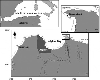
Materials used in this investigation represent three specimens collected from the coastal waters of Algiers Bay near the Tamentefoust area (3°13′E–36°48′N) (Figure 1).
Geographical location of the Tamentefoust area from where specimens of Holothuria (Roweothuria) arguinensis were collected (★).

The specimens were collected by SCUBA diving at 4.5 m depth, initially fixed in 10% formalin, but later washed and preserved in 90% ethanol. Digital photographs of live specimens were taken using a Canon EOS 1100 camera. The ossicles were photographed under a polarized light Leica DMLP microscope, equipped with a Nikon ME600 camera with DIC (differential interference contrast). The morphology, including the endoskeleton (ossicles), was studied according to conventional methods outlined by Mezali & Francour (Reference Mezali and Francour2012). For recognition and correct identification, descriptions of the species by Koehler & Vaney (Reference Koehler and Vaney1906) and Hérouard (Reference Hérouard1929) and the illustration given by Massin (Reference Massin1993) were considered. The identification was verified by Dr F.W.E. Rowe. Measurements were made of each type of ossicles (tables, buttons and perforated elongated plates) from the dorsal and ventral body wall and rods from the tube feet.
RESULTS
SYSTEMATICS
Class HOLOTHUROIDEA de Blaineville, 1834
Order ASPIDOCHIROTIDA Grube, 1840
Family HOLOTHURIIDAE Ludwig, 1894
Genus Holothuria Linnaeus, 1767
Sub-genus Roweothuria Thandar, Reference Thandar1988
Holothuria (Roweothuria) arguinensis Koehler & Vaney, Reference Koehler and Vaney1906
(Figures 2–3)
Holothuria arguinensis Koehler & Vaney, Reference Koehler and Vaney1906: 62–65, pl. 5, figures 5–13, pl. 6, figures 14–21; Gruvel, Reference Gruvel1909: 1018; Hérouard, Reference Hérouard1925: 7, figure 6; 1929: 48–52, figures 3A–K, pl. 1, figure 5; Cherbonnier, Reference Cherbonnier1950: 106–108, figure 3d.
Holothuria (Holothuria) arguinensis Panning, Reference Panning1935: 49, figure 44.
Holothuria (Lessonothuria) arguinensis Rowe, Reference Rowe1969: 149–150.
Holothuria (Roweothuria) arguinensis Thandar, Reference Thandar1988: 48, 53; Massin, Reference Massin1993: 400, figure 1.
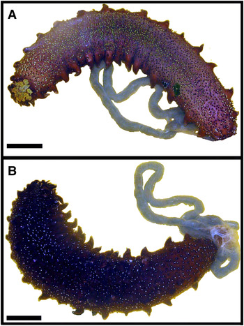
Specimens sub-cylindrical, with tough, leathery body wall, rough to the touch. Length of live animal about 350 mm, wet body weight 270 g. Dorsal and ventral surfaces well demarcated, the former arched, dark brown in coloration. Dorsal surface bears regularly arranged conical protuberances/warts, in two double rows, constituting an almost continuous border with 16 on the left and 15 on the right, each terminating in a white papilla. Other papillae, also white in colour, but borne on much smaller warts. Ventral surface, light brown in coloration, flattened, sole-like with numerous scattered tube feet (Figure 2). Mouth ventral, tentacles 20, lemon yellow. Anus also ventral, anal papillae present, few.
Holothuria (Roweothuria) arguinensis from Algiers Bay: (A) ventral view; (B) dorsal view (photograph: K. Mezali). Scale bar: 50 mm.

Calcareous ring well-developed. Polian vesicles 3–4; stone canals also multiple, six on right and five on left of dorsal mesentery. Cuvierian tubules absent.
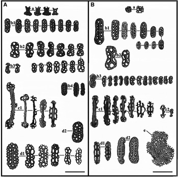
Ossicles (tables and buttons) of dorsal surface different from those of ventral surface (Figure 3). Dorsal tables more numerous with mostly a large, flat, spinose disc, sometimes appearing rectangular from lateral aspect (Figure 3A, a), diameter of disc 21.17 µm, spire of four straight to slightly curved pillars with a single, low cross-bar, ending in a crown resembling a ‘Maltese Cross’ when viewed from above, pillars not fusing apically, average height of spire 21.36 µm, teeth of crown unequal (two at end of each pillar). Ventral tables rare (Figure 3B, a).
Holothuria (Roweothuria) arguinensis from Algiers Bay. Ossicle assemblage of: (A) dorsal body wall; (B) ventral body wall and podia (light microscopy, scale bar: 50 µm). (a) Tables; (b1) rough, smooth and thorny buttons; (b2) buttons presenting shape of hourglass; (b3) pseudobuttons; (b4) buttons with obliterated holes; (c1) large, straight and curved rods; (c2) smaller rods; (d) perforated elongated plates; (e) reduced tube feet end plate.

Buttons of dorsal and ventral surfaces different, those of ventral surface more numerous, often smooth, and more densely packed (Figure 3B, b1), and with reduced holes; average length of dorsal buttons about 32 µm, average width about 16 µm, average length of ventral buttons about 34 µm, average width about 16 µm. All dorsal buttons irregular with somewhat crenulate, spiny or rugose margins (Figure 3A, b1); each perforated by an average of about eight rectangular to triangular to hexagonal holes of about 4.3 µm width. Ventral buttons with smaller holes. Some buttons characteristically constricted in the middle, often presenting the shape of the figure ‘8’ or that of an hourglass (Figure 3A, b2, B, b2).
Tube feet deposits include pseudobuttons (Figure 3B, b3) and straight rods (Figure 3B, c1), sometimes smaller (Figure 3B, c2). Straight or curved rods also exist in dorsal papillae, usually perforated at ends and sometimes also the middle (mean length 81.25 µm) (Figure 3A, c1). Perforated, elongated plates also present, accompanying buttons, in both dorsal and ventral body wall, but encountered especially in papillae (Figure 3A, d1,d2) and tube feet (Figure 3B, d1,d2). Such plates present usually an irregular contour (Figure 3AB, d1), average length 69.46 µm, average width 17.40 µm, holes average diameter 02.57 µm. Tube feet end plates reduced (Figure 3B, e).
REMARKS
In life, the species presents obvious warts/protuberances regularly arranged on both dorsal and lateral surfaces and white papillae as in Holothuria (R.) poli. This feature also corroborates with descriptions of H. (H.) stellati by Koehler (Reference Koehler and Lechevalier1921) and Gustato & Villari (Reference Gustato, Villari and Jangoux1979). Hence, the investigated specimens present similarities with both H. (H.) stellati (larger protuberances separating the bivium and the trivium) and H. (R.) poli (regularly arranged dorsal conical warts and white papillae) (Mezali, Reference Mezali2008; Mezali & Francour, Reference Mezali and Francour2012). At first glance, therefore, the current material was thought to represent H. (H.) stellati (see Mezali, Reference Mezali2008).
HABITAT AND DISTRIBUTION
Holothuria (R.) arguinensis is usually found on rocky or sandy substrate. It was formerly thought to be a species restricted to Mauritania, but sometimes extending to North Senegal and South Morocco. However, it is now also known from the Mediterranean Sea (Granada, southern Spain (Ocaña & Pérez-Ruzafa, Reference Ocaña and Pérez-Ruzafa2004) and El Mojon in the Alboran Sea (south-eastern Spain) (González-Wangüemert & Borrero-Pérez, Reference González-Wangüemert and Borrero-Pérez2012)) and now from Algiers Bay. The senior author also encountered the species in other localities on the Algerian coast (Figuier-plage (west Boumerdès) and Stidia (west Mostaganem)). The scarcity of its capture in the Mediterranean is perhaps linked to its low abundance, unless it has previously been confused with other species such as H. (H.) stellati or considered a hybrid species between H. (H.) stellati and H. (R.) poli (see Mezali, Reference Mezali2008, Reference Mezali2011).
DISCUSSION
The general body form, colour, the rough, thick, leathery integument, multiple Polian vesicles are in accordance with the descriptions of the species by Koehler & Vaney (Reference Koehler and Vaney1906) and Hérouard (Reference Hérouard1929). The live measurement of the examined specimen, however, far exceeds that recorded for the species. The abundance of button ossicles in the current specimens is in accordance with those figured by Massin (Reference Massin1993). The medially-constricted buttons which often present the shape of the figure ‘8’ or of an hourglass, are in accordance with the description of such ossicles by Hérouard (Reference Hérouard1929). Furthermore, the holes of the ventral buttons are also reduced, a character described for the species by various authors.
ACKNOWLEDGEMENTS
We thank Dr Frank W.E. Rowe, Honorary Associate, Division of Invertebrate Zoology, Australian Museum, for his valuable help and expertise in the confirmation of our identification and providing other pertinent information. The authors also thank the two anonymous referees for their helpful comments.